. 1998 Dec;180(23):6298-305.
doi: 10.1128/JB.180.23.6298-6305.1998.

Role and regulation of Bacillus subtilis glutamate dehydrogenase genes

Affiliations

Role and regulation of Bacillus subtilis glutamate dehydrogenase genes

B R Belitsky et al. J Bacteriol. 1998 Dec.

Abstract

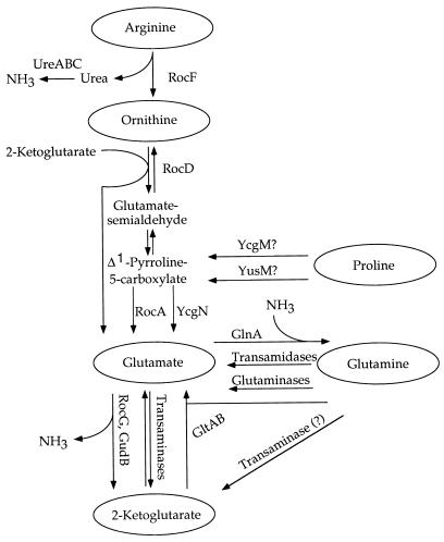
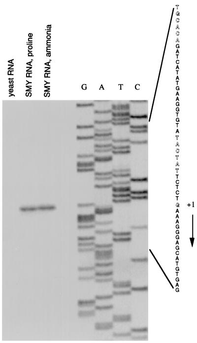
The complete Bacillus subtilis genome contains two genes with the potential to encode glutamate dehydrogenase (GlutDH) enzymes. Mutations in these genes were constructed and characterized. The rocG gene proved to encode a major GlutDH whose synthesis was induced in media containing arginine or ornithine or, to a lesser degree, proline and was repressed by glucose. A rocG null mutant was impaired in utilization of arginine, ornithine, and proline as nitrogen or carbon sources. The gudB gene was expressed under all growth conditions tested but codes for a GlutDH that seemed to be intrinsically inactive. Spontaneous mutations in gudB that removed a 9-bp direct repeat within the wild-type gudB sequence activated the GudB protein and allowed more-efficient utilization of amino acids of the glutamate family.


Figures

FIG. 1
FIG. 1
Pathways of utilization of amino acids of the glutamate family. Some enzymes are indicated by the names of their corresponding genes as follows UreABC, urease; RocF, arginase; RocD, ornithine aminotransferase; RocA and YcgN, Δ1-pyrroline-5-carboxylate dehydrogenases; YcgM and YusM, proline oxidases (proline dehydrogenases); RocG and GudB, glutamate dehydrogenases; GltAB, glutamate synthase; and GlnA, glutamine synthetase. Pathways of arginine and proline biosynthesis from glutamate are not shown.
FIG. 2
FIG. 2
Genetic map of the rocG region (19) and plasmids carrying different parts of this region. The restriction sites are abbreviated as follows: Bs, BstBI; C, ClaI; E, EcoRI; P, PstI; and X, XhoI. The ClaI and XhoI sites of the insert in pBB901 and derivative plasmids were constructed by PCR. All plasmids are derivatives of pBB544 (7). Transcription initiation sites for rocG (6) and rocABC (11) are shown by the arrows. The 1.4-kb ble cassette is not drawn to scale.
FIG. 3
FIG. 3
Genetic map of the gudB region (41) and plasmids carrying different parts of this region. The restriction sites are abbreviated as follows: Bg, BglII; Bt, BstYI; E, EcoRI; H, HindIII; K, KpnI; S, StyI; and V, EcoRV. Only relevant EcoRV, HindIII, and StyI sites are shown. The KpnI site of the insert in pBB902 and derivative plasmids was constructed by PCR. Plasmid pBB933 is a derivative of pJPM82 (7); other plasmids are derivatives of pBB544 (7). Construction of some plasmids is described in Materials and Methods; other plasmids were constructed by deleting or subcloning fragments of the gudB region. The location of the gudB transcription initiation site (see Results) is indicated by the arrow. The 1.9-kb tet cassette is not drawn to scale. x denotes the location of the gudB1 mutation within pBB928.
FIG. 4
FIG. 4
Partial alignment of the RocG, GudB, GudB1, and C. symbiosum GlutDH (45) sequences. Amino acids are numbered with respect to their positions in corresponding proteins. The amino acids comprising βc and α6 of C. symbiosum GlutDH (3) and the site of a deletion of 3 aa in GudB are underlined. Residues conserved in most GlutDHs (45) are in bold. C. symbiosum K89 and K113 are essential lysines involved in glutamate binding (42).
FIG. 5
FIG. 5
Primer extension analysis of gudB mRNA. Primer oBB60 was extended with reverse transcriptase by using total RNA from B. subtilis SMY grown in glucose minimal medium containing ammonia or proline as nitrogen source or from Saccharomyces cerevisiae (Sigma Chemical Company) as template. The sequence of the nontemplate strand of plasmid pBB917 deduced from sequencing reactions with oBB60 as primer is shown on the right. The apparent transcription start site of gudB and the −10 and the −35 regions of the likely gudB promoter are indicated by outlined letters. Primer oBB57 gave the same apparent gudB mRNA 5′ end (data not shown). The direction of transcription is shown by the arrow.

References

    1. Altschul S F, Madden T L, Schäffer A A, Zhang J, Zhang Z, Miller W, Lipman D J. Gapped BLAST and PSI-BLAST: a new generation of protein database search programs. Nucleic Acids Res. 1997;25:3389–3402. - PMC - PubMed
    1. Anagnastopoulos C, Spizizen J. Requirements for transformation in Bacillus subtilis. J Bacteriol. 1961;81:741–746. - PMC - PubMed
    1. Baker P J, Britton K L, Engel P C, Farrants G W, Lilley K S, Rice D W, Stillman T J. Subunit assembly and active site location in the structure of glutamate dehydrogenase. Proteins Struct Funct Genet. 1992;12:75–86. - PubMed
    1. Belitsky, B. R. Unpublished data.
    1. Belitsky B R, Sonenshein A L. Mutations in GltC that increase Bacillus subtilis gltA expression. J Bacteriol. 1995;177:5696–5700. - PMC - PubMed

Publication types

MeSH terms

LinkOut - more resources