. 1998 Nov 15;12(22):3528-40.
doi: 10.1101/gad.12.22.3528.

The Oct-1 POU domain activates snRNA gene transcription by contacting a region in the SNAPc largest subunit that bears sequence similarities to the Oct-1 coactivator OBF-1

Affiliations

The Oct-1 POU domain activates snRNA gene transcription by contacting a region in the SNAPc largest subunit that bears sequence similarities to the Oct-1 coactivator OBF-1

E Ford et al. Genes Dev. .

Abstract

The RNA polymerases II and III snRNA gene promoters contain an octamer sequence as part of the enhancer and a proximal sequence element (PSE) as part of the core promoter. The octamer and the PSE bind the POU domain activator Oct-1 and the basal transcription factor SNAPc, respectively. Oct-1, but not Oct-1 with a single E7R mutation within the POU domain, binds cooperatively with SNAPc and, in effect, recruits SNAPc to the PSE. Here, we show that SNAPc recruitment is mediated by an interaction between the Oct-1 POU domain and a small region of the largest subunit of SNAPc, SNAP190. This SNAP190 region is strikingly similar to a region in the B-cell-specific Oct-1 coactivator, OBF-1, that is required for interaction with octamer-bound Oct-1 POU domain. The Oct-1 POU domain-SNAP190 interaction is a direct protein-protein contact as determined by the isolation of a switched specificity SNAP190 mutant that interacts with Oct-1 POU E7R but not with wild-type Oct-1 POU. We also show that this direct protein-protein contact results in activation of transcription. Thus, we have identified an activation target of a human activator, Oct-1, within its cognate basal transcription complex.


Figures
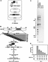
Figure 1
Figure 1
High-resolution mapping of the smallest amino-terminal SNAP190 truncation capable of interacting with Oct-1 in a one-hybrid screen in yeast. (A) Schematic diagram of the yeast one-hybrid screen used to isolate originally part of the SNAP190 cDNA (Wong et al. 1998). AT stands for Aminotriazole, a competitive inhibitor of the HIS3 gene product. (B) Schematic diagram of the yeast one-hybrid screen used to map the smallest amino-terminal SNAP190 truncation still capable of interacting with Oct-1. The primers used for the PCR amplification are indicated by small horizontal arrows. (C) DNA from yeast cells containing an integrated copy of the HIS3 reporter and the Oct-1 expression plasmid as diagrammed in A, as well as the SNAP190 deletion library diagrammed in B, was isolated, amplified in E. coli, and used as a template for PCR reactions with the primers depicted in B. The DNA template was isolated from yeast cells grown in synthetic medium lacking tryptophan and uracil, which selects for the retention of the two expression plasmids shown in A but not the expression of the HIS3 reporter gene (lane 1); yeast cells selected for one round (lane 2) or two rounds (lane 3) in synthetic medium lacking tryptophan, uracil, and histidine but containing galactose to induce expression of the SNAP190 deletion library and 10 mm AT to select for AT resistance. (Lanes 4,5) Gel purified DNA corresponding to the second to lowest (lane 4) and the lowest (lane 5) bands in lane 3 after PCR amplification. The amino-acid sequence corresponding to the DNA ladder is shown at left and was determined by: (1) Maxam–Gilbert sequencing of the PCR products in lanes 4 and 5, and (2) size of the DNA fragments. (D) Deletion constructs containing an HA tag between the VP16 activation domain and SNAP190 sequences and the SNAP190 sequences indicated were transformed into a tester yeast strain similar to that shown in A except that it contained a lacZ reporter gene bearing a TATA box driven by six reiterated octamer motifs. The resulting β-galactosidase activity was that measured in yeast strains expressing VP16-AD–SNAP190 fusion proteins and Oct-1 as indicated.
Figure 2
Figure 2
A 44 amino acid SNAP190 region is sufficient for association with the Oct-1 POU domain bound to an octamer sequence in vitro. An EMSA was performed with a DNA probe containing the IgH octamer and the proteins indicated above each lane. Equimolar amounts of each SNAP190 protein were added to each lane as determined by SDS-PAGE and autoradiography (data not shown). The positions of the free probe and the Oct-1 POU–DNA complex are indicated at left. (URL) Unprogrammed reticulocyte lysate; (GST) GST protein alone translated in vitro.
Figure 3
Figure 3
The regions of SNAP190 and OBF-1 that interact with the Oct-1 POU domain contain sequence similarity. The Oct-1 POU interacting regions of SNAP190 and OBF-1 are shown. Identical amino acids are shaded. The arrow marks amino acid 888, the amino-terminal boundary of the most extensively deleted fragment still capable of conferring growth in AT (deletion library boundary) (Fig. 1C). The smallest SNAP190 fragment (amino acids 869–912) that retained the ability to interact with the Oct-1 POU domain by EMSA (Fig. 2) is shown. The black and gray arrows indicate sequences still present in the most extensive amino-terminal (Fig. 1D) and carboxy-terminal (Fig. 2) truncations, respectively, that retained the ability to interact with the Oct-1 POU domain. The core region contains residues absolutely required for interaction with Oct-1 POU.
Figure 4
Figure 4
SNAP190 mutants K900E and K900E/E901E specifically disrupt activated but not basal transcription. (A–C). In vitro transcription assays were performed for the U6 (A; RNase T1 protection), 7SK (B; RNase T1 protection), and U2 (C; Gless cassette) templates containing an H2B octamer motif and a wild-type PSE (lanes 1–8) or a mutant octamer and a wild-type PSE (lanes 9–16). HeLa cell extracts (which contain Oct-1 but not Oct-2) were either mock depleted with protein A agarose beads (lanes 1,9) or depleted of SNAPc with anti-SNAP190–protein A–agarose beads (lanes 2–8 and 10–16; antibody 402). A total of 0.2 and 0.8 units (U6), 0.2 and 0.8 units (7SK), and 0.5 and 2 units (U2) of wild-type or mutant SNAPc was added to each reaction as indicated. Transcriptions were stopped and a nonspecific radiolabeled fragment of DNA (U6 and 7SK) or RNA (U2) was added to each reaction to normalize for nucleic acid recovery. Reactions were processed and fractionated on a denaturing polyacrylamide gel (data not shown). The internal control (I.C.) was quantitated and a second normalized gel was run. The U6, 7SK, and U2 transcription experiments were performed five, two, and four times, respectively, with similar results. (D–G). The ability of wild-type and mutant SNAPc complexes to support activated transcription correlates with their ability to be recruited to the PSE by the Oct-1 POU domain. (D) The fold enhancement of SNAPc binding in the presence of Oct-1 POU was determined by quantitating the EMSA in Fig. 5 and dividing the amount of SNAPc bound to DNA when Oct-1 POU was present in the reaction by the amount of SNAPc bound to DNA when Oct-1 POU was absent, i.e., (1) the signal in lane 3 divided by that in lane 2 (wt), (2) the signal in lane 6 divided by that in lane 5 (K900E), or (3) the signal in lane 9 divided by that lane 8 (K900E/R901E). (E–G). The fold activation of transcription was calculated by dividing the levels of transcription obtained with the H2B octamer-wild-type PSE templates by the corresponding levels obtained with the mutant octamer-wild-type PSE templates. For the U6 and U2 templates, only the fold activation obtained with the higher amount of SNAPc was calculated, because the levels of basal transcription obtained with the lower amount of SNAPc were not sufficiently above background. For the 7SK template, the fold activation obtained with both amounts of SNAPc was calculated and averaged.
Figure 5
Figure 5
SNAP190 mutants K900E and K900E/R901E form SNAP complexes that bind DNA and interact with switched or relaxed specificity with the wild-type Oct-1 POU domain and the Oct-1 POU domain containing the E7R mutation. An EMSA was performed with DNA probes containing the H2B octamer and the human U6 (hU6) PSE (lanes 1–10), a mutant octamer and the hU6 PSE (lanes 11–20), or a mutant octamer and a mutant PSE (lanes 21–30). Proteins were added as indicated above the gel. Wt, K900E, and K900E/R901E indicate the recombinant SNAPc complexes used. The positions of the free probe, single stranded probe (ss probe), and complexes containing Oct-1 POU (POU), SNAPc, and SNAPc together with Oct-1 POU (SNAPc/POU) are indicated at left.
Figure 6
Figure 6
Various examples of transcription activation by recruitment. (A) A typical activator on a mRNA promoter contains a DNA-binding domain (DBD) and an activation domain (AD). The activation domain may perform a number of functions, among them recruitment of core promoter-binding factors, such as TFIID and TFIIA, and recruitment of the holoenzyme. (B) The Oct-1 activator contains a DNA-binding domain consisting of a POU domain and an activation domain. On snRNA promoters, the Oct-1 POU domain recruits the core promoter-binding factor SNAPc through a direct protein–protein interaction with a small region of SNAP190. How the Oct-1 AD activates snRNA gene transcription is not known. (C). The λ cI repressor activates transcription from the λ PRM promoter by binding as a dimer close to the binding site for RNA polymerase and recruiting, through a direct protein–protein contact, the core promoter–recognizing subunit of the RNA polymerase, σ factor.

References

    1. Altschul SF, Madden TL, Schaffer AA, Zhang J, Zhang Z, Miller W, Lipman DJ. Gapped BLAST and PSI-BLAST: A new generation of protein database search programs. Nucleic Acids Res. 1997;25:3389–3402. - PMC - PubMed
    1. Assa-Munt N, Mortishire-Smith RJ, Aurora R, Herr W, Wright PE. The solution structure of the Oct-1 POU-specific domain reveals a striking similarity to the bacteriophage lamda repressor DNA-binding domain. Cell. 1993;73:193–205. - PubMed
    1. Aurora R, Herr W. Segments of the POU domain influence one another’s DNA-binding specificity. Mol Cell Biol. 1992;12:455–467. - PMC - PubMed
    1. Babb R, Cleary MA, Herr W. OCA-B is a functional analog of VP16 but targets a separate surface of the Oct-1 POU domain. Mol Cell Biol. 1997;17:7295–7305. - PMC - PubMed
    1. Beamer LJ, Pabo CO. Refined 1.8 A crystal structure of the lambda repressor-operator complex. J Mol Biol. 1992;227:177–196. - PubMed

Publication types

MeSH terms

LinkOut - more resources