. 1998 Nov 15;12(22):3541-50.
doi: 10.1101/gad.12.22.3541.

The molecular mechanism of mitotic inhibition of TFIIH is mediated by phosphorylation of CDK7

Affiliations

The molecular mechanism of mitotic inhibition of TFIIH is mediated by phosphorylation of CDK7

S Akoulitchev et al. Genes Dev. .

Abstract

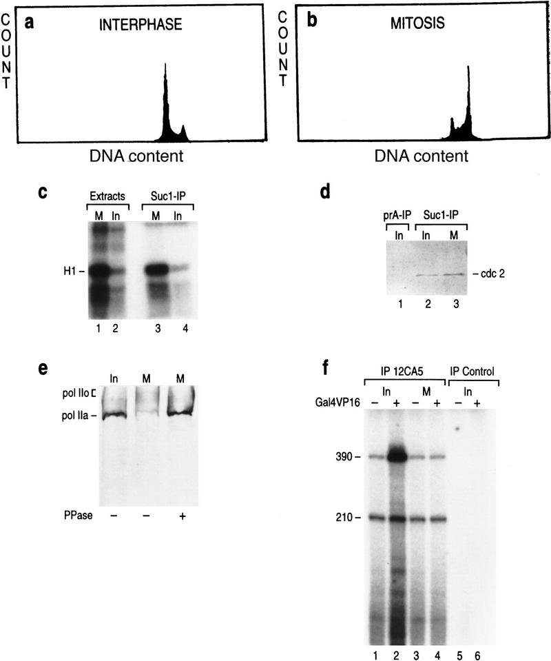
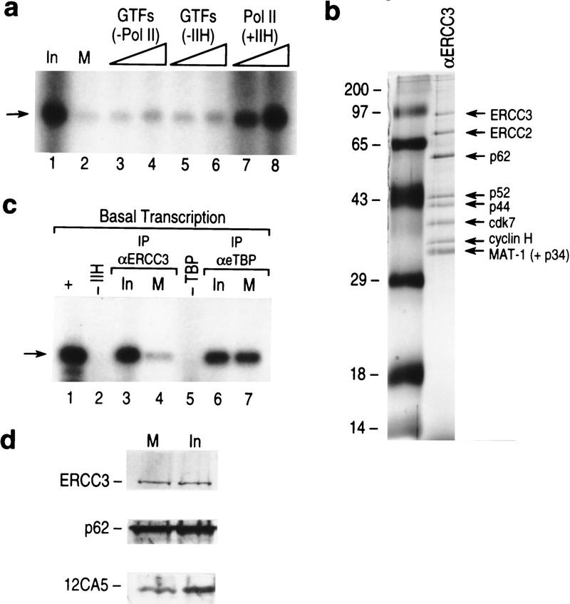
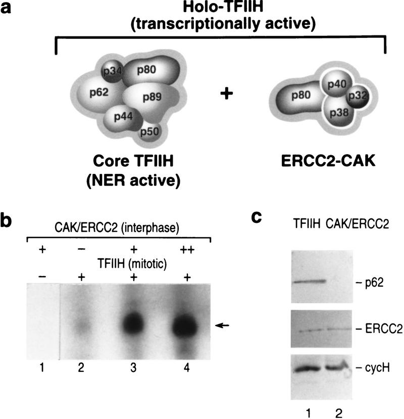
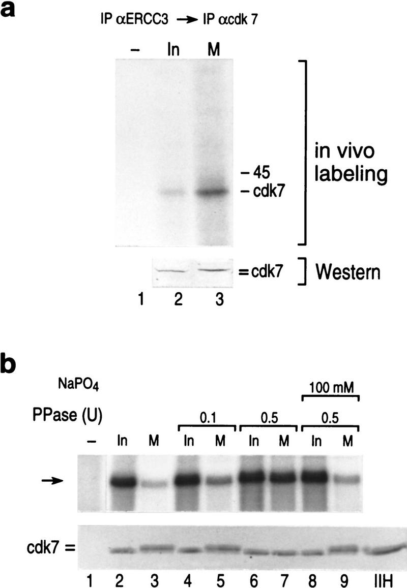
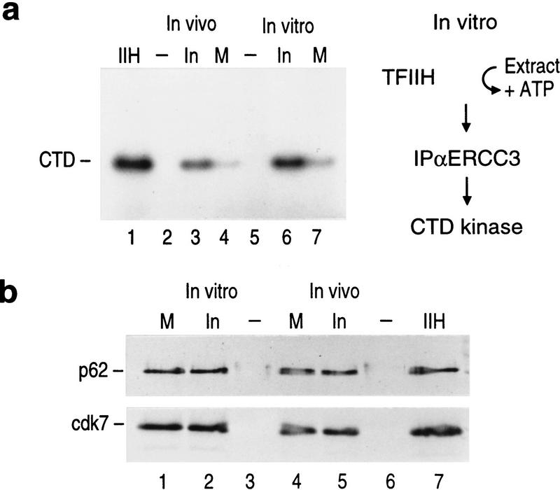
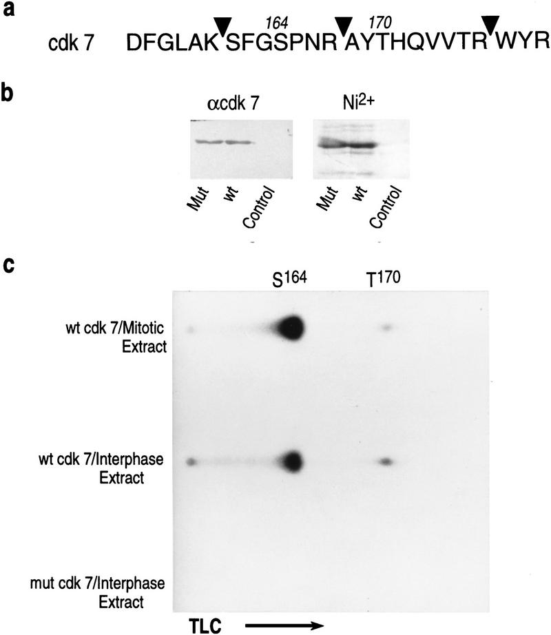
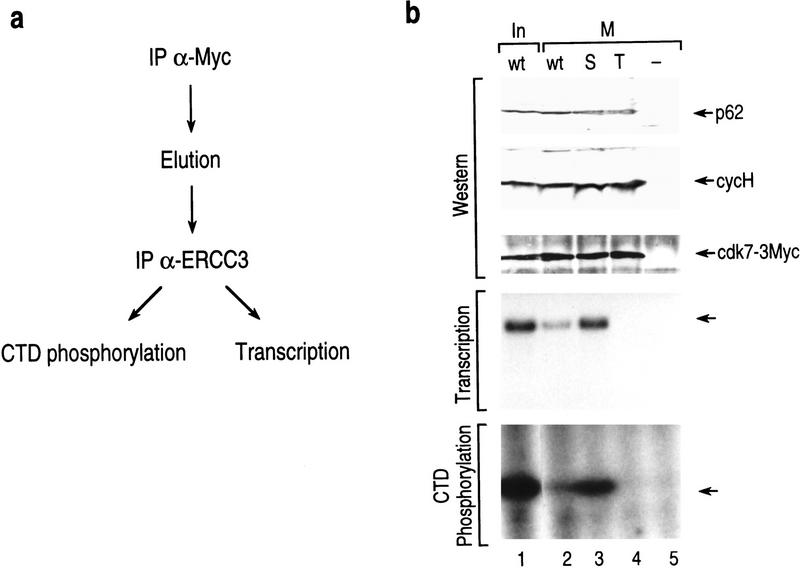
TFIIH is a multisubunit complex, containing ATPase, helicases, and kinase activities. Functionally, TFIIH has been implicated in transcription by RNA polymerase II (RNAPII) and in nucleotide excision repair. A member of the cyclin-dependent kinase family, CDK7, is the kinase subunit of TFIIH. Genetically, CDK7 homologues have been implicated in transcription in Saccharomyces cerevisiae, and in mitotic regulation in Schizosaccharomyces pombe. Here we show that in mitosis the CDK7 subunit of TFIIH and the largest subunit of RNAPII become hyperphosphorylated. MPF-induced phosphorylation of CDK7 results in inhibition of the TFIIH-associated kinase and transcription activities. Negative and positive regulation of TFIIH requires phosphorylation within the T-loop of CDK7. Our data establishes TFIIH and its subunit CDK7 as a direct link between the regulation of transcription and the cell cycle.


Figures

Figure 1
Figure 1
FACS analysis of the asynchronous (interphase) (a) and nocodazole-treated (mitotic) (b) HeLa cells: Distribution of cells in the G1, S, and G2/M phases of the cell cycle (Y axis) on the basis of the cellular DNA content (X axis) is plotted on the graph. (c) Histone H1 kinase assay with extracts derived from the mitotic and interphase HeLa cells (lanes 1,2) or with Suc1(p13)-affinity purified cdc2 kinase from mitotic and interphase extracts (lanes 3,4). (d) Western blot analysis of the Suc1(p13)-affinity purified cdc2 kinase (lanes 2,3) and affinity control (lane 1). (e) Western blot analysis of the CTD of RNAPII by use of 8WG16 antibodies (Parsons and Spencer 1997) in the interphase (lane 1) and mitotic extracts (lanes 2,3). (Lane 3) Extracts were pretreated with 10 units of alkaline phosphatase. (f) Reconstituted basal (210 nucleotides) and activated (390 nucleotides) transcription with eTFIID immunopurified from interphase (lanes 1,2) and mitotic (lanes 3,4) LTR HeLa cells (Zhou et al. 1992). Reactions contained the activator Gal4–VP16 and the coactivators PC4 and TFIIA (lanes 2,4; Ma et al. 1996). The templates used were pG5MLP and pMLP that contain G-less cassettes of different sizes (390 and 210 nucleotides). Transcription was directed by the AdML promoter. The 390-nucleotide transcript was derived from pG5MLP, which contains five Gal4-binding sites upstream of the TATA box. pMLP is devoid of Gal4-binding sites.
Figure 2
Figure 2
(a) Transcriptional activity of mitotic extracts (lane 2) as compared with the interphase extracts (lane 1) and complemented with different GTFs and RNAPII (lanes 38). Titration included all GTFs without RNAPII (lanes 3,4 ), GTFs and RNAPII without TFIIH (lanes 5,6), TFIIH and RNAPII (lanes 7,8). (Arrow) The specific transcript derived from the pMLP template. (b) Silver-staining of immunoaffinity purified TFIIH (LeRoy et al. 1998) (lane 2). (Lane 1) Molecular weight markers. (c) Reconstituted basal transcription (lane 1), dependent on TFIIH (lane 2) and TBP (lane 5). Reactions were reconstituted with immunoaffinity purified TFIIH or TFIID from interphase (lanes 3,6) or mitotic (lanes 4,7) extracts. (d) Western blot of the ERCC3 and p62 subunits of immunopurified TFIIH and of eTBP (Zhou et al. 1992), used in the reconstituted transcription in c.
Figure 3
Figure 3
(a) Schematic representation of the composition of core TFIIH, the ERCC2–CAK complex, and holo-TFIIH. (b) Basal transcription by the AdML promoter was reconstituted with affinity purified mitotic TFIIH (lanes 24), CAK/ERCC2 affinity purified from interphase extract (lane 1), or with increasing amounts of the CAK/ERCC2 complex in the presence of mitotic TFIIH (lanes 3,4). (Arrow) The specific transcript derived from the pMLP template. (c) Western blot analysis of the immunoaffinity preparation of mitotic TFIIH and interphase CAK/ERCC2 complexes.
Figure 4
Figure 4
(a) In vivo-labeled CDK7 immunoprecipitated from affinity purified TFIIH and analyzed by Western blot (bottom) after autoradiography (top). (=) The resolved shift in the mobility of mitotic CDK7. (b). (Top) Basal transcription from the AdML promoter was reconstituted with affinity purified interphase TFIIH (lane 2), mitotic TFIIH (lane 3), mitotic or interphase TFIIH pretreated with alkaline phosphatase in the absence or presence of sodium phosphate as indicated. (Lane 1) The result using a mock affinity purification procedure. (Bottom) Western blot analysis of the samples analyzed in transcription at top using anti-CDK7 antibodies.
Figure 5
Figure 5
Mitotic TFIIH is deficient in CTD kinase activity. (a) CTD kinase activity of TFIIH directly purified from mitotic and interphase cells (in vivo, lanes 3,4). The kinase activity of conventionally purified TFIIH is shown in lane 1. Purified TFIIH was pretreated with mitotic or interphase extracts in the presence of ATP (in vitro, lanes 6–7, see diagram at right). (b) Western blot analysis of TFIIH subunits (p62 and CDK7) from samples analyzed in a. This blot shows that similar amounts of TFIIH were used in the assay performed in a. The electrophoretic mobility of CDK7 caused by phosphorylation in mitosis is not resolved in this analysis. The samples were separated on a Bio-Rad mini-gel.
Figure 6
Figure 6
(a) Amino acid sequence of a fragment of CDK7 spanning the sites of phosphorylation in vivo (Ser-164 and Thr-170). (Arrowheads) Positions of cleavage by trypsin. (b) Purified recombinant wild-type and mutant [S164D, T170D] CDK7 containing a carboxy-terminal-His-tag as detected by Western blot using antibodies against CDK7 (α-CDK7) and by Coomassie-blue staining (Ni2+). (c) Recombinant wild-type and mutant CDK7 were incubated with interphase or mitotic extracts in the presence of [γ-32]ATP. Labeled proteins were affinity purified, digested with trypsin, and resolved by one-dimension thin layer chromatography. Positions of Ser-164 and Thr-170 containing fragments are indicated.
Figure 7
Figure 7
(a) Schematic representation of the procedure used to isolate TFIIH from transfected cells containing mutated subunits of CDK7. TFIIH containing c-Myc-tagged CDK7 was isolated from interphase and mitotic cells. After cell lysis, the transfected CDK7 subunit was recovered by immunoprecipitation by use of antibodies recognizing the c-myc tag. The IPs were eluted and the transfected myc-tagged CDK7 subunits were selected for those subunits that were incorporated into TFIIH by performing a second immunoprecipitation with anti-ERCC3 monoclonal antibodies. The IPs were functionally analyzed in transcription and CTD phosphorylation. (b) TFIIH isolated from mitotic cells and containing a mutation in Ser-164 [Ser164A (S)] or a mutation in Thr-170 [Thr170A (T)] or a control vector were assayed for transcription and kinase activities. The amount of TFIIH recovered and used in the functional analysis was analyzed by Western blot by use of antibodies against the p62, cyclin H, and CDK7-[3xMyc] subunits (top). The samples were separated on a Bio-Rad minigel; therefore, the change in electrophoretic mobility due to phosphorylation of CDK7-[3xMyc] could not be resolved. Functionally, the mutant proteins were assayed in basal transcription (middle), and for CTD-kinase (bottom) activities. Minus (−) above the lane denotes the activity present in cells that were transfected with the Myc tag but devoid of the CDK7 coding sequences (empty vector).

References

    1. Adamczewski JP, Rossignol M, Tassan J-P, Nigg EA, Moncollin V, Egly JM. MAT1, cdk7 and cyclin H form a kinase complex which is UV light-sensitive upon association with TFIIH. EMBO J. 1996;15:1877–1844. - PMC - PubMed
    1. Akoulitchev S, Mäkelä TP, Weinberg RA, Reinberg D. Requirement for TFIIH kinase activity in transcription by RNA polymerase II. Nature. 1995;377:557–560. - PubMed
    1. Bellier S, Dubois MF, Nishida E, Almouzni G, Bensaude O. Phosphorylation of the RNA polymerase II largest subunit during Xenopus laevis oocyte maturation. Mol Cell Biol. 1997;17:1434–1440. - PMC - PubMed
    1. Buck V, Russel P, Millar JBA. Identification of a cdk-activating kinase in fission yeast. EMBO J. 1995;14:6173–6183. - PMC - PubMed
    1. Cisek LJ, Corden JL. Phosphorylation of RNA polymerase by the murine homologue of the cell-cycle control protein cdc2. Nature. 1989;339:679–684. - PubMed

Publication types

MeSH terms

Substances