. 1998 Nov 15;12(22):3551-63.
doi: 10.1101/gad.12.22.3551.

Synaptonemal complex morphogenesis and sister-chromatid cohesion require Mek1-dependent phosphorylation of a meiotic chromosomal protein

Affiliations

Synaptonemal complex morphogenesis and sister-chromatid cohesion require Mek1-dependent phosphorylation of a meiotic chromosomal protein

J M Bailis et al. Genes Dev. .

Abstract

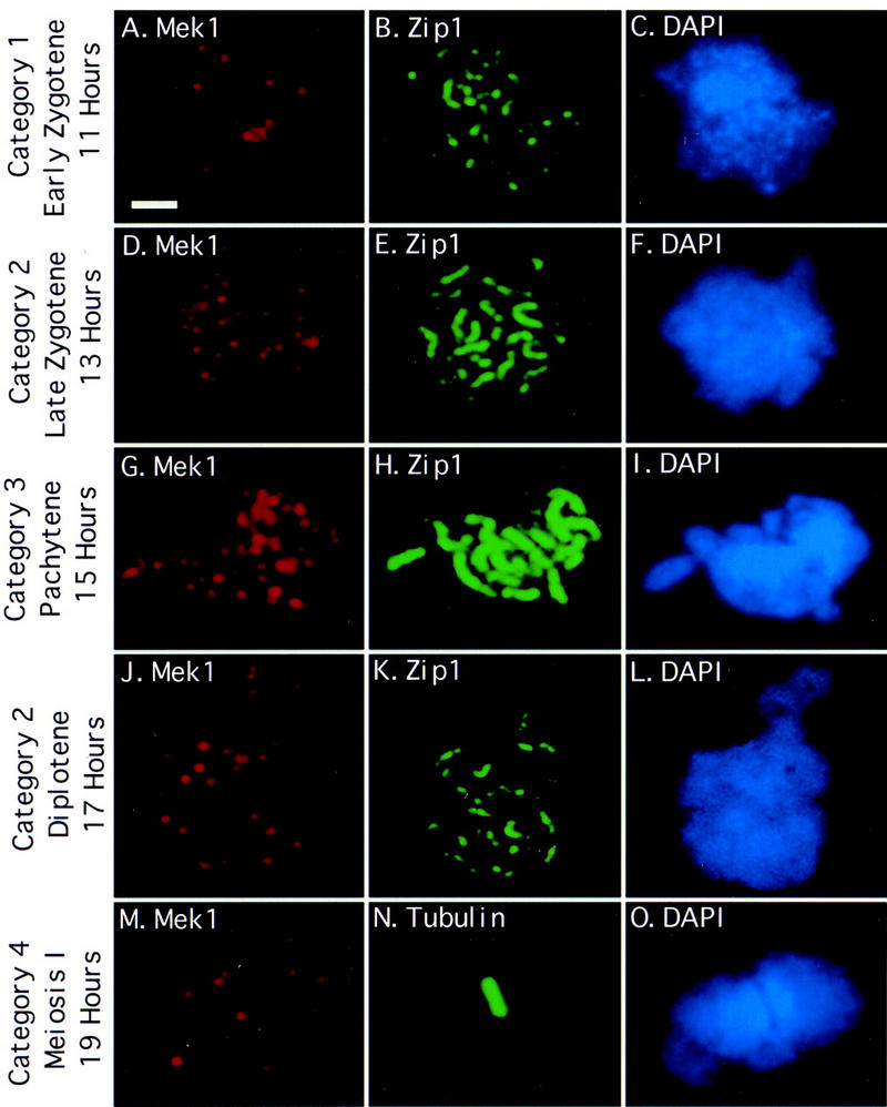
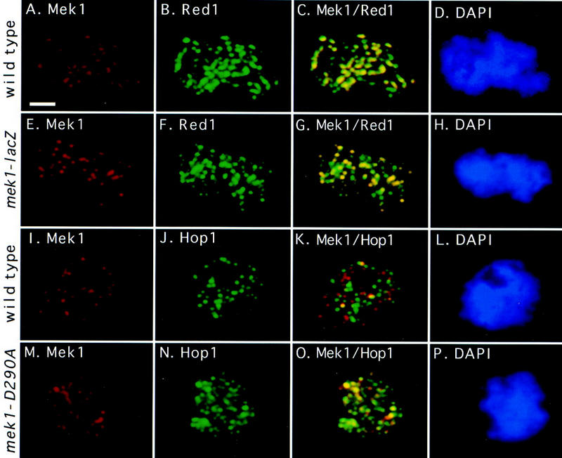
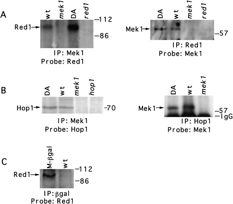
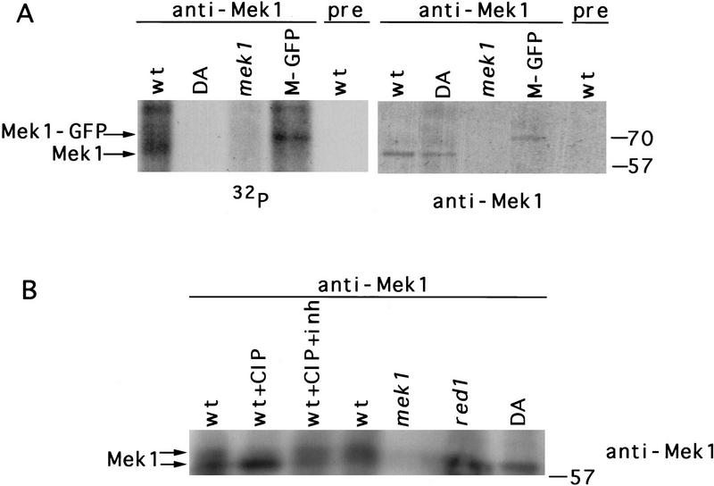
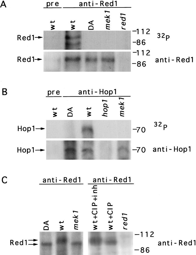
Development of yeast meiotic chromosome cores into full-length synaptonemal complexes requires the MEK1 gene product, a meiosis-specific protein kinase homolog. The Mek1 protein associates with meiotic chromosomes and colocalizes with the Red1 protein, which is a component of meiotic chromosome cores. Mek1 and Red1 interact physically in meiotic cells, as demonstrated by coimmunoprecipitation and the two-hybrid protein system. Hop1, another protein associated with meiotic chromosome cores, also interacts with Mek1 but only in the presence of Red1. Red1 displays Mek1-dependent phosphorylation, both in vitro and in vivo, and Mek1 kinase activity is necessary for Mek1 function in vivo. Fluorescent in situ hybridization analysis indicates that Mek1-mediated phosphorylation of Red1 is required for meiotic sister-chromatid cohesion, raising the possibility that cohesion is regulated by protein phosphorylation.


Figures

Figure 1
Figure 1
Mek1 localizes to chromosomes during and after synapsis. Spread meiotic chromosomes from wild type (BR2495) were stained with antibodies to Mek1 (A,D,G,J,M), antibodies to Zip1 (B,E,H,K) or tubulin (N), and DAPI (C,F,I,L,O). Categories of Mek1 staining are indicated at left (see text for explanation). Scale bar, 2 μm.
Figure 2
Figure 2
Time course analysis of Mek1 and Zip1 localization. Spread nuclei from wild type (BR2495) were prepared hourly during meiosis and double stained with antibodies to Mek1 and Zip1. The frequencies of the four categories of Mek1 localization are indicated; at least 100 nuclei were examined per time point. (Category 1) Early zygotene, punctate Zip1 staining; (category 2) Zip1 dots and linear stretches; first peak, late zygotene; second peak, diplotene; (category 3) pachytene, continuous Zip1 staining; (category 4) meiosis I (MI). The average number of Mek1 foci per nucleus in categories 1–4 is 24, 30, 40 and 17, respectively.
Figure 3
Figure 3
Mek1 localization pattern compared to Red1 and Hop1. A wild-type spread nucleus (BR2495) was stained with antibodies to Mek1 (A) and Red1 (B) and with DAPI (D); (C) fusion of A and B. A spread nucleus from the mek1–lacZ mutant (JM198) was stained with antibodies to β-galactosidase (E) and Red1 (F) and with DAPI (H); (G) fusion of E and F. A wild-type spread nucleus (BR2495) was stained with antibodies to Mek1 (I) and Hop1 (J) and with DAPI (L); (K) fusion of I and J. A spread nucleus from the mek1–D290A mutant (JM191) was stained with antibodies to Mek1 (M) and Hop1 (N) and with DAPI (P); (O) fusion of M and N. All nuclei are from a 13-hr time point. Areas of yellow in the merged images indicate overlap. Scale bar, 2 μm.
Figure 4
Figure 4
Association of Mek1 with Red1 and Hop1 in meiotic cells. (A) Anti-Mek1 antibodies coprecipitate Red1, and anti-Red1 antibodies coprecipitate Mek1. (B) Coimmunoprecipitation of Mek1 and Hop1. (C) Red1 coprecipitates with the Mek1–β-gal protein, which contains just the amino-terminal region of Mek1. Proteins were immunoprecipitated from 1 liter of cells that had been sporulated for 13 hr and then analyzed by SDS-PAGE and immunoblotting. Antibodies used for immunoprecipitation (IP) and for immunoblotting (Probe) are indicated on the bottom of each blot. When antibodies to Hop1 are used to coimmunoprecipitate Mek1, as in B, Mek1 is not detected in the hop1 mutant (data not shown). Molecular mass markers are shown at the right of each blot. (IgG) Immunoglobulin G. Strains used are wild type (BR2495), mek1–D290A (JM191), mek1::LYS2 (JM82), red1::URA3 (MY231), hop1::TRP1 (BR2498), and mek1–lacZ (JM198). (wt) Wild type; (DA) mek1–D290A; (M-βgal) Mek1–β-galactosidase.
Figure 5
Figure 5
Mek1 is a phosphoprotein that displays kinase activity. (A) Mek1 kinase activity in vitro. Anti-Mek1 immunoprecipitates were incubated with [γ-32P]ATP, separated by an 8% SDS–polyacrylamide gel, and analyzed by autoradiography and immunoblotting with anti-Mek1 antibodies. (B) Gel mobility shift analysis of Mek1. Anti-Mek1 immunoprecipitates were untreated, treated with CIP, or treated with CIP plus phosphatase inhibitor (CIP + inh). Conditions of electrophoresis (20 V for 16 hr) were slower than those used in A. The Mek1 protein was detected by immunoblotting. Molecular mass markers (in kD) are at right. Strains used are wild type (BR2495), mek1–D290A (JM191), mek1:: LYS2 (JM82), MEK1::GFP (JM98), and red1 (MY231). (wt) Wild type; (DA) mek1–D290A; (M-GFP) Mek1–GFP; (pre) preimmune serum.
Figure 6
Figure 6
Mek1-dependent phosphorylation of Red1. (A) In vitro phosphorylation of Red1 protein immunoprecipitated with anti-Red1 antibodies. (B) In vitro phosphorylation of Hop1 immunoprecipitated with antibodies to Hop1. Immunoprecipitates were incubated with [γ-32P]ATP, then analyzed by SDS-PAGE, autoradiography, and immunoblotting with antibodies to either Red1 (A) or Hop1 (B). Additional phosphorylated bands may represent other proteins in the immune complex. (C) Gel mobility shift analysis of Red1. Anti-Red1 immunoprecipitates were untreated, treated with CIP, or treated with CIP plus inhibitor (CIP + inh). Immunoprecipitates were analyzed by slower conditions of electrophoresis (20 V for 16 hr) than used in A. The Red1 protein was detected by immunoblotting. Molecular mass markers (in kD) are at right. Strains used are wild type (BR2495), mek1–D290A (JM191), mek1::LYS2 (JM82), and red1::URA3 (MY231). (wt) Wild type; (DA) mek1–D290A; (pre) preimmune serum.

References

    1. Bai C, Elledge SJ. Gene identification using the yeast two-hybrid system. Methods Enzymol. 1997;283:141–156. - PubMed
    1. Bishop D, Park D, Xu L, Kleckner N. DMC1: A meiosis-specific yeast homolog of E. coli recA required for recombination, synaptonemal complex formation, and cell cycle progression. Cell. 1992;69:439–456. - PubMed
    1. Brunelli JP, Pall ML. A series of yeast shuttle vectors for the expression of cDNAs and other DNA sequences. Yeast. 1993;9:1299–1308. - PubMed
    1. Burns N, Grimwade B, Ross-Macdonald PB, Choi E-Y, Finberg K, Roeder GS, Snyder M. Large-scale analysis of gene expression, protein localization and gene disruption in Saccharomyces cerevisiae. Genes & Dev. 1994;8:1087–1105. - PubMed
    1. Chua PR, Roeder GS. Zip2, a meiosis-specific protein required for the initiation of chromosome synapsis. Cell. 1998;93:349–359. - PubMed

Publication types

MeSH terms