. 1998 Nov 15;12(22):3591-602.
doi: 10.1101/gad.12.22.3591.

Dorsoventral patterning in the Drosophila central nervous system: the intermediate neuroblasts defective homeobox gene specifies intermediate column identity

Affiliations

Dorsoventral patterning in the Drosophila central nervous system: the intermediate neuroblasts defective homeobox gene specifies intermediate column identity

J B Weiss et al. Genes Dev. .

Abstract

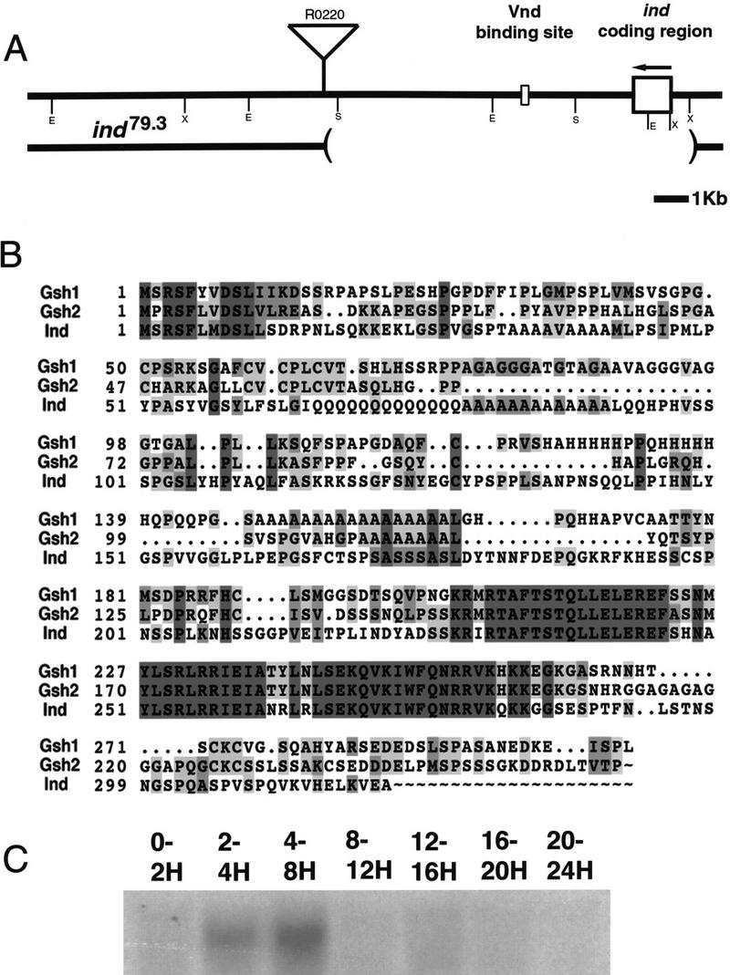
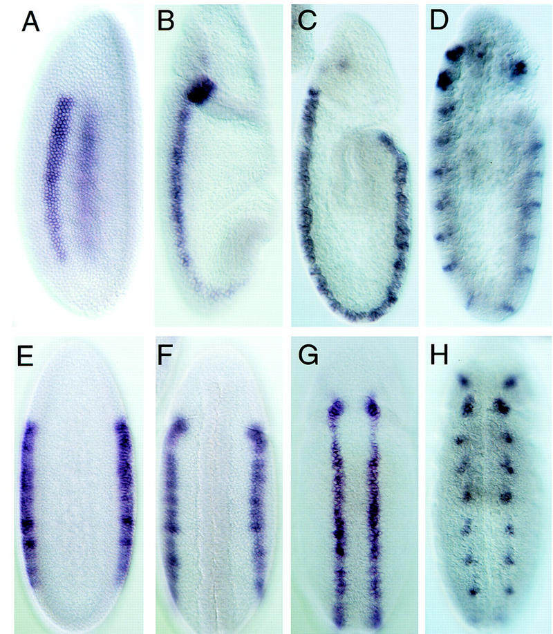
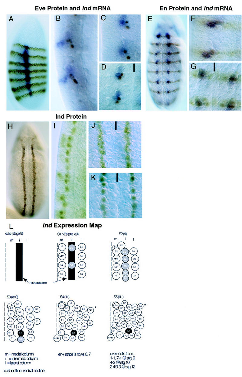
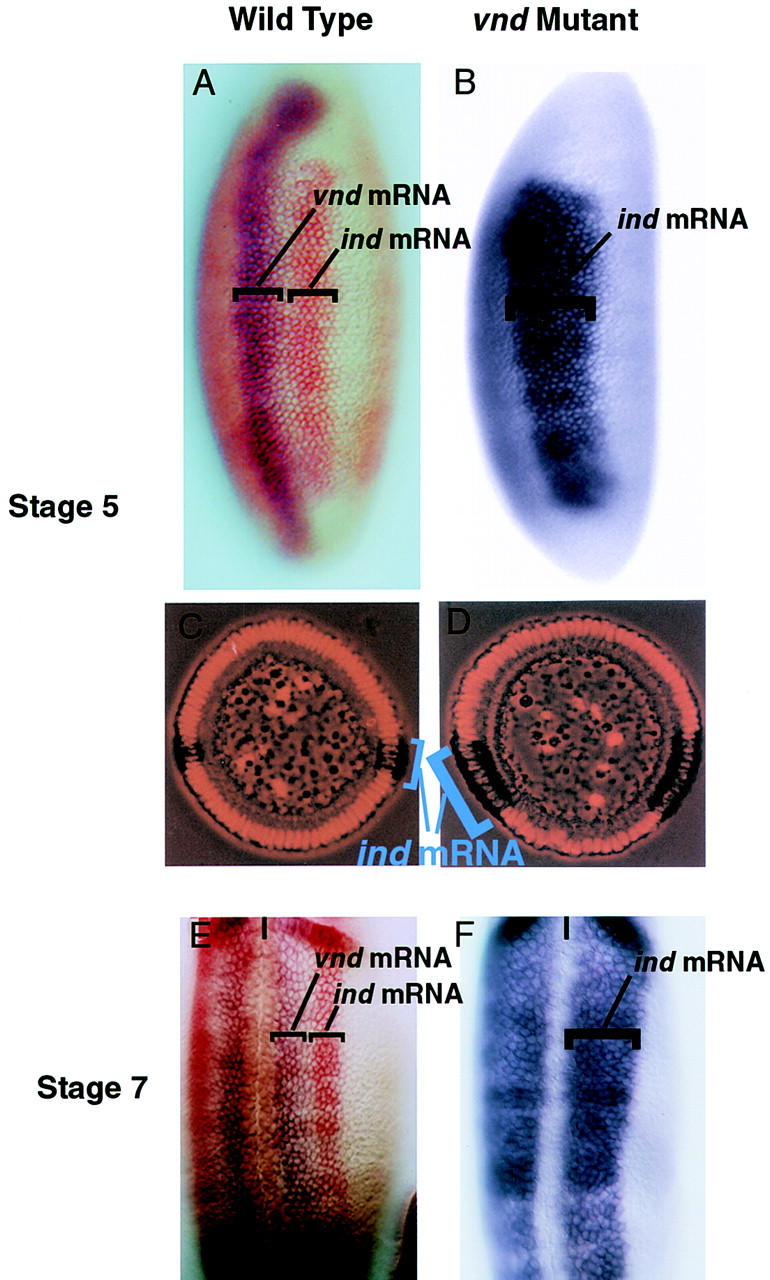
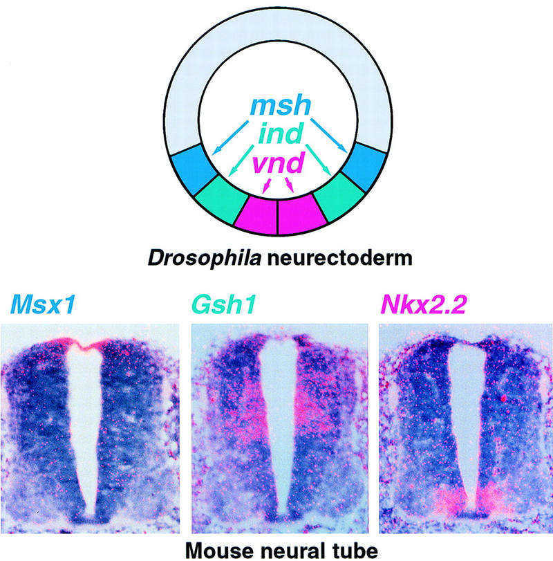
One of the first steps in neurogenesis is the diversification of cells along the dorsoventral axis. In Drosophila the central nervous system develops from three longitudinal columns of cells: ventral cells that express the vnd/nk2 homeobox gene, intermediate cells, and dorsal cells that express the msh homeobox gene. Here we describe a new Drosophila homeobox gene, intermediate neuroblasts defective (ind), which is expressed specifically in the intermediate column cells. ind is essential for intermediate column development: Null mutants have a transformation of intermediate to dorsal column neuroectoderm fate, and only 10% of the intermediate column neuroblasts develop. The establishment of dorsoventral column identity involves negative regulation: Vnd represses ind in the ventral column, whereas ind represses msh in the intermediate column. Vertebrate genes closely related to vnd (Nkx2.1 and Nkx2.2), ind (Gsh1 and Gsh2), and msh (Msx1 and Msx3) are expressed in corresponding ventral, intermediate, and dorsal domains during vertebrate neurogenesis, raising the possibility that dorsoventral patterning within the central nervous system is evolutionarily conserved.


Figures

Figure 1
Figure 1
The structure of the ind genomic region, amino acid sequence of Ind, and developmental expression of ind. (A) The structure of the ind gene in wild-type and a mutant allele, ind79.3. (Top) The structure of the wild-type allele. The coding sequence for ind is contained in a single exon, shown as a large box. ind is transcribed in the direction indicated by the arrow. The binding site for Vnd, marked by a smaller box, lies 3 kb 3′ to the coding sequence. The insertion site of a P element, R0220, in relation to the gene is shown. Restriction endonuclease sites are marked with single letters: (E) EcoRI, (S) SalI, (X) XhoI. (Bottom) The extent of a deletion that was generated by mobilization of the R0220 P element. The deletion is 7 kb long and removes the coding sequence for ind. (B) The predicted amino acid sequence of Ind aligned with two closely related mouse genes, Gsh1 and Gsh2. The alignment shows conservation primarily within the homeodomain (amino acids 227–286 in Ind) though there is also a short region of homology at the amino terminus. Identities are marked in dark gray, similarities in light gray. Numbers at the left indicate the amino acid positions in the respective proteins. (C) An RNA blot of staged embryos hybridized with a radiolabeled ind cDNA clone. The times in hours postfertilization of the embryo collections are marked above the lanes. All lanes were loaded with 20 μg of total RNA.
Figure 2
Figure 2
ind expression in wild-type embryos. Shown are lateral and ventral views of whole-mount in situ hybridizations with the ind cDNA to a developmental series of wild-type embryos. The embryos on the top (A–D) are shown in lateral views with anterior up and ventral to the left. The embryos on the bottom (E–H) are ventral views again with anterior up. The embryos are approximately stage matched and represent a developmental series starting at the left. (A,E) Stage 5, cellular blastoderm, embryos in which ind is expressed in two longitudinal stripes five cells wide in the intermediate neurectoderm. (B,F) Stage 7 embryos, following ventral furrow formation and ventral migration of the neurectoderm. ind expression in these embryos is narrowed to longitudinal stripes three cells wide. (C,G) Early stage 9 embryos in which ind is expressed in the intermediate neurectoderm and S1, intermediate column neuroblasts. (D,H) Late stage 9 to early stage 10 embryos. At this stage ind mRNA is progressively restricted to neuroblasts 6-2 and 7-2.
Figure 3
Figure 3
ind expression monitored with antibodies. (A–D) DV localization of ind-expressing Nbs. Anti-Eve antibody staining is brown, hybridization to ind mRNA is blue. Anterior is up, ventral to the left. (A) Lateral view of a stage 5, cellular blastoderm embryo showing ind expression in two parallel bands in intermediate neurectoderm, orthogonal to Eve. (B) Higher magnification, lateral view of late stage 9 embryos showing delamination of the Eve positive medial Nbs 1-1 and 7-1 prior to delamination of the ind-expressing intermediate Nbs 6-2 and 7-2. (C) Lateral view of midstage 10 embryo showing delamination of the ind expressing Nbs 6-2 and 7-2. (D) Ventral view of midstage 10 embryo showing medial Eve positive Nbs 1-1 and 7-1 adjacent to intermediate ind-expressing Nbs 6-2 and 7-2. The ventral midline is marked with a black line. (E–G) AP localization of ind-expressing Nbs. Anti-Engrailed (En) antibody staining is in brown, hybridization to ind mRNA is in blue. All embryos are at stage 10. (E) Whole embryo showing expression of ind in register with En in the AP axis. (F,G) Lateral and ventral views, respectively, of delaminated ind expressing Nbs showing the identity of En- and Ind-positive intermediate Nbs. The ventral midline is marked with a black line. (H–K) Ind protein expression in the neurectoderm and intermediate Nbs. (H) Ventral view of stage 8 embryo showing two parallel stripes of neurectodermal Ind protein. (I) Higher magnification, lateral view of late stage 9 embryo showing persistent accumulation of Ind protein in all delaminated intermediate column NBs. (J) Higher magnification, ventral view of stage 8 embryo showing intermediate neurectoderm presence of Ind. (K) Higher magnification ventral view of stage 9 embryo showing Ind in all intermediate-column Nbs. The ventral midline is marked with a black line. (L) ind expression map diagram of neuroblast formation in one hemisegment stages 8–11. Ventral view, anterior is up, the midline is at the left of each panel. ind expression is shown in gray and black. Neuroblasts are designated by their final row number–column number.
Figure 4
Figure 4
ind and vnd expression in wild-type embryos. Ventral derepression of ind in vnd mutant embryos. (A–D) Lateral (A,B) and cross sectional (C,D) views of wild-type (A,C) and vnd mutant (B,D) stage 5 embryos. Anterior is up, ventral to the left in A and B. In C and D ventral is down. (A) ind (red) is dorsal and adjacent to vnd (blue) in wild-type embryos. (B) In vnd mutant embryos ind (blue) expression is broadened ventrally to encompass the vnd expression domain. (C,D) Cross sectional views of wild-type (C) and vnd mutant (D) stage 5 embryos stained with ind (blue-black) and propidium iodide to mark nuclei. The ventral expansion of ind in vnd mutants is seen clearly in these cellular blastoderm embryos. (E,F) Ventral views of wild-type and vnd mutant stage 7 embryos. Anterior is up, the ventral midline is marked with a black line. (E) As above ind (red) is dorsal to vnd (blue). (F) In the vnd mutant ind (blue) expands ventrally to encompass the neurectoderm and the vnd expression domain.
Figure 5
Figure 5
Binding of the Vnd protein to ind sequences. (A) DNase I footprints of both strands of the ind DNA. Three major protected regions become visible as the amount of recombinant Vnd homeodomain is increased. (B) Electrophoresis mobility-shift assay using ind DNA nucleotides 5–135, which includes all three Vnd-binding regions. A DNA/protein complex is observed upon addition of Vnd. This complex can be competed out with increasing amounts of the Vnd-I oligonucleotide but not with an irrelevant oligonucleotide (H2A). (C) Sequence showing the Vnd-binding regions as determined by DNase I footprinting. The protected regions are shaded in light gray, whereas a candidate consensus sequence is shaded in dark gray. The Vnd-I oligonucleotide sequence is underlined.
Figure 6
Figure 6
Loss of ind function results in loss of intermediate-column neuroblasts and intermediate-column-derived neurons. All views are ventral except A and B which are lateral. Anterior is up, ventral to the left in A and B. The ventral midline is marked with a black line. (A–F) Embryos doubly labeled for msh protein (blue) and Vnd protein (brown). (A,C,E) Wild-type embryos, blank space delineates the domain of Ind expression. (B,D,F). ind16.2 mutant embryos show Msh is expanded in the neurectoderm at stage 7 (B). This persists until stage 9 when the neuroblasts delaminate (D,F). (G–J) Achaete (Ac) is repressed in the neurectoderm and neuroblasts by Ind. In wild-type embryos Ac is expressed in four clusters per hemisegment in the neurectoderm (G) and in four neuroblasts per hemisegment (I). In ind mutant embryos Ac expression is derepressed in the intermediate column (H). When a neuroblast forms at these positions it expresses Ac inappropriately (J). (K,L) Hunchback, En double labels showing loss of intermediate-column neuroblasts in ind mutant embryos. Pan neural expression of Hunchback in wild-type late stage 9 embryos shows there are three columns of neuroblasts (K). In ind16.2 embryos only two columns of S1 neuroblasts are observed. Ind is therefore required for formation of intermediate-column neuroblasts (L). (M,N) Ind is required for neurons derived from intermediate-column neuroblasts. Even-skipped staining is shown in brown. The embryos are stage 13 and two types of Even-skipped positive cells are seen per hemisegment in this focal plane. The larger clusters are neurons derived from medial column neuroblasts. They are unaffected in ind mutants (N). The smaller clusters, highlighted by arrows, are RP2/RP2sib neurons that are derived from intermediate-column neuroblasts. These neurons are absent in the ind mutants (N).
Figure 7
Figure 7
DV domains of expression of Drosophila and related mouse homeobox genes in the developing CNS. On the left a schematic cross section of a stage 7 Drosophila embryo showing the expression of msh (blue), ind (green), and vnd (red) in symmetric columns of neurectoderm. On the right, three panels show in situ hybridization with three related mouse genes performed on serial cross sections of the neural tube at the midthoracic level of an 11.5 day mouse embryo. Dorsal is up, ventral is down.

Similar articles

Cited by

References

    1. Adams B, Dorfler P, Aguzzi A, Kozmik Z, Urbanek P, Maurer-Fogy I, Busslinger M. Pax-5 encodes the transcription factor BSAP and is expressed in B lymphocytes, the developing CNS, and adult testis. Genes & Dev. 1992;6:1589–1607. - PubMed
    1. Altschul SF, Madden TL, Schaffer AA, Zhang J, Zhang Z, Miller W, Lipman DJ. Gapped BLAST and PSI-BLAST: A new generation of protein database search programs. Nucleic Acids Res. 1997;25:3389–3402. - PMC - PubMed
    1. Angerer L, Angerer R. In situ hybridization to cellular RNA with radiolabelled RNA probes. In: Wilkinson DG, editor. In situ hybridization: A practical approach. Oxford, UK: Oxford University Press; 1992. pp. 15–32.
    1. Ausubel FM, Brent R, Kingston RE, Moore DD, Seidman JG, Smith JA, Struhl K. Current protocols in molecular biology. New York, NY: John Wiley & Sons; 1994.
    1. Azpiazu N, Frasch M. tinman and bagpipe: Two homeobox genes that determine cell fates in the dorsal mesoderm of Drosophila. Genes & Dev. 1993;7:1325–1340. - PubMed

Publication types

MeSH terms

Associated data