. 1998 Nov 15;12(22):3603-12.
doi: 10.1101/gad.12.22.3603.

Dorsoventral patterning in the Drosophila central nervous system: the vnd homeobox gene specifies ventral column identity

Affiliations

Dorsoventral patterning in the Drosophila central nervous system: the vnd homeobox gene specifies ventral column identity

J A McDonald et al. Genes Dev. .

Abstract

The Drosophila CNS develops from three columns of neuroectodermal cells along the dorsoventral (DV) axis: ventral, intermediate, and dorsal. In this and the accompanying paper, we investigate the role of two homeobox genes, vnd and ind, in establishing ventral and intermediate cell fates within the Drosophila CNS. During early neurogenesis, Vnd protein is restricted to ventral column neuroectoderm and neuroblasts; later it is detected in a complex pattern of neurons. We use molecular markers that distinguish ventral, intermediate, and dorsal column neuroectoderm and neuroblasts, and a cell lineage marker for selected neuroblasts, to show that loss of vnd transforms ventral into intermediate column identity and that specific ventral neuroblasts fail to form. Conversely, ectopic vnd produces an intermediate to ventral column transformation. Thus, vnd is necessary and sufficient to induce ventral fates and repress intermediate fates within the Drosophila CNS. Vertebrate homologs of vnd (Nkx2.1 and 2.2) are similarly expressed in the ventral CNS, raising the possibility that DV patterning within the CNS is evolutionarily conserved.


Figures
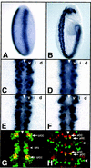
Figure 1
Figure 1
Vnd protein pattern in wild-type embryos. (A) Cellular blastoderm, ventro-lateral view. Vnd is detected in two stripes adjacent to the mesoderm anlagen and extending ∼0%–90% of egg length. (B) Early stage 9, ventro-lateral view. Vnd is detected in the ventral column of neuroectoderm. (C) Stage 9, ventral view. Vnd labels all ventral column neuroectodermal cells; the ventral midline CNS cells are unstained at this stage. (D–F) Vnd labels all ventral column neuroblasts at stage 9 (D), stage 10 (E), and stage 11 (F). (G,H) Vnd is detected in a complex pattern of GMCs and neurons at stage 11 (G) and stage 15 (H). Vnd, green; Eve, red; coexpression, yellow. (G) Vnd is detected in the progeny of the ventral column neuroblasts, including the U and aCC/pCC (CC) neurons (yellow, arrow) derived from NBs 7-1 and 1-1. Vnd is not detected in the RP2 neuron (arrowhead) derived from the intermediate column NB 4-2. (H) Vnd is observed in a subset of neurons derived from ventral neuroblasts. For example, it is detected in pCC but not the sibling aCC neuron (arrows) derived from NB 1-1. In addition, Vnd is detected in neurons lying near the lateral edge of the CNS (arrowhead), likely derived from intermediate and/or dorsal column neuroblasts. Anterior is up; (triangle) ventral midline; (v) ventral column; (i) intermediate column; (d) dorsal column.
Figure 2
Figure 2
Vnd is necessary and sufficient to activate ventral column gene expression. Expression of the ventral column markers Achaete (Ac), Odd, and Prospero (Pros) in wild-type, vnd, and hs–vnd embryos. (Arrow) Ventral column identity; (*) intermediate column identity; (arrowhead) dorsal column identity. (NE) Neuroectoderm; (NB) neuroblast. For other symbols and orientation, see legend to Fig. 1. (A–F) Achaete staining in neuroectoderm (A–C, stage 8) or neuroblasts (D–F, early stage 9). (A,D) In wild-type embryos, Achaete is detected in ventral and dorsal neuroectoderm and neuroblasts of rows 3 and 7, and is completely repressed in the intermediate column. (B,E) In vnd embryos, Achaete is not detected in ventral column neuroectoderm and neuroblasts; expression is normal in the dorsal column. (C,F) In hs–vnd embryos, Achaete is ectopically detected in the intermediate column of neuroectoderm and neuroblasts of rows 3 and 7; expression is normal in the ventral and dorsal columns. (G–I) Odd staining. (G) In wild-type embryos, Odd labels a ventral column neuroblast (NB 1-1; arrow) and a dorsal column neuroblast (NB 2-5; arrowhead), but not the adjacent intermediate column neuroblast (NB 3-2;*). (H) In vnd embryos, Odd is not detected in the ventral column NB 1-1; dorsal column expression is normal. (I) In hs–vnd embryos, there is ectopic Odd in an intermediate column neuroblast (probably NB 3-2; middle arrow); ventral and dorsal column Odd expression is normal. (J–L) Prospero staining. (J) In wild-type embryos, Prospero labels the large ventral column MP2 nucleus (arrow) as well as all smaller GMC nuclei. (K) In vnd embryos, Pros is not detected in MP2, because of a failure in MP2 formation. (L) In hs–vnd embryos, ectopic Prospero is detected in a large nucleus of an intermediate column neuroblast (right arrow) in addition to the ventral column MP2 nucleus (left arrow).
Figure 3
Figure 3
Vnd represses intermediate and dorsal column gene expression. Expression of the intermediate column markers Ind and Huckebein (Hkb) and the dorsal column marker Msh in wild-type, vnd, and hs–vnd embryos. (Arrow) Ventral column identity; (*) intermediate column identity; (arrowhead) dorsal column identity. For other symbols and orientation see legends to Figs. 1 and 2. (A–F) Ind staining in neuroectoderm (A–C, stage 8) or neuroblasts (D–F, stage 9). (A,D) In wild-type embryos, Ind expression is restricted to the intermediate column neuroectoderm and neuroblasts. (B,E) In vnd embryos, Ind is ectopically expressed in the ventral column neuroectoderm and neuroblasts; note that many Ind+ intermediate column neuroblasts shift ventrally because of loss of ventral neuroblasts. (C,F) In hs–vnd embryos, Ind is repressed in the intermediate column neuroectoderm and neuroblasts. (G–I) Huckebein staining in the neuroectoderm, stage 10; row numbers are indicated. (G) In wild-type embryos, Huckebein labels the intermediate column of row 3 and the ventral and intermediate columns of rows 1 and 5. (H) In vnd embryos, Huckebein is ectopically expressed in the ventral column of row 3. (I) In hs–vnd embryos, Huckebein is repressed in the intermediate column of row 3, and ectopically detected in the dorsal column of row 1. (J–L) Msh staining in the neuroectoderm, stage 9. (J) In wild-type embryos, Msh expression is restricted to the dorsal column. (K) In vnd embryos, the pattern of Msh is identical to wild type. (L) In hs–vnd embryos, Msh is partially repressed, resulting in small patches of Msh+ dorsal column neuroectoderm (arrowhead) and neuroblasts (data not shown).
Figure 4
Figure 4
vnd regulates ventral column cell morphology. Camera lucida tracings of neuroectoderm in wild-type and vnd embryos. Cells with an asymmetry ratio (long axis divided by short axis) of 1.5 or greater are black and shown as a percentage below and the number of cells counted are indicated. Ind+ cells are shown in gray and their boundary is indicated by the black line. Ventral midline, dotted line. For other symbols and orientation see legend to Fig. 1. (A) In wild-type embryos at late stage 8, 64% of the ventral column neuroectodermal cells are elongated (black; asymmetry ratio of 1.5 or more), whereas only 25% of the intermediate column neuroectodermal cells are elongated (black), the remainder being round (asymmetry ratio of 1.0–1.5). (B) In vnd embryos at late stage 8, both ventral and intermediate columns are Ind+ (gray) and both columns now have only 24% elongated cells (black).
Figure 5
Figure 5
Vnd regulates neuroblast cell lineages. (A) In wild-type embryos, Eve is detected in the aCC/pCC/U/CQ neurons derived from ventral column neuroblasts 1-1 and 7-1 (v) and in the RP2/RP2sib neurons derived from the intermediate column neuroblast 4-2 (i). (B) In vnd embryos, the Eve+ RP2/RP2sib neurons are duplicated (i; the two large cells are RP2, the two small cells are RP2sib), whereas the Eve+ aCC/pCC/U/CQ neurons are not detected. (C) In hs–vnd embryos, the Eve+ aCC/pCC/U/CQ neurons are duplicated (v); the fate of the Eve+ RP2/RP2sib neurons was not scored in this experiment. For other symbols and orientation see legend to Fig. 1.
Figure 6
Figure 6
Vnd represses ind and msh expression in the procephalic neuroectoderm. (A) In wild-type embryos Vnd (brown) and Msh (blue) are detected in nonoverlapping domains within the procephalic neuroectoderm at stage 9. Ind is expressed in three domains (see E); domain 1 coexpresses Vnd, whereas domains 2 and 3 do not express either Vnd or Msh (data not shown). (B–D) Msh expression. In each panel, the approximate wild-type boundary is indicated by a broken line. (B) In wild-type embryos, Msh is detected in dorsal/posterior neuroectoderm. (C) In vnd embryos, Msh expands into the ventral/anterior ectoderm. (D) In hs-vnd embryos, Msh is partially repressed, most frequently in the most ventral portion of the cluster. (E–G) Ind expression. (E) In wild-type embryos, Ind labels three small cell clusters (1, 2, 3). Cluster 1 coexpresses Vnd and the staining is primarily restricted to neuroblasts; clusters 2 and 3 do not express Vnd or Msh and the staining is equally strong in neuroectoderm and neuroblasts. Cluster 3 is an extension of the intermediate column expression in the germ band. (F) In vnd embryos, cluster 1 is missing, cluster 2 is unaffected, and cluster 3 expands ventrally and anteriorly. (G) In hs–vnd embryos, Ind is repressed in cluster 2 and 3, whereas cluster 1 is unaffected. For other symbols and orientation see legend to Fig. 1.
Figure 7
Figure 7
Vnd is necessary and sufficient to establish ventral column identity in the CNS. Cartoon summary of gene expression patterns along the DV axis in wild-type, vnd and hs–vnd embryos. (Rectangles) Neuroectoderm; (circles) neuroblasts; (vm) ventral midline cells; (d ecto) dorsal ectoderm. In wild-type embryos, the ventral column (black, v) expresses Vnd, and the Achaete, Odd, and Prospero markers; the intermediate column (gray, i) expresses Ind and Huckebein; and the dorsal column (white, d) expresses Msh, Achaete, and Odd. In vnd embryos, the ventral column is transformed into an intermediate column identity; the dorsal column is unaffected. Neuroblasts in the ventral column occasionally do not form (broken line). In hs–vnd embryos, the intermediate column is transformed into a ventral column identity, and the dorsal column shows a mixture of dorsal and ventral markers (stipple) suggesting a partial dorsal to ventral column transformation.

Similar articles

Cited by

References

    1. Bhat KM. The patched signaling pathway mediates repression of gooseberry allowing neuroblast specification by wingless during Drosophila neurogenesis. Development. 1996;122:921–932. - PubMed
    1. Bhat KM, Schedl P. Requirement for engrailed and invected genes reveals novel regulatory interactions between engrailed/invected, patched, gooseberry and wingless during Drosophila neurogenesis. Development. 1997;124:1675–1688. - PubMed
    1. Bier E. Anti-neural inhibition: A conserved mechanism for neural induction. Cell. 1997;89:681–684. - PubMed
    1. Broadus J, Skeath JB, Spana EP, Bossing T, Technau G, Doe CQ. New neuroblast markers and the origin of the aCC/pCC neurons in the Drosophila central nervous system. Mech Dev. 1995;53:393–402. - PubMed
    1. Buescher M, Chia W. Mutations in lottchen cause cell fate transformations in both neuroblast and glioblast lineages in the Drosophila embryonic central nervous system. Development. 1997;124:673–681. - PubMed

Publication types

MeSH terms