Comparative Study
. 1998 Nov 30;143(5):1215-25.
doi: 10.1083/jcb.143.5.1215.

Unc-45 mutations in Caenorhabditis elegans implicate a CRO1/She4p-like domain in myosin assembly

Affiliations
Comparative Study

Unc-45 mutations in Caenorhabditis elegans implicate a CRO1/She4p-like domain in myosin assembly

J M Barral et al. J Cell Biol. .

Abstract

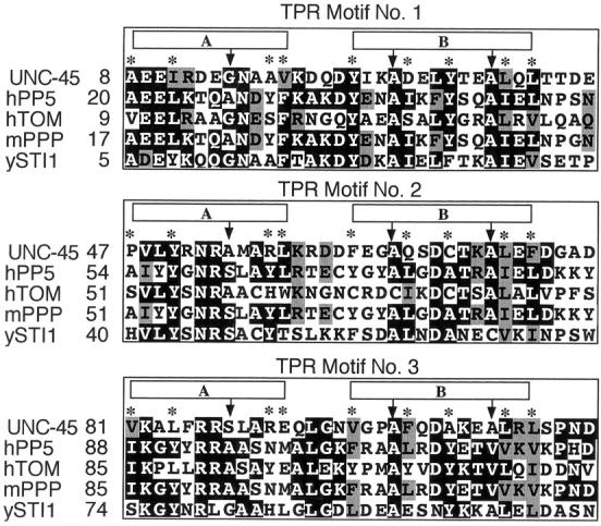
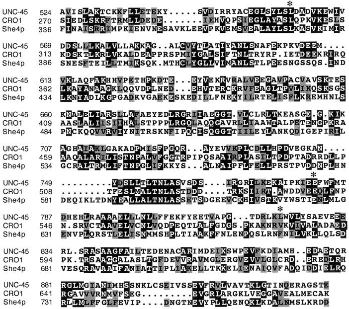
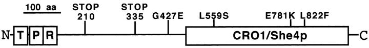
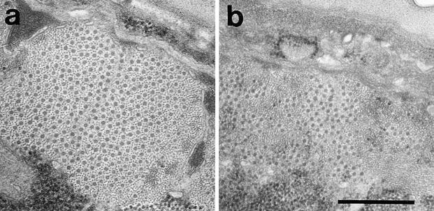
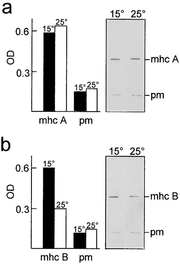
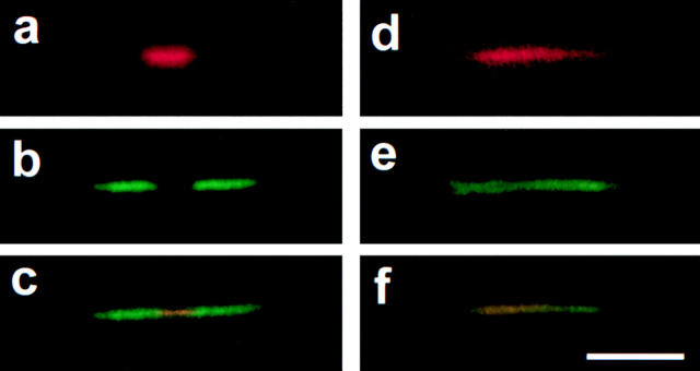
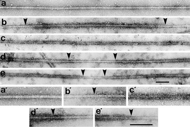
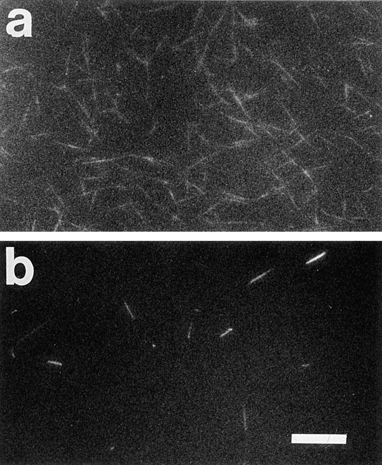
The Caenorhabditis elegans unc-45 locus has been proposed to encode a protein machine for myosin assembly. The UNC-45 protein is predicted to contain an NH2-terminal domain with three tetratricopeptide repeat motifs, a unique central region, and a COOH-terminal domain homologous to CRO1 and She4p. CRO1 and She4p are fungal proteins required for the segregation of other molecules in budding, endocytosis, and septation. Three mutations that lead to temperature-sensitive (ts) alleles have been localized to conserved residues within the CRO1/She4p-like domain, and two lethal alleles were found to result from stop codon mutations in the central region that would prevent translation of the COOH-terminal domain. Electron microscopy shows that thick filament accumulation in vivo is decreased by approximately 50% in the CB286 ts mutant grown at the restrictive temperature. The thick filaments that assemble have abnormal structure. Immunofluorescence and immunoelectron microscopy show that myosins A and B are scrambled, in contrast to their assembly into distinct regions at the permissive temperature and in wild type. This abnormal structure correlates with the high degree of instability of the filaments in vitro as reflected by their extremely low yields and shortened lengths upon isolation. These results implicate the UNC-45 CRO1/She4p-like region in the assembly of myosin isoforms in C. elegans and suggest a possible common mechanism for the function of this UCS (UNC-45/CRO1/She4p) protein family.


Figures

Figure 1
Figure 1
Multiple sequence alignments on which the UNC-45 TPR domain designation is based. The UNC-45 NH2-terminal sequence is aligned with four other TPR-containing sequences. Identical residues have a black background; residues closely similar in charge or polarity, a gray one. Boxes A and B represent the anti-parallel α-helices of each motif. Consensus positions are indicated by arrows (small hydrophobic amino acid residues) and asterisks (large hydrophobic amino acid residues). Proteins listed are hPP5, human protein phosphatase 5 (U25174); hTOM, human putative outer mitochondrial membrane 34-kD translocase (U58970); mPPP, Mus musculus phosphoprotein phosphatase (U12204); ySTI1, Saccharomyces cerevisiae heat shock protein 1 (M28486).
Figure 2
Figure 2
Multiple sequence alignments on which the UNC-45 CRO1/She4p-like domain designation is based. The UNC-45 COOH-terminal half is aligned with region 2 (see text) of the two related proteins CRO1 (Y16261), from Podospora anserina, and She4p (X93598), from Saccharomyces cerevisiae. Identical residues have a black background; residues closely similar in charge or polarity, a gray one. Asterisks indicate positions where mutations were found.
Figure 8
Figure 8
Model of UNC-45 predicted structure. The TPR and CRO1/She4p-like domains are represented by boxes; the central region, with no similarities to any known protein, by a horizontal line connecting the boxes. The positions of the mutant amino acid substitutions identified are indicated by vertical lines. Putative functions of each region are discussed in the text.
Figure 3
Figure 3
Thick filament accumulation in strain CB286 at 15 vs. 25°C. Electron micrographs of region II cross sections (Mackenzie et al., 1978) from nematodes grown at 15° (a) or 25°C (b). Bar, 0.5 mm.
Figure 4
Figure 4
mhc and paramyosin accumulation in strain CB286 at 15 vs. 25°C. Immunoblot of mhc A (a) and mhc B (b) content using paramyosin (pm) as an internal control. The same membrane was used in both experiments. The black (15°C) and white (25°C) bars represent the OD readings from the corresponding bands shown.
Figure 5
Figure 5
Immunofluorescence microscopy of myosin isoform distribution in strain CB286. Thick filaments isolated from nematodes grown at 15° (a–c) or 25°C (d–f) were reacted simultaneously with rhodamine-conjugated mAb 5-6 (specific for mhc A) and fluorescein-conjugated mAb 5-8 (specific for mhc B). (a and d) Rhodamine emission; (b and e) fluorescein emission; (c and f) simultaneous emission of both fluorochromes. Bar, 5 μm.
Figure 6
Figure 6
Immunoelectron microscopy of myosin isoform distribution in strain CB286. (a) Negatively stained 15°C filament; (b) mAb 5-6 (mhc A specific) labeling of 15°C filament, reaction can be seen inside arrowheads; (c) mAb 5-6 labeling of 25°C filament, reaction can be seen throughout its length; (d) mAb 28.2 (mhc B specific) labeling of 15°C filament, reaction can be seen outside arrowheads; (e) mAb 28.2 labeling of 25°C filament, reaction can be seen outside arrowheads. Zones of interest in a through e can be observed at higher magnification in a′ through e′, respectively, and show the presence (b′, d′, and e′) or absence (a′ and c′) of labeling transitions due to reacting and nonreacting regions along individual filaments. a′ is unreacted and c′ is labeled homogeneously by anti-mhc A antibody. Bars: (a–e and a′–e′) 0.2 μm.
Figure 7
Figure 7
Immunofluorescence microscopy of thick filament isolation in strain CB286. Undiluted samples of 6.2 K supernatant thick filament enriched fractions (Deitiker and Epstein, 1993) from equivalent amounts of starting materials from nematodes grown at 15 or 25°C were reacted simultaneously with rhodamine-conjugated mAb 5-6 (specific for mhc A) and fluorescein-conjugated mAb 5-8 (specific for mhc B). (a) Simultaneous emission of both fluorochromes in 15°C sample; (b) simultaneous emission of both fluorochromes in 25°C sample. Bar, 10 μm.

References

    1. Altschul SF, Lipman DJ. Protein database searches for multiple alignments. Proc Natl Acad Sci USA. 1990;87:5509–5513. - PMC - PubMed
    1. Altschul SF, Gish W, Miller W, Myers EW, Lipman DJ. Basic local alignment search tool. J Mol Biol. 1990;215:403–410. - PubMed
    1. Barstead RJ, Waterston RH. Vinculin is essential for muscle function in the nematode. J Cell Biol. 1991;114:715–724. - PMC - PubMed
    1. Bernstein SI, Mogami K, Donady JJ, Emerson CP., Jr Drosophilamuscle myosin heavy chain encoded by a single gene in a cluster of muscle mutations. Nature. 1983;302:393–397. - PubMed
    1. Berteaux-Lecellier V, Zickler D, Debuchy R, Panvier-Adoutte A, Thompson-Coffe C, Picard M. A homologue of the yeast SHE4 gene is essential for the transition between the syncytial and cellular stages during sexual reproduction of the fungus Podospora anserina. . EMBO (Eur Mol Biol Organ) J. 1998;17:1248–1258. - PMC - PubMed

Publication types

MeSH terms