. 1998 Nov 30;143(5):1283-94.
doi: 10.1083/jcb.143.5.1283.

The kakapo mutation affects terminal arborization and central dendritic sprouting of Drosophila motorneurons

Affiliations

The kakapo mutation affects terminal arborization and central dendritic sprouting of Drosophila motorneurons

A Prokop et al. J Cell Biol. .

Abstract

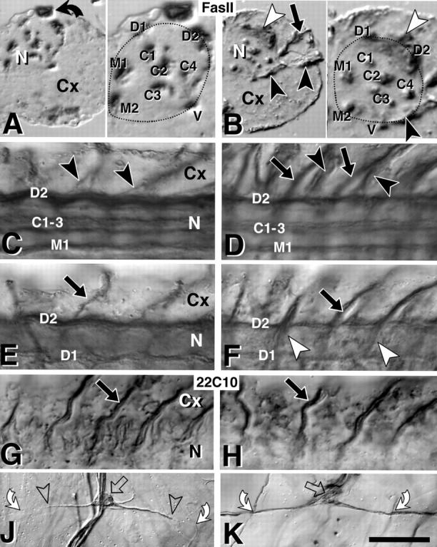
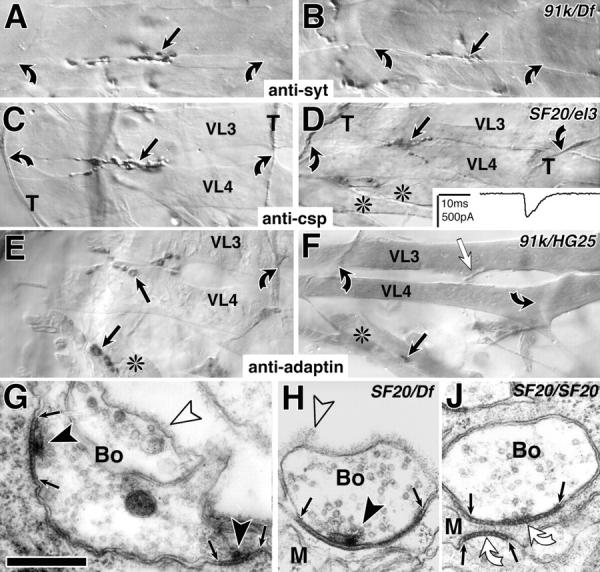
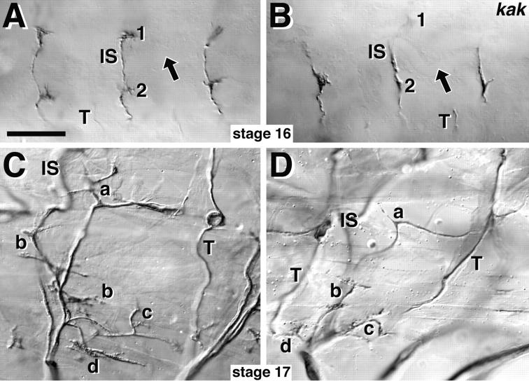
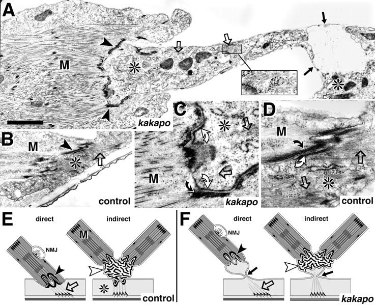
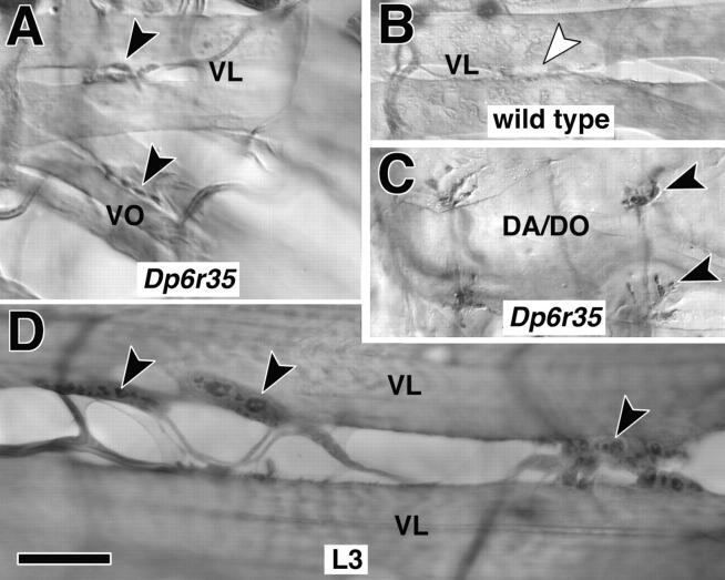
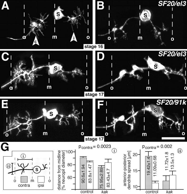
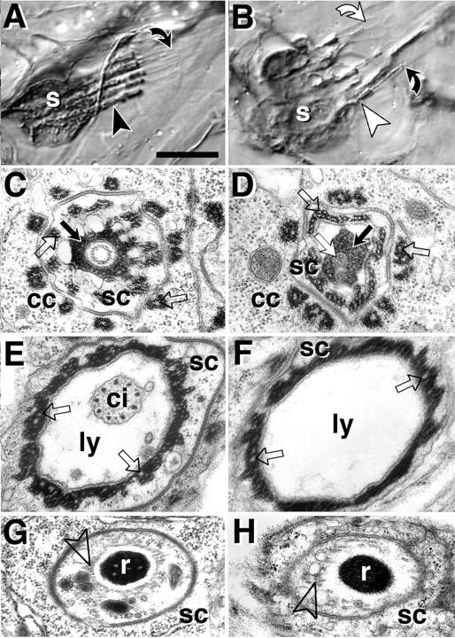
The lethal mutation l(2)CA4 causes specific defects in local growth of neuronal processes. We uncovered four alleles of l(2)CA4 and mapped it to bands 50A-C on the polytene chromosomes and found it to be allelic to kakapo (. Genetics. 146:275- 285). In embryos carrying our kakapo mutant alleles, motorneurons form correct nerve branches, showing that long distance growth of neuronal processes is unaffected. However, neuromuscular junctions (NMJs) fail to form normal local arbors on their target muscles and are significantly reduced in size. In agreement with this finding, antibodies against kakapo (Gregory and Brown. 1998. J. Cell Biol. 143:1271-1282) detect a specific epitope at all or most Drosophila NMJs. Within the central nervous system of kakapo mutant embryos, neuronal dendrites of the RP3 motorneuron form at correct positions, but are significantly reduced in size. At the subcellular level we demonstrate two phenotypes potentially responsible for the defects in neuronal branching: first, transmembrane proteins, which can play important roles in neuronal growth regulation, are incorrectly localized along neuronal processes. Second, microtubules play an important role in neuronal growth, and kakapo appears to be required for their organization in certain ectodermal cells: On the one hand, kakapo mutant embryos exhibit impaired microtubule organization within epidermal cells leading to detachment of muscles from the cuticle. On the other, a specific type of sensory neuron (scolopidial neurons) shows defects in microtubule organization and detaches from its support cells.


Figures

Figure 5
Figure 5
Axonal markers are misexpressed in kak mutant embryos. All nerve cords are late stage 17, control embryos on the left, kakel3/kakSF20 mutant embryos on the right. (A) Right half of a transverse section through a Fas II–labeled ventral nerve cord (Cx, cortex; N, neuropile; bent arrow, β-Gal labeled cell due to blue balancer), and a close-up of the neuropile on the right (stippled line). Dorsal (D1 and D2), ventral (V), median (M1 and M2), and central (C1–C4) Fas II–positive longitudinal fascicles are indicated. (B) In kak mutant nerve cords all longitudinal fascicles are present but partly reduced in size (M1 + M2 in B reduced to 70% compared with A), as is the whole neuropile (stippled area; B/A = 80%). The split into subfascicles seen in C3 occurs similarly in fascicles C1, 2 and 3 also in wild type (data not shown). Nerve roots strongly express FasII only in the mutant (black and white arrowheads; black arrow in B). (C–H) Dorsal views of the right half of ventral nerve cords (3–4 hemisegments shown; anterior to the left; C and D are more ventral, E–H are more dorsal) stained with anti–Fas II (C–F) or 22C10 (G and H); symbols correspond to those in A and B. Ventral (black arrowheads; only shown for Fas II) and dorsal (black arrows) nerve roots in the mutant embryos (right) stain more strongly for Fas II but with similar strength for 22C10, when compared with control embryos (left). (J) Processes of the dorsal bipolar dendrite neurons of the peripheral nervous system (open arrows, cell bodies) span a whole epidermal segment (bent arrows, crossing points with transverse nerve at segment border), but 22C10 expression is restricted to the proximal part (between open arrowheads). (K) In kak mutant embryos 22C10 extends along the entire process. Note that the profile of soma and dendrites is less sharp and more irregular than in the control. Bars: (A–H) 18 μm; (J and K) 40 μm.
Figure 1
Figure 1
kak mutant NMJs are reduced in size at stage 17. (A–F) Light microscopic view of NMJs (black arrows) on ventral longitudinal muscles VL3 and 4 (nomenclature as in Bate, 1993) or ventral oblique muscles (*) in the central abdomen (A3 to 5) of control (left) and kak mutant embryos (right; allelic combination indicated top right corner) labeled with anti-synaptotagmin (anti-syt in A and B), anti-cysteine string protein (anti-csp in C and D), or anti–α-adaptin (E and F; control and mutants were stained together in each case); muscle tips (bent arrows), in C and D also indicated by csp-labeled transverse nerve (T). kak mutant NMJs are reduced in size (right), but synaptic transmission occurs (D, inset, electrophysiological trace recorded from the muscle in D). (F) In kak91k/kakHG25 mutant embryos NMJs are extremely reduced (black arrow) and often synaptic markers fail to detect them, although the incoming nerve can be seen (white arrow). (G–J) Ultrastructure of control (G) and kak mutant embryos (H and J). kak mutant NMJs exhibit junctions between nerve terminal (Bo) and muscle (M), and synapses with regularly structured material in the synaptic cleft (between small arrows), T-shaped dense bars (black arrowheads), and clustered vesicles (H). Occasionally T-bars are missing (J). Open arrowheads, basement membrane. Bars: (A–F) 9 μm; (G–J) 300 nm.
Figure 2
Figure 2
Nerve branches can form and be maintained in kak mutant embryos. Control embryos on the left, kakel3/kakSF20 mutant embryos on the right, anterior is to the left and dorsal up, all preparations are labeled with anti–Fas II antibodies. (A) Three segments in dorsolateral view at stage 16 with the dorsal tips of transverse nerves (T) and intersegmental nerves (IS), which form NMJs on muscles DA1/DO1 (1) and DA2/DO2 (2; muscle nomenclature according to Bate, 1993). Arrows, dorsoventral position of the trachea. (B) In kak mutant embryos intersegmental and transverse nerves reach their target area. (C and D) At stage 17 all segmental nerve branches (a–d, Thomas et al., 1984) can be identified in kak mutant embryos, but are mislocated, most likely secondarily due to the muscle detachment phenotype (Fig. 6). Bar, 30 μm.
Figure 6
Figure 6
At muscle attachment sites kak mediates anchoring of microtubules to the membrane. (A–D) Direct muscle attachment sites at stage 17 in kakapo mutant (A and C) and wild-type embryos (B and D; genotypes indicated at bottom right corner). Muscles (M) and epidermal cells (*) are closely apposed at connecting hemiadherens junctions in control and mutant embryos (C and D and arrowheads in A and B), but the epidermal cells rupture in the mutant embryos (black arrows in A and F). (C and D) At the hemiadherens junctions membrane-associated material in the muscle (bent black arrows) anchors thin filaments in control and mutant embryos. Similar dense material in the epidermal cell (bent white arrows) anchors microtubules (open arrows) in control embryos, but fails to do so in kak mutant embryos, in spite of the presence of microtubules (open arrows in A and C; inset in A). The schematic representation shows direct and indirect muscle attachments in wild-type (E) and kak mutant embryos (F). At indirect muscle attachments extracellular tendon matrix (white arrowheads) connects muscles to each other and to the epidermis (Prokop et al., 1998). Rupture of the epidermis at kak mutant indirect muscle attachments (arrow in F) allows muscles to remain connected to each other via tendon matrix but causes detachment from the epidermis (data not shown). Microtubules have been drawn to connect to dense material on the apical cell surface (Tepass and Hartenstein, 1994) although we are not certain about this fact in kak mutant embryos. Bars: (A and B) 1.5 μm; (C and D) 500 nm.
Figure 3
Figure 3
kakapo is localized at NMJs. (A–C) At stage 17 anti-kakapo staining is found at NMJs (arrowheads; shown NMJs are on ventral longitudinal [VL], ventral oblique [VO], and dorsal acute and oblique muscles [DA/DO]; muscle nomenclature according to Bate, 1993). In embryos carrying four copies of kak (indicated as Dp6r35) anti-kak staining is stronger and more reliable than in embryos carrying one (data not shown) or two copies (wild type) of kak (compare black arrowheads in A and C with white arrowhead in B; A and B were dissected and processed in the same drop of solution). (D) At the late larval NMJ (L3) kakapo staining appears concentrated in the presynaptic boutons (black arrowheads). Bar, 9 μm.
Figure 4
Figure 4
Central dendrites are affected in kak mutant embryos. (A–F) DiI-labeled RP3 neurons in control (left) and kak mutant embryos (right; allelic combination indicated top right corner) at late stage 16 (A and B) or stage 17 (C–F). Anterior is up in all panels. Midline (m) and outer neuropile borders (o) are indicated. The soma (S) of RP3 lies close to the midline sending an ipsilateral process (i in arrowhead) and a contralateral axon (c in arrowhead), both of which have extensive dendritic arborizations. Branching of dendrites is affected mainly on the contralateral side of kak mutant embryos from stage 16 onwards. Note that the somata of kak mutant RP3 neurons appear malformed. (G) Schematic presentation of the modes (box on left) and results (i and ii) of measurements: mediolateral spread of dendrites measured from the midline (i) and their largest spread in anterior-posterior direction (ii) show significant differences on the contralateral side (grey columns, contralateral; white columns, ipsilateral; pcontra, significance for contralateral measurements). Bar, 10 μm.
Figure 7
Figure 7
kak is required for the differentiation of scolopidial sensory neurons. All images are taken from late stage 17 embryos or freshly hatched larvae. Wild-type on the left, kak mutant scolopidia on the right. (A and B) Light microscope images of 22C10-labeled pentascolopidial organs (group of five scolopidia). (A) Cell bodies of scolopidial neurons (s) send out thick dendrites (arrowhead) extending into thin cilia, which have their ends attached to the tip of hollow lymph-filled capsules (bent black arrow). (B) In kak mutant scolopidia (tested alleles: SF20/91k, SF20/el3) the thick dendrites appear collapsed towards the somata (white arrowheads) and cilia frequently appear detached from the tip of the capsule (bent white arrow). (C–H) Ultrastructural phenotypes (tested alleles: SF20/SF20, 91k/91k, SF20/91k). (C) At the tip of the capsule (bent black arrow in A), the cilium is anchored in extracellular matrix (black arrow) attached to the scolopale cell (sc) which is surrounded by processes of the cap cell (cc). (D) In kak mutant scolopidia grey inclusions (white arrow) can be found in the extracellular matrix, which might represent remnants of the cilium. (E) Further proximal the cilium (ci) is seen in the lymph-filled capsule (ly), surrounded by the scolopale cell. (F) The cilium is often missing at this position in kak mutant scolopidia. (G) Further proximal (black arrowhead in A) the dendrite contains the ciliary rootlet (r) surrounded by a circle of microtubules (open arrowhead). (H) kak mutant rootlets either lack a circle of microtubules or it is poorly developed. Intracellular dense material in cap and scolopale cells shows deeply embedded microtubules in control and mutant embryos (white stipples, open arrows in C–F). Note that microtubules are differently anchored at hemiadherens junctions, where they are not embedded in but associated with the dense material (Fig. 6 D). Bars: (A and B) 10 μm; (C–H) 480 nm.

References

    1. Angau-Petit D, Toth P, Rogero O, Faille L, Tejedor FJ, Ferrús A. Enhanced neurotransmitter release is associated with reduction of neuronal branching in a Drosophilamutant overexpressing frequenin. Eur J Neurosci. 1998;10:423–434. - PubMed
    1. Arcaro KF, Lnenicka GA. Intrinsic differences in axonal growth from crayfish fast and slow motorneurons. Dev Biol. 1995;168:272–283. - PubMed
    1. Ashburner M, Faithfull J, Littlewood T, Richards G, Smith S, Velissariou V, Woodruff RC. Report of new mutants—Drosophila melanogaster. . Dros Info Service. 1980;55:193–195.
    1. Avila J, Domínguez J, Díaz-Nido J. Regulation of microtubule dynamics by microtubule-associated protein expression and phosphorylation during neuronal development. Int J Dev Biol. 1994;38:13–25. - PubMed
    1. Bate, M. 1993. The mesoderm and its derivatives. In The Development of Drosophila melanogaster. M. Bate and A. Martínez Arias, editors. Cold Spring Harbor Laboratory Press, Cold Spring Harbor, NY. 1013–1090.

Publication types

MeSH terms