. 1998 Dec;42(12):3256-65.
doi: 10.1128/AAC.42.12.3256.

An Escherichia coli expression assay and screen for human immunodeficiency virus protease variants with decreased susceptibility to indinavir

Affiliations

An Escherichia coli expression assay and screen for human immunodeficiency virus protease variants with decreased susceptibility to indinavir

L Melnick et al. Antimicrob Agents Chemother. 1998 Dec.

Abstract

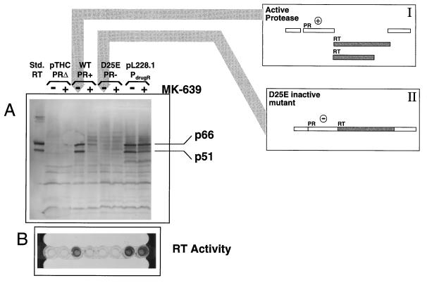
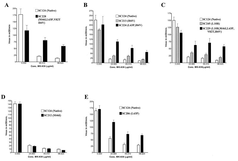
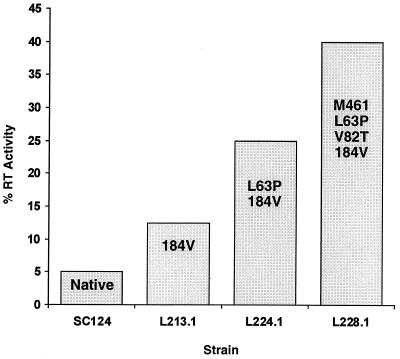
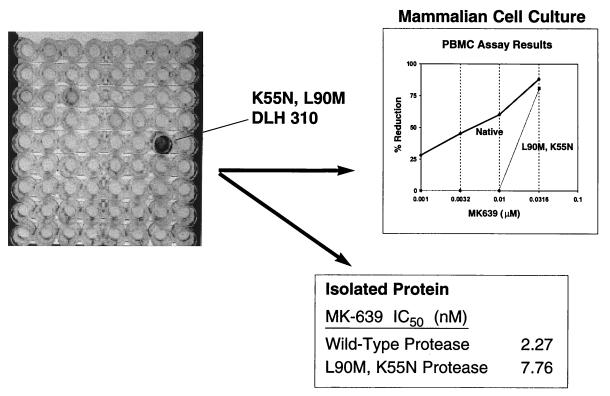
We have developed a recombinant Escherichia coli screening system for the rapid detection and identification of amino acid substitutions in the human immunodeficiency virus (HIV) protease associated with decreased susceptibility to the protease inhibitor indinavir (MK-639; Merck & Co.). The assay depends upon the correct processing of a segment of the HIV-1 HXB2 gag-pol polyprotein followed by detection of HIV reverse transcriptase activity by a highly sensitive, colorimetric enzyme-linked immunosorbent assay. The highly sensitive system detects the contributions of single substitutions such as I84V, L90M, and L63P. The combination of single substitutions further decreases the sensitivity to indinavir. We constructed a library of HIV protease variant genes containing dispersed mutations and, using the E. coli recombinant system, screened for mutants with decreased indinavir sensitivity. The discovered HIV protease variants contain amino acid substitutions commonly associated with indinavir resistance in clinical isolates, including the substitutions L90M, L63P, I64V, V82A, L24I, and I54T. One substitution, W6R, is also frequently found by the screen and has not been reported elsewhere. Of a total of 12,000 isolates that were screened, 12 protease variants with decreased sensitivity to indinavir were found. The L63P substitution, which is also associated with indinavir resistance, increases the stability of the isolated protease relative to that of the native HXB2 protease. The rapidity, sensitivity, and accuracy of this screen also make it useful for screening for novel inhibitors. We have found the approach described here to be useful for the detection of amino acid substitutions in HIV protease that have been associated with drug resistance as well as for the screening of novel compounds for inhibitory activity.


Figures

FIG. 1
FIG. 1
HIV protease (PR) processing of the E. coli-expressed polyprotein segment, reverse transcriptase (RT) activity, and inhibition by indinavir (MK-639). The basis for the E. coli expression assay for HIV protease activity is that, to achieve a high level of reverse transcriptase activity, HIV protease must be active to excise reverse transcriptase from the E. coli-expressed polyprotein segment. Extracts from E. coli-expressed protease-reverse transcriptase polyprotein segments were examined by Western blot analysis, and reverse transcriptase activity was examined by ELISA. The lanes of the Western immunoblot are labeled as follows: pTHC, pTrcHisC (Invitrogen), the expression plasmid without HIV DNA sequences; WT, plasmid pL124.23, which is pTrcHisC containing native HXB2 HIV protease-reverse transcriptase sequences; D25E, plasmid pLD25E, which contains the HIV protease portion of the protease-reverse transcriptase polyprotein that expresses a D25E-substituted, inactive HIV protease; pL228.1, which expresses a protease region with four substitutions encoding M46I, L63P, V82T, and I84V. Plasmids pLD25E and pL228.1 are identical to pL124.23 except for mutations within the protease-encoding DNA. Western blot analysis and the ELISA for reverse transcriptase activity are described in the Materials and Methods section of the text. (A) Western blot analysis. Polyclonal anti-HIV reverse transcriptase antibody was used to detect E. coli-expressed antigen. The reverse transcriptase standard displays the expected two antibody-reacting bands, p51 and p66, in approximately equimolar amounts. Samples to which indinavir was not added display these bands for E. coli extracts from cells expressing native protease (wild type) and drug-resistant (pL228.1) protease. The bands are not detected for the polyprotein segment containing the D25E inactive protease. For native (wild-type) protease (WT lane), the addition of indinavir prevents the appearance of the p51 and p66 bands. In contrast, for the drug-resistant genotype sample, pL228.1, the p51 and p66 bands appear, despite incubation of expressing cells in the presence of MK-639. (B) Reverse transcriptase ELISA. Cells containing the described plasmids were grown in 20-μl aliquots of medium with or without indinavir and were induced with IPTG. Following permeabilization of the cells by freeze-thaw cycles and centrifugation, the 20-μl supernatants were assayed for RT activity. Reverse transcriptase activity is indicated by dark coloration in the photograph. Clones containing the pTrcHisC construct lacking the HIV sequences (plasmid without insert) did not exhibit reverse transcriptase activity. A solution of buffer (data not shown) also gave a negative result. Assay of the supernatant from induced cells containing pL124.23 with the described HIV sequences and grown in medium without indinavir revealed high levels of reverse transcriptase activity. Supernatants from these cells grown in the presence of indinavir contained little detectable reverse transcriptase activity. However, indinavir does not prevent the indication of reverse transcriptase activity from pL228 expressing a drug-resistant protease. For the inactive protease mutant D25E, no reverse transcriptase activity is detected. High levels of RT activity correspond with the appearance in the Western immunoblot (A) of the p51 and p66 RT heterodimer bands.
FIG. 2
FIG. 2
E. coli expression assay: sensitivities of HIV protease variants to indinavir (MK-639). HIV protease variants obtained by site-directed mutagenesis and library-discovered HIV protease variants were assayed for the resistance contribution of single and combined mutations. Mutant HIV protease genes are configured to be a portion of a polyprotein gene segment as described for plasmid pL124.23. A reverse transcriptase ELISA was used to determine reverse transcriptase activity as an indirect indication of HIV protease processing efficiency, as described in the Materials and Methods section. HIV protease-expressing E. coli cells were grown in different concentrations of indinavir as indicated on the x axes. Reverse transcriptase activity is represented as Vmax. Six separate determinations for each protease construct at each inhibitor concentration were averaged to derive the results described above. (A) Native HIV protease and HIV protease with the M46I, L63P, V82T, and I84V substitutions. (B) Native HIV protease I84V-substituted HIV protease and L63P-I84V-substituted HIV protease. (C) Native HIV protease, L10R-substituted HIV protease, and HIV protease with the L10R, M46I, L63P, V82T, and I84V substitutions. (D) Native HIV protease and M46I-substituted HIV protease. (E) Native HIV protease and L63P-substituted HIV protease.
FIG. 3
FIG. 3
E. coli expression assay: effect of additive substitutions on the susceptibility to indinavir of HIV protease variants. Site-directed mutagenesis was used to construct HIV HXB2 protease variant genes which were inserted into the E. coli expression vectors as a portion of a polyprotein gene segment as described in the text for the plasmid pL124.23. Cells were grown in the presence of 99 mg of indinavir per ml, as described in the text. The reverse transcriptase ELISA was used to determine activity as described in Materials and Methods. The reverse transcriptase (RT) activity from drug-treated cells was calculated as a percentage of the activity from cells grown without drug. Each assay was repeated at least six times, and averaged readings from a Molecular Devices plate reader are depicted. Incremental loss of sensitivity clearly coincides with additive effects for this set of mutations.
FIG. 4
FIG. 4
Construction and screening of a library of HIV protease (PR) variants. Error-prone PCR was used to introduce dispersed mutations within the HIV protease gene. This mutagenized population of DNAs was ligated into the E. coli expression vector pTrcHisC (Invitrogen) along with unmutagenized DNA encoding the entire HIV reverse transcriptase (RT) gene. This plasmid library was used to transform E. coli Top10 cells (Invitrogen), and single colonies were distributed into individual microplate wells containing growth media and the protease inhibitor indinavir. IPTG was added to induce expression, cells were made permeable by freezing and thawing, and reverse transcriptase activity was assayed as described in Materials and Methods. DrugS, drug susceptible; DrugR, drug resistant.
FIG. 5
FIG. 5
Identification of the HIV protease variant with K55N and L90M substitutions from a library of protease variant genes by the reverse transcriptase ELISA-based screen. The reverse transcriptase ELISA was carried out in a 96-well microplate format. Each well contains material expressed by an individual E. coli colony that expresses a plasmid from a library of HIV protease variant genes. Individual colonies from the library were grown in microplate wells in the presence of indinavir (see Materials and Methods). For the E. coli-expressed protease-reverse transcriptase polyprotein segments, efficient activation of reverse transcriptase requires the activity of the HIV protease (Fig. 1). One microplate well shown above indicates a high level of reverse transcriptase activity. Since indinavir is expected to inhibit HIV protease activation of reverse transcriptase, only a protease variant with lowered susceptibility to inhibitor is expected to efficiently activate reverse transcriptase in the presence of inhibitor. The identified HIV protease variant was determined to contain the substitutions K55N and L90M. The selected protease gene containing the K55N- and L90M-encoding mutations was transferred to the vector pET21c for expression of protein for isolated protease experiments and to the vector pNL4-3 for virus-infected cell culture studies. The IC50 for the protease isolated from the variant with the K55N and L90M substitution was heightened, and by using pLN4-3 infected PBMCs, the variant showed decreased susceptibility to indinavir (MK-639) (PBMC culture studies were done in the laboratory of Douglas Richman).
FIG. 6
FIG. 6
Isolated HIV protease containing the L63P polymorphism displays increased thermostability. The L63P polymorphism contributes to high levels of resistance to indinavir in clinical and cell culture studies (4, 5). Using site-directed mutagenesis we constructed HIV protease containing the L63P substitution. The variant protease as well as the native protease and the L90M-K55N variants were expressed by using the pET21c vector, and isolated protein was examined for activity after incubation at 37°C. The graph indicates the greater stability of the L63P variant compared to the stabilities of both the wild-type and the L90M and K55N double mutant. It is interesting that an autolytic junction within the HIV protease is located between residues L63 and I64 (29). This suggests the possibility that the L63P polymorphism could contribute to drug resistance by extending the protein half-life through decreased autolysis at the amino acid junction at positions 63 and 64.

Similar articles

Cited by

References

    1. Cadwell R C, Joyce G F. Mutagenic PCR. PCR Methods Appl. 1994;3:S136–S140. - PubMed
    1. Cadwell R C, Joyce G F. Randomization of genes by PCR mutagenesis. PCR Methods Appl. 1992;2:28–33. - PubMed
    1. Cheng Y S, McGowan M H, Kettner C A, Schloss J V, Erickson-Viitanen S, Yin F H. High-level synthesis of recombinant HIV-1 protease and the recovery of active enzyme from inclusion bodies. Gene. 1990;87:243–248. - PubMed
    1. Condra J H, Holder D J, Schleif W A, Blahy O M, Danovich R M, Gabryelski L J, Graham D J, Laird D, Quintero J C, Rhodes A, Robbins H L, Roth E, Shivaprakash M, Yang T, Chodakewitz J A, Deutsch P J, Leavitt R Y, Massari F E, Mellors J W, Squires K E, Steigbigel R T, Teppler H, Emini E A. Genetic correlates of in vivo viral resistance to indinavir, a human immunodeficiency virus type 1 protease inhibitor. J Virol. 1996;70:8270–8276. - PMC - PubMed
    1. Condra J H, Schleif W A, Blahy O M, Gabryelski L J, Graham D J, Quintero J C, Rhodes A, Robbins H L, Roth E, Shivaprakash M, Titus D, Yang T, Teppler H, Squires K E, Deutsch P J, Emini E A. In vivo emergence of HIV-1 variants resistant to multiple protease inhibitors. Nature. 1995;374:569–571. - PubMed

MeSH terms

LinkOut - more resources