

doi: 10.1128/AAC.42.12.3296.
An ampD gene in Pseudomonas aeruginosa encodes a negative regulator of AmpC beta-lactamase expression
Affiliations
- PMID: 9835532
- PMCID: PMC106040
- DOI: 10.1128/AAC.42.12.3296
Item in Clipboard
An ampD gene in Pseudomonas aeruginosa encodes a negative regulator of AmpC beta-lactamase expression
Antimicrob Agents Chemother.
1998 Dec.
Abstract
The ampD and ampE genes of Pseudomonas aeruginosa PAO1 were cloned and characterized. These genes are transcribed in the same orientation and form an operon. The deduced polypeptide of P. aeruginosa ampD exhibited more than 60% similarity to the AmpD proteins of enterobacteria and Haemophilus influenzae. The ampD product transcomplemented Escherichia coli ampD mutants to wild-type beta-lactamase expression.
Figures
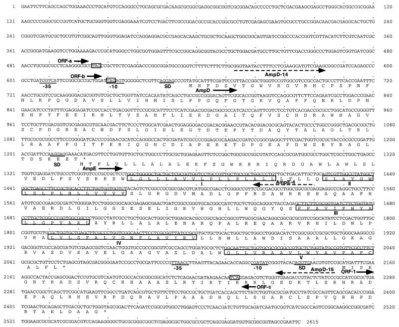
Nucleotide sequence of the P. aeruginosa ampD and ampE genes with ORF-1 and predicted amino acids. The amino acids are presented according to the one-letter code. The putative SD sequences and the potential −10 and −35 regions of promoters are underlined. The stop codons are shown by asterisks. The five transmembrane domains of AmpE are boxed and named I, II, III, IV, and V. The stop codons for the potential ORF-a, ORF-b, and ORF-c are boxed. The oligonucleotide primers used for PCR amplification are shown by dashed arrows.
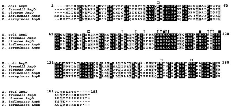
Alignment of the amino acid sequence of P. aeruginosa PAO1 AmpD with those of the E. coli, C. freundii, E. cloacae, and putative H. influenzae AmpD proteins. The similar and identical amino acids are lightly and darkly shaded, respectively. The crosses and the open squares indicate the amino acids conserved in the core and outside region of the Bacillus cell wall hydrolases, respectively. The solid squares show the amino acids strictly conserved in various cell wall hydrolases (12).
References
-
- Ambler R P. The structure of β-lactamases. Philos Trans R Soc London Ser B. 1980;289:321–331. - PubMed
Publication types
MeSH terms
Substances
Associated data
- Actions
LinkOut - more resources
Full Text Sources
Molecular Biology Databases
