. 1998 Dec;64(12):4650-7.
doi: 10.1128/AEM.64.12.4650-4657.1998.

A polyphasic approach To study the diversity and vertical distribution of sulfur-oxidizing thiomicrospira species in coastal sediments of the german wadden Sea

Affiliations

A polyphasic approach To study the diversity and vertical distribution of sulfur-oxidizing thiomicrospira species in coastal sediments of the german wadden Sea

T Brinkhoff et al. Appl Environ Microbiol. 1998 Dec.

Abstract

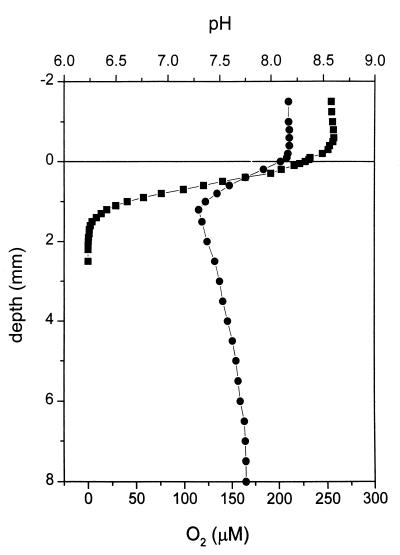
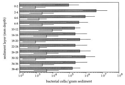
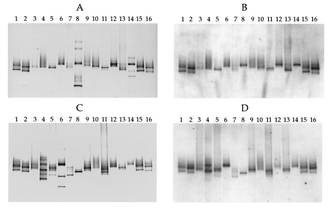
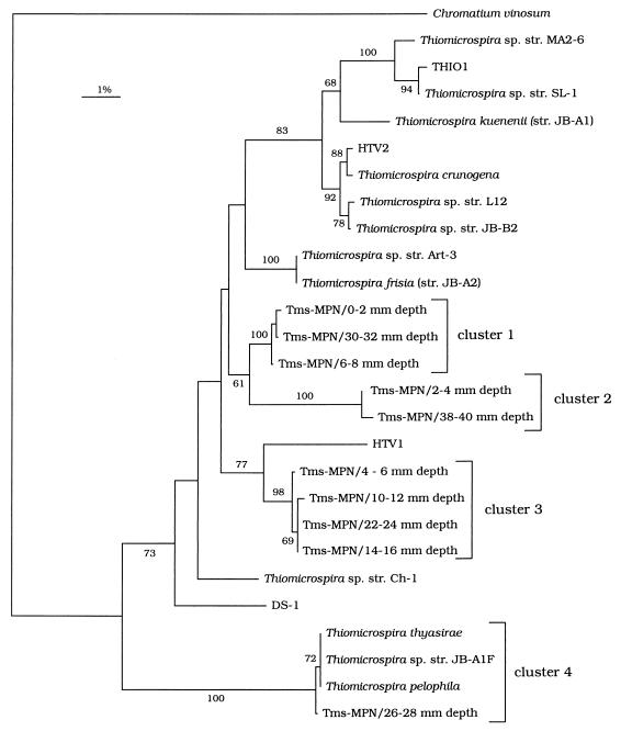
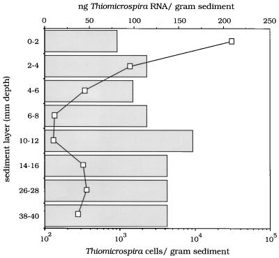
Recently, four Thiomicrospira strains were isolated from a coastal mud flat of the German Wadden Sea (T. Brinkhoff and G. Muyzer, Appl. Environ. Microbiol. 63:3789-3796, 1997). Here we describe the use of a polyphasic approach to investigate the functional role of these closely related bacteria. Microsensor measurements showed that there was oxygen penetration into the sediment to a depth of about 2.0 mm. The pH decreased from 8.15 in the overlaying water to a minimum value of 7.3 at a depth of 1.2 mm. Further down in the sediment the pH increased to about 7.8 and remained constant. Most-probable-number (MPN) counts of chemolithoautotrophic sulfur-oxidizing bacteria revealed nearly constant numbers along the vertical profile; the cell concentration ranged from 0.93 x 10(5) to 9.3 x 10(5) cells per g of sediment. A specific PCR was used to detect the presence of Thiomicrospira cells in the MPN count preparations and to determine their 16S rRNA sequences. The concentration of Thiomicrospira cells did not decrease with depth. It was found that Thiomicrospira strains were not dominant sulfur-oxidizing bacteria in this habitat. Denaturing gradient gel electrophoresis (DGGE) of PCR-amplified 16S ribosomal DNA fragments followed by hybridization analysis with a genus-specific oligonucleotide probe revealed the diversity of Thiomicrospira strains in the MPN cultures. Sequence analysis of the highest MPN dilutions in which the genus Thiomicrospira was detected revealed that there were four clusters of several closely related sequences. Only one of the 10 Thiomicrospira sequences retrieved was related to sequences of known isolates from the same habitat. Slot blot hybridization of rRNA isolated from different sediment layers showed that, in contrast to the concentration of Thiomicrospira cells, the concentration of Thiomicrospira-specific rRNA decreased rapidly in the region below the oxic layer of the sediment. This study revealed the enormous sequence diversity of closely related microorganisms present in one habitat, which so far has been found only by sequencing molecular isolates. In addition, it showed that most of the Thiomicrospira populations in the sediment studied were quiescent.


Figures

FIG. 1
FIG. 1
Oxygen (■) and pH (•) microprofiles for a sediment core from Jadebusen Bay. Note that oxygen was found only in the top 2.0 mm of the sediment. The sulfide content was also examined, but sulfide was not detected. The sediment surface was defined as a depth of 0 mm. Measurements for negative depth values are measurements for the water phase overlaying the sediment; positive depth values indicate the depth in the sediment.
FIG. 2
FIG. 2
MPN counts for chemolithoautotrophic sulfur-oxidizing bacteria (dark bars) in sediment samples from Jadebusen Bay, an intertidal coastal mud flat area of the German Wadden Sea. The numbers of Thiomicrospira cells (light bars) were determined by enzymatic amplification of the 16S rDNA with genus-specific primers. The bars indicate 95% confidence intervals.
FIG. 3
FIG. 3
Hybridization analysis of DGGE profiles of 16S rDNA fragments obtained with primers specific for the domain Bacteria and template DNA from MPN cultures and Thiomicrospira isolates from coastal sediments of the Wadden Sea. (A) DGGE patterns. Lanes 1 and 15, T. pelophila (lower band) and Thiomicrospira sp. strain JB-B2 (upper band); lanes 2 and 16, T. kuenenii JB-A1 (lower band) and T. frisia JB-A2 (upper band); lane 3, lowest-dilution MPN culture of the sediment layer at a depth of 0 to 2 mm; lane 4, highest-dilution MPN culture of the sediment layer at a depth of 0 to 2 mm in which Thiomicrospira spp. could still be detected; lanes 5 and 6, equivalent MPN cultures from a depth of 2 to 4 mm; lanes 7 and 8, equivalent MPN cultures from a depth of 4 to 6 mm; lanes 9 and 10, equivalent MPN cultures from a depth of 6 to 8 mm; lanes 11 and 12, equivalent MPN cultures from a depth of 10 to 12 mm; lanes 13 and 14, equivalent MPN cultures from a depth of 14 to 16 mm. (B) Hybridization analysis of the DGGE pattern in panel A performed with the Thiomicrospira-specific, digoxigenin-labeled oligonucleotide, whose target sequence is located within the rDNA amplified. (C) DGGE patterns. Lanes 1, 2, 15, and 16, Thiomicrospira standards (see the explanation above for panel A); lanes 3 and 4, equivalent MPN cultures from a depth of 18 to 20 mm; lanes 5 and 6, equivalent MPN cultures from a depth of 22 to 24 mm; lanes 7 and 8, equivalent MPN cultures from a depth of 26 to 28 mm; lanes 9 and 10, equivalent MPN cultures from a depth of 30 to 32 mm; lanes 11 and 12, equivalent MPN cultures from a depth of 34 to 36 mm; lanes 13 and 14, equivalent MPN cultures from a depth of 38 to 40 mm. (D) Hybridization analysis of the DGGE pattern in panel C. Comparison of panels A and B and panels C and D shows that chemolithoautotrophic bacteria other than Thiomicrospira spp. were present in some of the MPN cultures.
FIG. 4
FIG. 4
Unrooted tree showing the phylogenetic relationships of Thiomicrospira strains present in the highest MPN dilutions containing Thiomicrospira cells. The tree is based on partial 16S rRNA sequences and was produced by using the neighbor-joining algorithm with maximum-likelihood correction. The sequences determined in this study are indicated as follows: Tms-MPN/x-y mm depth, where x-y mm depth indicates the depth of the sediment from which a sequence was obtained. The sequence of Chromatium vinosum was used as an outgroup. The numbers on the branches are bootstrap values (1,000 replicates); only values greater than 50% are shown.
FIG. 5
FIG. 5
Amounts of Thiomicrospira-specific rRNA (line) in sediment samples from different depths and MPN counts of Thiomicrospira cells (bars) for these samples. The results indicate that a small but active Thiomicrospira population was present in the oxic surface layer of the sediment.

References

    1. Alm E W, Oerther D B, Larsen N, Stahl D A, Raskin L. The Oligonucleotide Probe Database. Appl Environ Microbiol. 1996;62:3557–3559. - PMC - PubMed
    1. Amann R I, Binder B J, Olson R J, Chisholm S W, Devereux R, Stahl D A. Combination of 16S rRNA-targeted oligonucleotide probes with flow cytometry for analyzing mixed microbial populations. Appl Environ Microbiol. 1990;56:1919–1925. - PMC - PubMed
    1. Amann R I, Ludwig W, Schleifer K-H. Phylogenetic identification and in situ detection of individual microbial cells without cultivation. Microbiol Rev. 1995;59:143–169. - PMC - PubMed
    1. Amann R I, Kühl M. In situ methods for assessment of microorganisms and their activities. Curr Opin Microbiol. 1998;1:352–358. - PubMed
    1. American Public Health Association. Standard methods for the examination of water and wastewater, including bottom sediments and sludge. Washington, D.C: American Public Health Association; 1969. pp. 604–609.

LinkOut - more resources