Review
. 1998 Dec;62(4):1079-93.
doi: 10.1128/MMBR.62.4.1079-1093.1998.

Multimodular penicillin-binding proteins: an enigmatic family of orthologs and paralogs

Affiliations
Review

Multimodular penicillin-binding proteins: an enigmatic family of orthologs and paralogs

C Goffin et al. Microbiol Mol Biol Rev. 1998 Dec.

Abstract

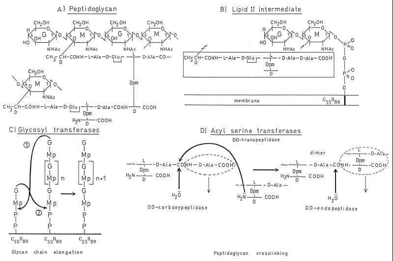
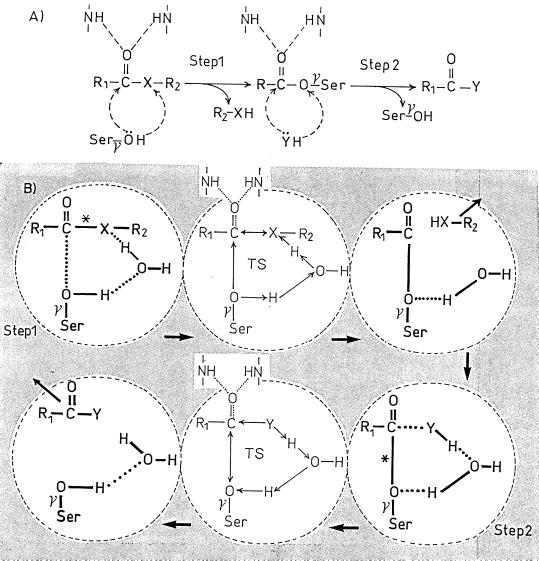
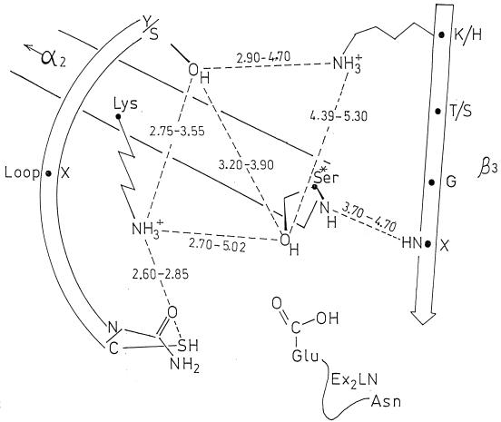
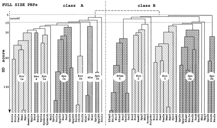
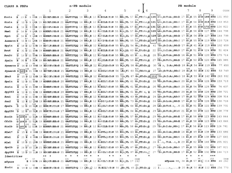
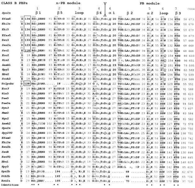
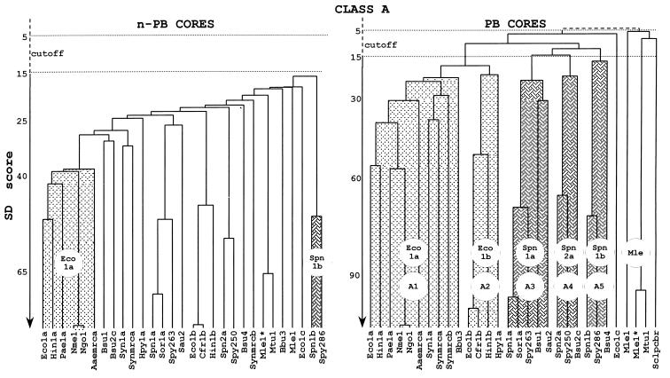
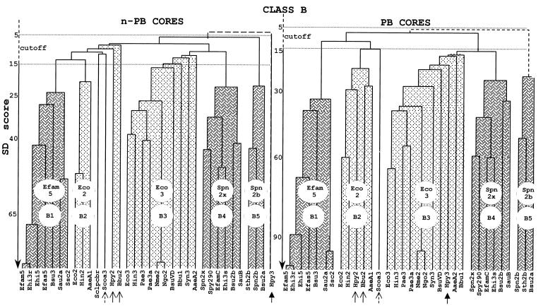
The monofunctional penicillin-binding DD-peptidases and penicillin-hydrolyzing serine beta-lactamases diverged from a common ancestor by the acquisition of structural changes in the polypeptide chain while retaining the same folding, three-motif amino acid sequence signature, serine-assisted catalytic mechanism, and active-site topology. Fusion events gave rise to multimodular penicillin-binding proteins (PBPs). The acyl serine transferase penicillin-binding (PB) module possesses the three active-site defining motifs of the superfamily; it is linked to the carboxy end of a non-penicillin-binding (n-PB) module through a conserved fusion site; the two modules form a single polypeptide chain which folds on the exterior of the plasma membrane and is anchored by a transmembrane spanner; and the full-size PBPs cluster into two classes, A and B. In the class A PBPs, the n-PB modules are a continuum of diverging sequences; they possess a five-motif amino acid sequence signature, and conserved dicarboxylic amino acid residues are probably elements of the glycosyl transferase catalytic center. The PB modules fall into five subclasses: A1 and A2 in gram-negative bacteria and A3, A4, and A5 in gram-positive bacteria. The full-size class A PBPs combine the required enzymatic activities for peptidoglycan assembly from lipid-transported disaccharide-peptide units and almost certainly prescribe different, PB-module specific traits in peptidoglycan cross-linking. In the class B PBPs, the PB and n-PB modules cluster in a concerted manner. A PB module of subclass B2 or B3 is linked to an n-PB module of subclass B2 or B3 in gram-negative bacteria, and a PB module of subclass B1, B4, or B5 is linked to an n-PB module of subclass B1, B4, or B5 in gram-positive bacteria. Class B PBPs are involved in cell morphogenesis. The three motifs borne by the n-PB modules are probably sites for module-module interaction and the polypeptide stretches which extend between motifs 1 and 2 are sites for protein-protein interaction. The full-size class B PBPs are an assortment of orthologs and paralogs, which prescribe traits as complex as wall expansion and septum formation. PBPs of subclass B1 are unique to gram-positive bacteria. They are not essential, but they represent an important mechanism of resistance to penicillin among the enterococci and staphylococci. Natural evolution and PBP- and beta-lactamase-mediated resistance show that the ability of the catalytic centers to adapt their properties to new situations is limitless. Studies of the reaction pathways by using the methods of quantum chemistry suggest that resistance to penicillin is a road of no return.


Figures

FIG. 1
FIG. 1
(A and B) Structure of the wall peptidoglycan of E. coli (A) and assembly from the lipid II intermediate (B). G, N-acetylglucosamine; M, N-acetylmuramic acid; Dpm, meso-diaminopimelic acid; transmembrane bar, C55H89 isoprenoid alcohol carrier; Mp, N-acetylmuramyl pentapeptide. (C) Reaction 1, glycan chain elongation at the nonreducing end of the chain; reaction 2, glycan chain elongation at the reducing end of the chain. (D) Overall reactions catalyzed by the dd-transpeptidases, dd-carboxypeptidases, and dd-endopeptidases.
FIG. 2
FIG. 2
Acyl serine transferase-catalyzed reaction on R1—CO—X—R2 carbonyl donors. (A) Overall reaction. (B) Role of a water molecule as proton transmitter. X is O, S, or NH (in penicillin, the CO—N bond is endocyclic). HY is an acceptor, i.e., water or an amino compound. The carbonyl of the donor and that of the acyl enzyme are polarized by NH groups of the enzyme polypeptide backbone. TS, transition state; ∗, scissile bond of the carbonyl donor (step 1) and scissile bond of the acyl enzyme (step 2). The shaded area symbolizes the active-site environment.
FIG. 3
FIG. 3
Amino acid sequence signature of the penicilloyl serine transferases superfamily. S. K15, Streptomyces strain K15; S. R61, Streptomyces strain R61; Eco, E. coli; Bsu, B. subtilis; Spn, S. pneumoniae; Eclo, E. cloacae; Tgase, transglycosylase; Tpase, transpeptidase. PBPs and β-lactamases marked by an asterisk are of known three-dimensional structure (at 3.5-Å resolution only for Spn PBP2x). Motifs 1, 2, and 3 are at the top, and secondary structures are at the bottom. The monofunctional PBPs and β-lactamases are synthesized with a cleavable signal peptide. The R61 PBP and the β-lactamases are secreted; E. coli PBP4 is, somehow, loosely attached to the membrane; the Streptomyces strain K15 PBP is membrane associated via an internal hydrophobic segment; E. coli PBP5 and probably B. subtilis PBP5 are membrane associated via a carboxy-terminal amphiphilic helix. The multimodular PBPs are synthesized with a noncleavable signal peptide that serves as membrane anchor.
FIG. 4
FIG. 4
Schematic representation of the catalytic center of the penicilloyl serine transferases. Average distances in angstroms between heavy atoms (O-O, O-N, S-N, and N-N) of side chains of the active-site-defining motif 1 (on helix α2), motif 2 (on a loop), and motif 3 (on strand β3) are given. The values derived from X-ray data apply to the proteins marked by an asterisk in Fig. 3. Motif 1 is, invariably, Sx2K. Motif 2, SxN, is replaced by SxC in Streptomyces strain K15 PBP and by YxN in Streptomyces strain R61 PBP and class C β-lactamases. Motif 3, K[T/S]G, is replaced by HTG in Streptomyces strain R61 PBP.
FIG. 5
FIG. 5
Hierarchical analysis of full-size, multimodular PBPs. PBP codes are given in Table 1. Each cluster (or subclass) is identified by a prototypic PBP. For SD score values and more details, see the text.
FIG. 6
FIG. 6
Amino acid sequence analysis of full-size, multimodular class A PBPs. Conserved motifs, intermodule junction sites, inserts, and amino- and carboxy-terminal extensions are shown. mTgase, putative monofunctional transglycosylase of E. coli (Swiss-Prot accession no. P46022). mTpase, monofunctional dd-transpeptidase/PBP of Streptomyces strain K15 (Swiss-Prot accession no. P39042); Mle1∗, high-affinity, thermolabile class A PBP of M. leprae (43). Asterisks at the bottom of the figure highlight identities defining the amino acid sequence signatures of the modules. Amino acid residues that do not obey the consensus are in underlined italics. For more details, see the text.
FIG. 7
FIG. 7
Amino acid sequence analysis of full-size, multimodular class B PBPs. Conserved motifs, intermodule junction sites, inserts, and carboxy-terminal extensions are shown. Secondary structures of the S. pneumoniae PBP2x are shown at the top. Identities (asterisks) define the amino acid sequence signatures of the modules. Amino acid residues that do not obey the consensus are in underlined italics. For more details, see the text.
FIG. 8
FIG. 8
Hierarchical analysis of the n-PB cores and PB cores of the multimodular class A PBPs. PBP codes are given in Table 1.
FIG. 9
FIG. 9
Hierarchical analysis of the n-PB cores and PB cores of the multimodular class B PBPs. PBP codes are given in Table 1.

References

    1. Adam M, Fraipont C, Rhazi N, Nguyen-Distèche M, Lakaye B, Frère J M, Devreese B, Van Beeumen J, van Heijenoort Y, van Heijenoort J, Ghuysen J M. The bimodular G57-V577 polypeptide chain of the class B penicillin-binding protein 3 of Escherichia coli catalyses peptide bond formation from thiolesters and does not catalyze glycan chain polymerization from lipid II intermediate. J Bacteriol. 1997;179:6005–6009. - PMC - PubMed
    1. Barton G J. Protein multiple sequence alignment and flexible pattern matching. Methods Enzymol. 1990;183:403–428. - PubMed
    1. Basu J, Mahapatra S, Kundu M, Mukhopadhyay S, Nguyen-Distèche M, Dubois P, Joris B, Van Beeumen J, Cole S T, Chakrabarti P, Ghuysen J M. Identification and overexpression in Escherichia coli of a Mycobacterium leprae gene, pon1, encoding high-molecular-mass class A penicillin-binding protein PBP1. J Bacteriol. 1996;178:1707–1711. - PMC - PubMed
    1. Berger-Bächi B. Expression of resistance to methicillin. Trends Microbiol. 1994;2:389–393. - PubMed
    1. Blattner F R, et al. The complete genome sequence of Escherichia coli K12. Science. 1997;277:1453–1462. - PubMed

Publication types

MeSH terms