Review
. 1998 Dec;62(4):1094-156.
doi: 10.1128/MMBR.62.4.1094-1156.1998.

Molecular biology and pathogenicity of mycoplasmas

Affiliations
Review

Molecular biology and pathogenicity of mycoplasmas

S Razin et al. Microbiol Mol Biol Rev. 1998 Dec.

Abstract

The recent sequencing of the entire genomes of Mycoplasma genitalium and M. pneumoniae has attracted considerable attention to the molecular biology of mycoplasmas, the smallest self-replicating organisms. It appears that we are now much closer to the goal of defining, in molecular terms, the entire machinery of a self-replicating cell. Comparative genomics based on comparison of the genomic makeup of mycoplasmal genomes with those of other bacteria, has opened new ways of looking at the evolutionary history of the mycoplasmas. There is now solid genetic support for the hypothesis that mycoplasmas have evolved as a branch of gram-positive bacteria by a process of reductive evolution. During this process, the mycoplasmas lost considerable portions of their ancestors' chromosomes but retained the genes essential for life. Thus, the mycoplasmal genomes carry a high percentage of conserved genes, greatly facilitating gene annotation. The significant genome compaction that occurred in mycoplasmas was made possible by adopting a parasitic mode of life. The supply of nutrients from their hosts apparently enabled mycoplasmas to lose, during evolution, the genes for many assimilative processes. During their evolution and adaptation to a parasitic mode of life, the mycoplasmas have developed various genetic systems providing a highly plastic set of variable surface proteins to evade the host immune system. The uniqueness of the mycoplasmal systems is manifested by the presence of highly mutable modules combined with an ability to expand the antigenic repertoire by generating structural alternatives, all compressed into limited genomic sequences. In the absence of a cell wall and a periplasmic space, the majority of surface variable antigens in mycoplasmas are lipoproteins. Apart from providing specific antimycoplasmal defense, the host immune system is also involved in the development of pathogenic lesions and exacerbation of mycoplasma induced diseases. Mycoplasmas are able to stimulate as well as suppress lymphocytes in a nonspecific, polyclonal manner, both in vitro and in vivo. As well as to affecting various subsets of lymphocytes, mycoplasmas and mycoplasma-derived cell components modulate the activities of monocytes/macrophages and NK cells and trigger the production of a wide variety of up-regulating and down-regulating cytokines and chemokines. Mycoplasma-mediated secretion of proinflammatory cytokines, such as tumor necrosis factor alpha, interleukin-1 (IL-1), and IL-6, by macrophages and of up-regulating cytokines by mitogenically stimulated lymphocytes plays a major role in mycoplasma-induced immune system modulation and inflammatory responses.


Figures

FIG. 1
FIG. 1
Biosynthetic pathways genes in the genomes of H. influenzae, M. pneumoniae, and M. genitalium. Numbers above the bars indicate the percentages of the total putatively identified genes. Based on data from Fleischmann et al. (130), Fraser et al. (139), and Himmelreich et al. (181, 182).
FIG. 2
FIG. 2
Cellular processes, metabolic pathways, and regulatory functions genes in the genomes of H. influenzae, M. pneumoniae, and M. genitalium. Numbers above the bars indicate the percentages of total putatively identified genes. Based on data from Fleischmann et al. (130), Fraser et al. (139), and Himmelreich et al. (181, 182).
FIG. 3
FIG. 3
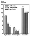
DNA replication, transcription, and translation genes in the genomes of H. influenzae, M. pneumoniae, and M. genitalium. Numbers above the bars indicate the percentages of total putatively identified genes. Based on data from Fleischmann et al. (130), Fraser et al. (139), and Himmelreich et al. (181, 182).
FIG. 4
FIG. 4
Selective hemadsorption of human erythrocytes to a distinct sector of a M. gallisepticum colony (16).
FIG. 5
FIG. 5
Schematic representation and structural features of the M. bovis VspA protein. The VspA ORF is shown as a rectangle consisting of internal blocks delineating various features of the VspA protein, aligned from the 5′ end to the 3′ end of the vspA gene. The solid block, labeled Signal, contains 25 amino acids of a putative lipoprotein signal peptide. Different hatched or shaded blocks designated RA1, RA2, RA3, and RA4 represent four in-frame repetitive regions encoding distinctive periodic amino acid sequences of 6, 6, 8, and 8 amino acids, respectively. Subrepetitive units within the RA4 region designated RA4.1 and RA4.2 are also shown. Based on data from Lysnyansky et al. (267).
FIG. 6
FIG. 6
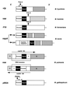
Schematic representation of regulatory and structural features of antigenic variation systems in mycoplasmas. Representative genes of distinct antigenic variation systems of six mycoplasma species are shown (not to scale) as rectangles consisting of internal blocks aligned from the 5′ end to the 3′ end of each gene. The system designation and the corresponding mycoplasma species are indicated on the left and on the right, respectively. The solid black block, labeled Signal, contains the species-specific amino acid sequence of a putative lipoprotein signal peptide. Different hatched blocks represent system-specific in-frame reiterated sequences. The location of a homopolymeric tract of contiguous adenines (Poly-A) or of oligonucleotide repeats (GAA)n, within the promoter region (vlp, pMGA) (159, 485) or within the coding-gene region (vaa, P78) (451, 491) is shown by an arrow. Two vsp genes from two M. bovis clonal isolates, exhibiting ON or OFF expression states of the variable surface lipoprotein VspA, are shown. A genomic fragment that was inserted within the vspA promoter region, leading to its OFF expression state, is shown by a hatched box labelled Insertion sequence. Two vsa genes isolated from two M. pulmonis variants displaying the VsaHA or the VsaHA+ phenotypes are shown (421). A chromosomal fragment that inverted during phase transition (VsaHA ↔ VsaHA+) is indicated by brackets. The direction of expression of the vsa gene from each variant is marked by an arrow.

Similar articles

Cited by

References

    1. Aguila H N, Lai W C, Lu Y S, Pakes S P. Experimental Mycoplasma pulmonis infection of rats suppresses humoral but not cellular immune response. Lab Anim Sci. 1988;38:138–142. - PubMed
    1. Al-Daccak R, Mehindate K, Hebert J, Rink L, Mecheri S, Mourad W. Mycoplasma arthritidis-derived superantigen induces proinflammatory monokine gene expression in the THP-1 human monocytic cell line. Infect Immun. 1994;62:2409–2416. - PMC - PubMed
    1. Al-Daccak R, Mehindate K, Poubelle P E, Mourad W. Signalling via MHC class II molecules selectively induces IL-1 beta over IL-1 receptor antagonist gene expression. Biochem Biophys Res Commun. 1994;201:855–860. - PubMed
    1. Almeida R A, Wannemuehler M J, Rosenbusch R F. Interaction of Mycoplasma dispar with bovine alveolar macrophages. Infect Immun. 1992;60:2914–2919. - PMC - PubMed
    1. Amikam D, Glaser G, Razin S. Mycoplasmas (Mollicutes) have a low number of rRNA genes. J Bacteriol. 1984;158:376–378. - PMC - PubMed