Review
. 1998 Dec;62(4):1315-52.
doi: 10.1128/MMBR.62.4.1315-1352.1998.

The virulence plasmid of Yersinia, an antihost genome

Affiliations
Review

The virulence plasmid of Yersinia, an antihost genome

G R Cornelis et al. Microbiol Mol Biol Rev. 1998 Dec.

Abstract

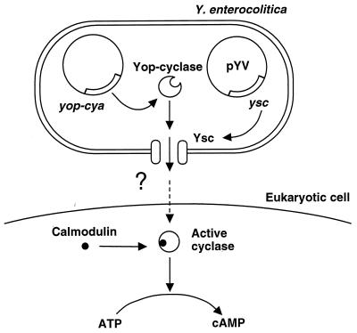
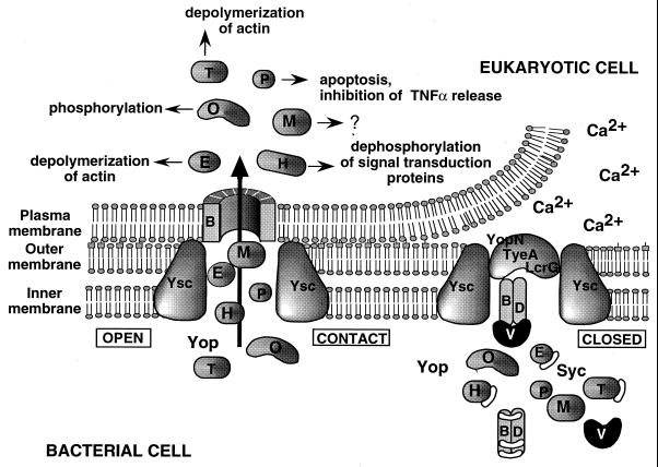
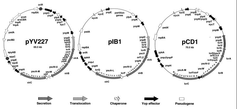
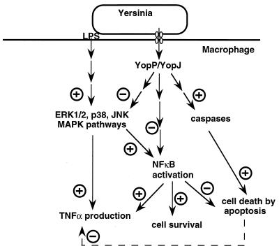
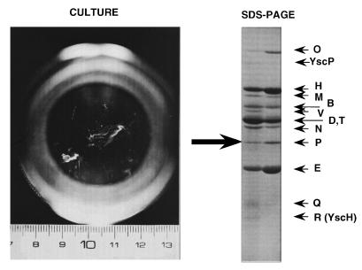
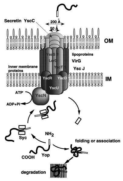
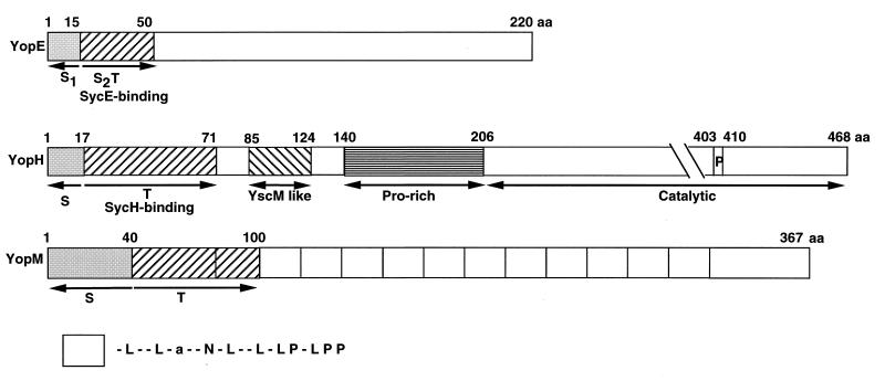
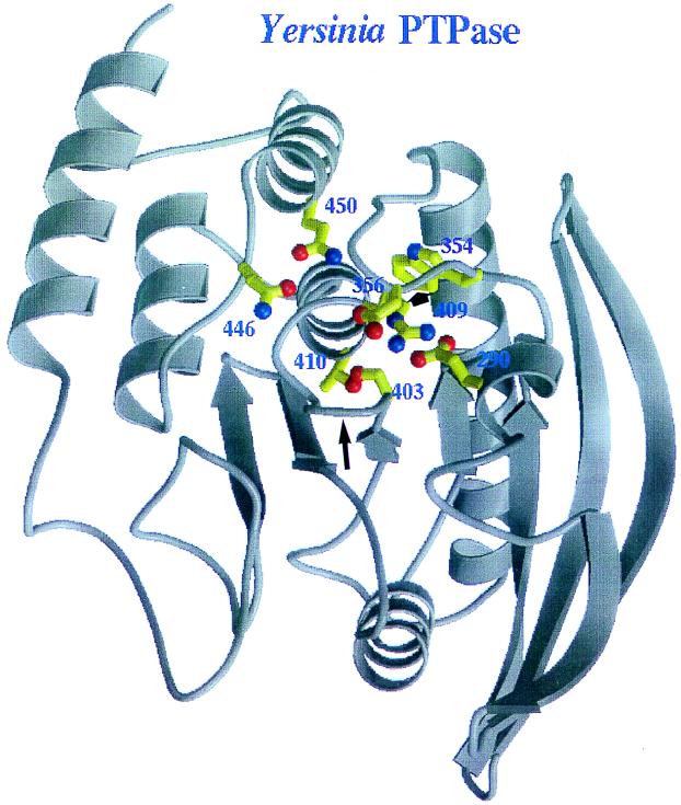
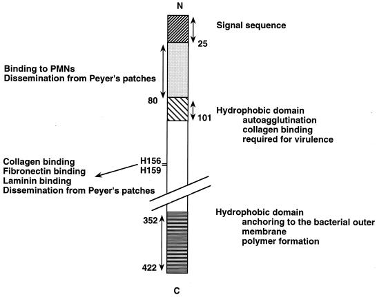
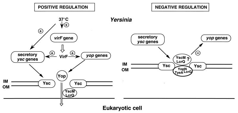
The 70-kb virulence plasmid enables Yersinia spp. (Yersinia pestis, Y. pseudotuberculosis, and Y. enterocolitica) to survive and multiply in the lymphoid tissues of their host. It encodes the Yop virulon, an integrated system allowing extracellular bacteria to disarm the cells involved in the immune response, to disrupt their communications, or even to induce their apoptosis by the injection of bacterial effector proteins. This system consists of the Yop proteins and their dedicated type III secretion apparatus, called Ysc. The Ysc apparatus is composed of some 25 proteins including a secretin. Most of the Yops fall into two groups. Some of them are the intracellular effectors (YopE, YopH, YpkA/YopO, YopP/YopJ, YopM, and YopT), while the others (YopB, YopD, and LcrV) form the translocation apparatus that is deployed at the bacterial surface to deliver the effectors into the eukaryotic cells, across their plasma membrane. Yop secretion is triggered by contact with eukaryotic cells and controlled by proteins of the virulon including YopN, TyeA, and LcrG, which are thought to form a plug complex closing the bacterial secretion channel. The proper operation of the system also requires small individual chaperones, called the Syc proteins, in the bacterial cytosol. Transcription of the genes is controlled both by temperature and by the activity of the secretion apparatus. The virulence plasmid of Y. enterocolitica and Y. pseudotuberculosis also encodes the adhesin YadA. The virulence plasmid contains some evolutionary remnants including, in Y. enterocolitica, an operon encoding resistance to arsenic compounds.


Figures

FIG. 1
FIG. 1
The Yop-Cya reporter strategy used to study translocation of Yop proteins into the cytosol of eukaryotic cells. Reprinted from reference with permission of the publisher.
FIG. 2
FIG. 2
A tentative model for the interaction between Yersinia and a macrophage. When Yersinia is placed at 37°C in a rich environment, the Ysc secretion apparatus is installed and a stock of Yop proteins is synthesized. Some of these proteins are capped with their specific Syc chaperones, which presumably prevent premature associations. As long as there is no contact with a eukaryotic cell, the YopN-TyeA-LcrG plug blocks the Ysc secretion channel. Upon Ca2+ depletion or contact with the eukaryotic target cell, the secretion channel opens and the YopB translocator inserts in the eukaryotic cell with the help of YopD and LcrV. The Yop effectors (YopE, YopH, YopM, YopO/YpkA, YopP/YopJ, and YopT) are then transported through the secretion channel and translocated across the plasma membrane, guided by the translocators. YopE and YopT act on the cytoskeleton, while YopP/YopJ induces apoptosis and inhibits the release of TNF-α.
FIG. 3
FIG. 3
The genetic maps of pYV227 from Y. enterocolitica W227 (serotype O:9) (redrawn from reference 170), pIB1 from Y. pseudotuberculosis YPIII (redrawn from reference 259), and pCD1 from Y. pestis KIM (redrawn from references and 257a). Note that none of these maps is complete. For pCD1, the plasmid has been sequenced twice (165a, 257a) and only the genes that are identified in the two sequences or in one sequence and in Y. enterocolitica are shown. Plasmid pYV227 has also been completely sequenced (171), but only the genes described in this review are included here. Shading of the genes has been done on the basis of the data presented in this review.
FIG. 4
FIG. 4
YopP-induced apoptosis. Semithin sections were stained with toluidine blue and examined by light microscopy (a to d). (a) Wild-type Y. enterocolitica E40. Apoptotic nuclei (arrows) and cell surface-associated bacteria (brown particles, arrowhead) are visible. (b) yscN secretion mutant. No apoptotic cells are detected. Internalized bacteria either in tight (single arrowhead) or spacious (double arrowhead) phagosomes are abundant. (c) yopP effector mutant. No apoptotic cells are detected. Bacteria are seen at the cell surface (arrowhead). (d) yopP+++ (yopP cloned in a multicopy vector). Apoptotic cells are visible (arrows). (e and f) Ultrastructural analysis of cells infected with yopP+++ from panel d is shown. Typical features of apoptosis include (i) peripheral chromatin condensation in crescents, except in the vicinity of nuclear pores (large arrows); (ii) bulging of nuclear crescents into the cytoplasm (best seen in panel e); and (iii) appearance of central clusters of small particles of unknown nature, typical of apoptosis (small arrow in panel f). Nuclear and plasma membrane alterations contrast with a good ultrastructural preservation of cytoplasm, particularly of endoplasmic reticulum and mitochondria. Bars, 10 μm (a to d) and 2 μm (e and f). Reprinted from reference with permission of the publisher.
FIG. 5
FIG. 5
Model showing the effects of Yersinia spp. on the macrophage intracellular cascades. Lipopolysaccharide (LPS) activates the ERK1/2, JNK, and p38 MAPK pathways, leading to increased TNF-α production. Activated MAPKs can lead to NF-κB activation; activated NF-κB can, in turn, enhance TNF-α transcription. Translocated YopP/YopJ induces macrophage apoptosis by a mechanism involving caspase activation. It also downregulates MAPKs and impairs NF-κB activation, two effects that could explain the YopP/YopJ-induced reduction of TNF-α production. See the text for details and references.
FIG. 6
FIG. 6
Yops secreted by Y. enterocolitica W22703. Bacteria were grown at 28°C in a conical flask (seen from the top) containing oxalated brain heart infusion and then transferred to 37°C. The photograph showing the Yop filaments was taken 4 h after the temperature shift. SDS-PAGE of the filaments (right lane) and of Yops precipitated from the supernatant by ammonium sulfate (left lane) is shown on the right. Adapted from reference .
FIG. 7
FIG. 7
Model for Yop secretion. OM, outer membrane; IM, inner membrane.
FIG. 8
FIG. 8
Schematic representation of YopE, YopH, and YopM. S1, first secretion domain; S2/T, second secretion domain and translocation domain. The catalytic domain of YopH includes the P-loop (P). The LRR motifs (193) in YopM are represented by open boxes, and their composition is given below.
FIG. 9
FIG. 9
Catalytic domain of YopH. A ribbon diagram of the (C403S) Yersinia PTPase is shown. The sulfate anion has been left out of the active site for clarity. A few critical conserved residues are depicted with stick bonds in yellow and are labeled. The P-loop is depicted by a long arrow, and the flexible loop is indicated by a short arrow. The general acid on the flexible loop, aspartic acid at position 356, can be seen in the “closed” conformation over the active site. Serine 403 is seen in place of the catalytic cysteine, although the two residues have very similar conformations. Diagram courtesy of J. Dixon (University of Michigan Medical School).
FIG. 10
FIG. 10
Representation of the YadA protein of Y. enterocolitica O:8 showing the mapped domains and their functions (277, 278, 314, 316, 342).
FIG. 11
FIG. 11
Schematic representation of the two circuits regulating yop gene transcription. IM, inner membrane; OM, outer membrane.

References

    1. Achtman M, Kennedy N, Skurray R. Cell-cell interactions in conjugating Escherichia coli: role of traT protein in surface exclusion. Proc Natl Acad Sci USA. 1977;74:5104–5108. - PMC - PubMed
    1. Aguero M E, Aron L, DeLuca A G, Timmis K N, Cabello F C. A plasmid-encoded outer membrane protein, TraT, enhances resistance of Escherichia coli to phagocytosis. Infect Immun. 1984;46:740–746. - PMC - PubMed
    1. Albertini A M, Caramori T, Crabb W D, Scoffone F, Galizzi A. The flaA locus of Bacillus subtilis is part of a large operon coding for flagellar structures, motility functions, and an ATPase-like polypeptide. J Bacteriol. 1991;173:3573–3579. - PMC - PubMed
    1. Alfano J R, Collmer A. The type III (Hrp) secretion pathway of plant pathogenic bacteria: trafficking harpins, Avr proteins, and death. J Bacteriol. 1997;179:5655–5662. - PMC - PubMed
    1. Alfano J R, Kim H S, Delaney T P, Collmer A. Evidence that the Pseudomonas syringae pv. syringae hrp-linked hrmA gene encodes an Avr-like protein that acts in an hrp-dependent manner within tobacco cells. Mol Plant-Microbe Interact. 1997;10:580–588. - PubMed

Publication types

Substances

LinkOut - more resources