Review
. 1998 Dec;62(4):1371-414.
doi: 10.1128/MMBR.62.4.1371-1414.1998.

Staphylococcal cell wall: morphogenesis and fatal variations in the presence of penicillin

Affiliations
Review

Staphylococcal cell wall: morphogenesis and fatal variations in the presence of penicillin

P Giesbrecht et al. Microbiol Mol Biol Rev. 1998 Dec.

Abstract

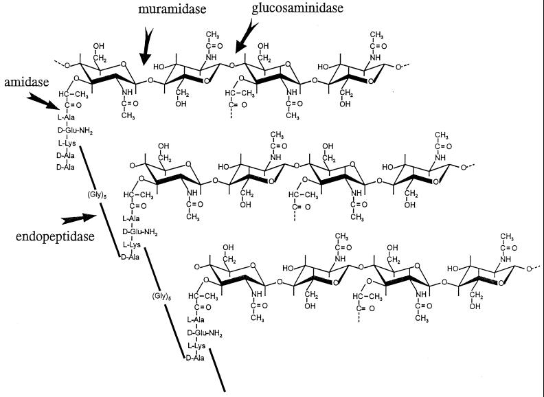
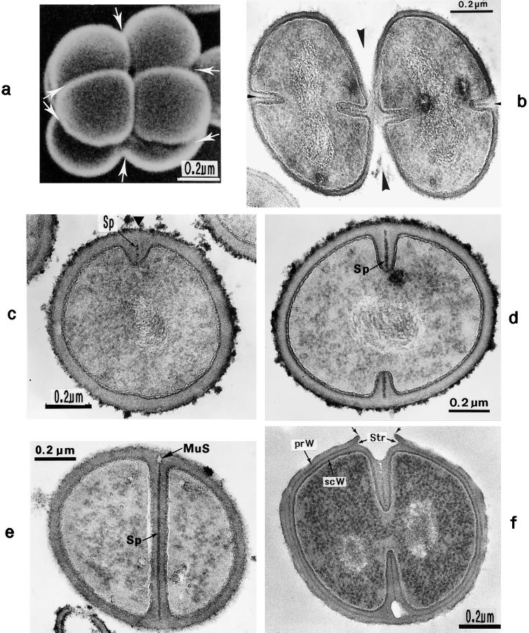
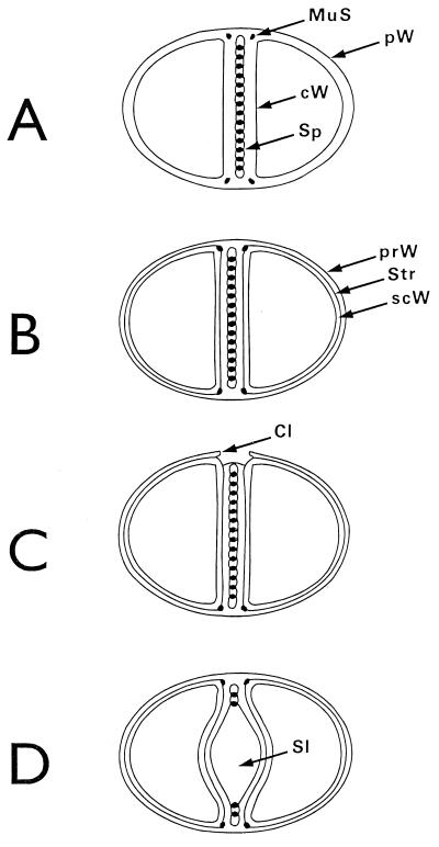
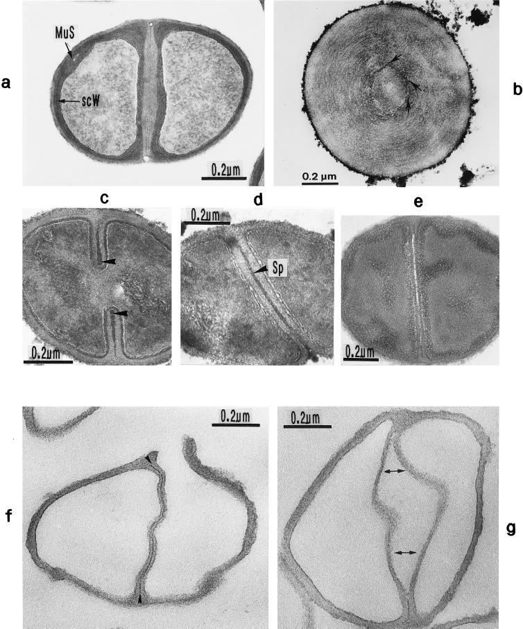
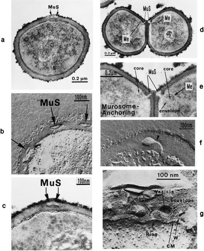
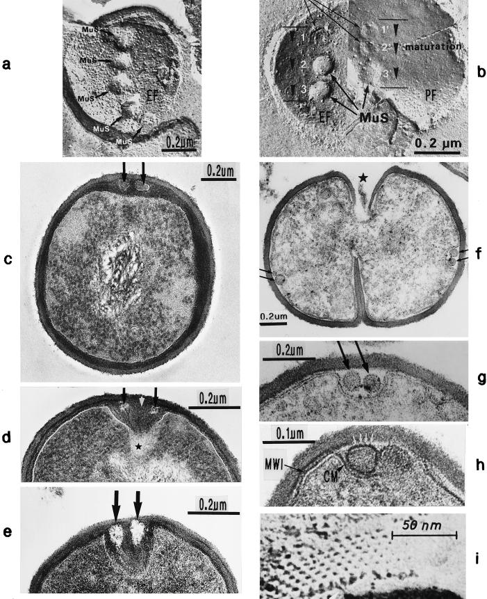
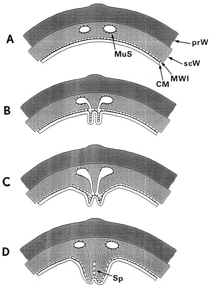
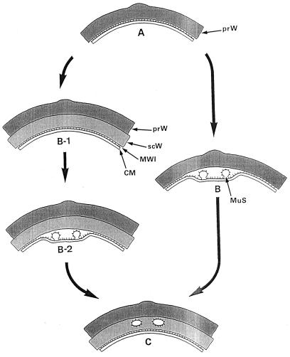
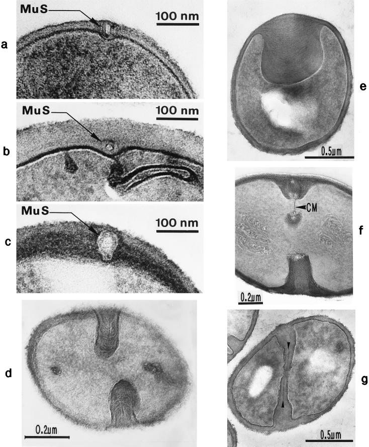
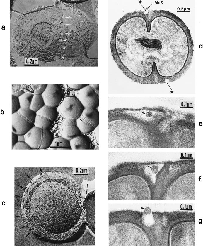
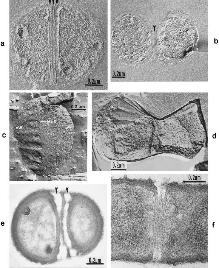
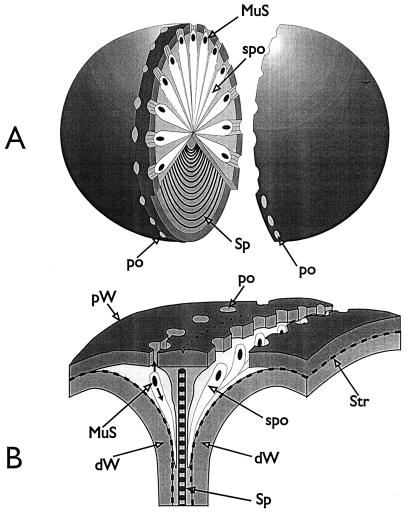
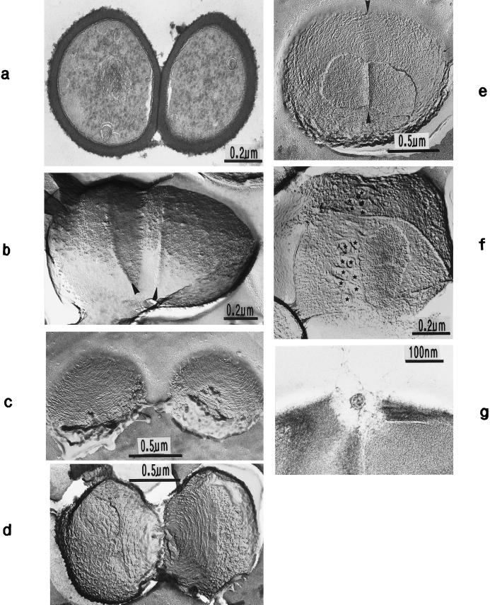
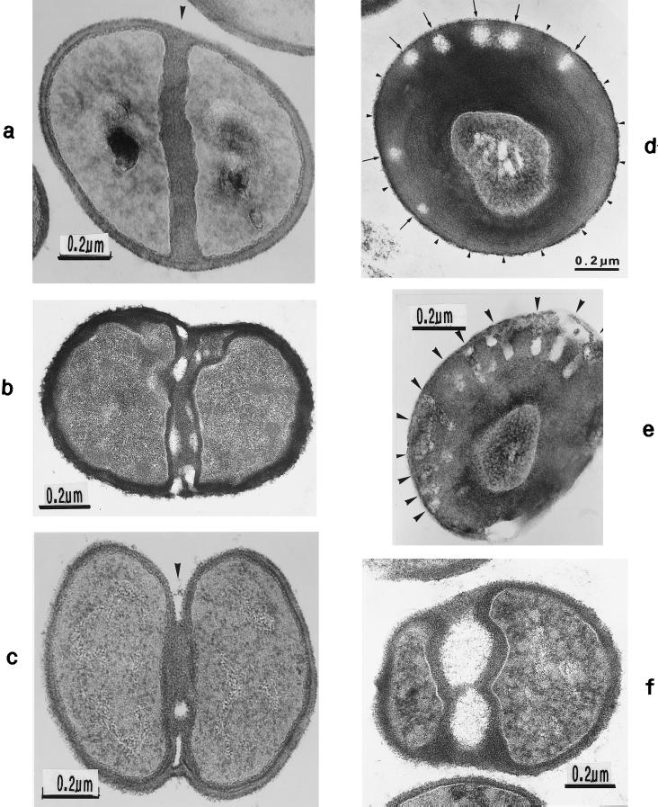
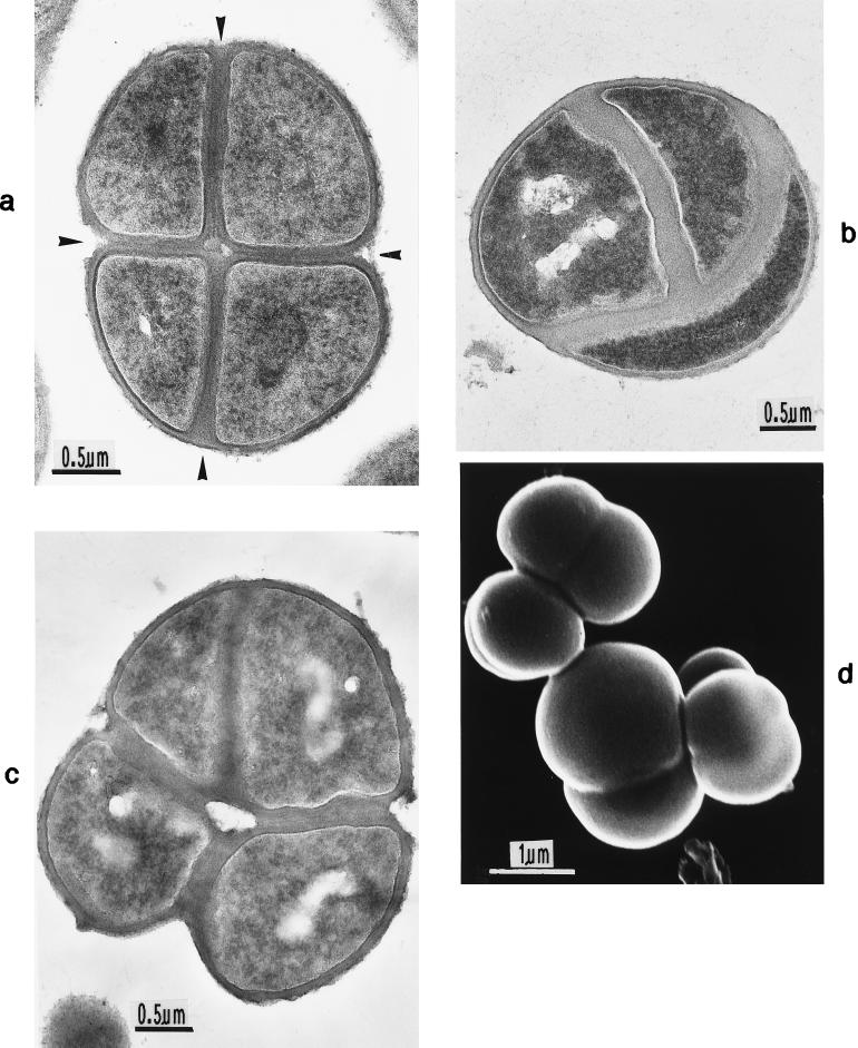
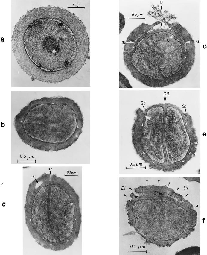
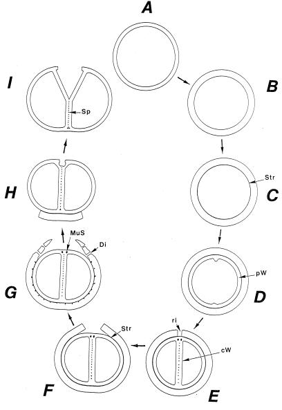
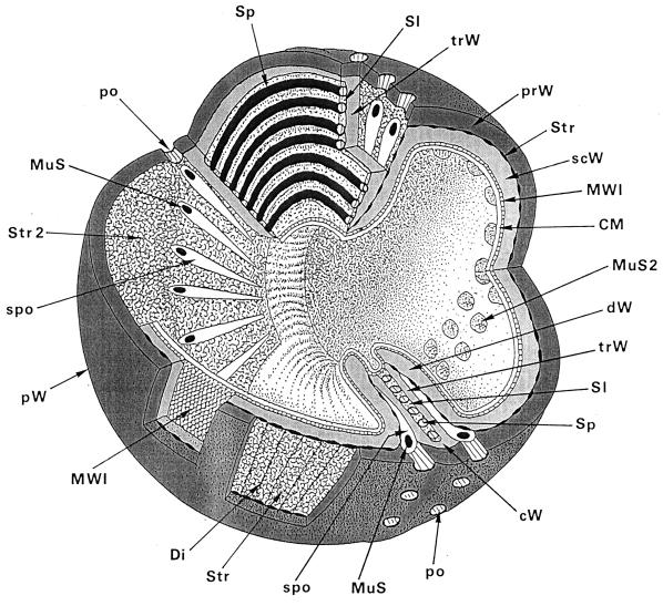
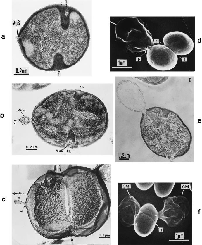
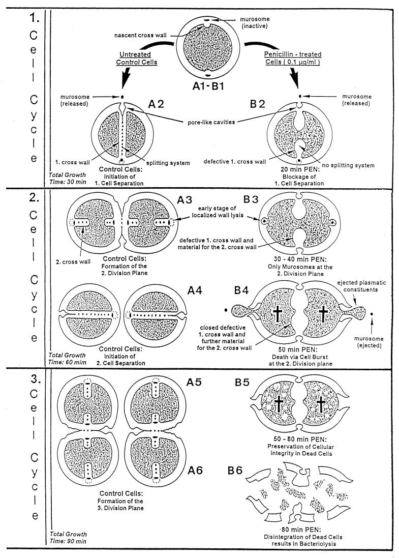
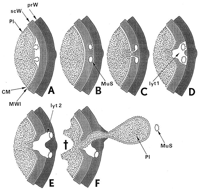
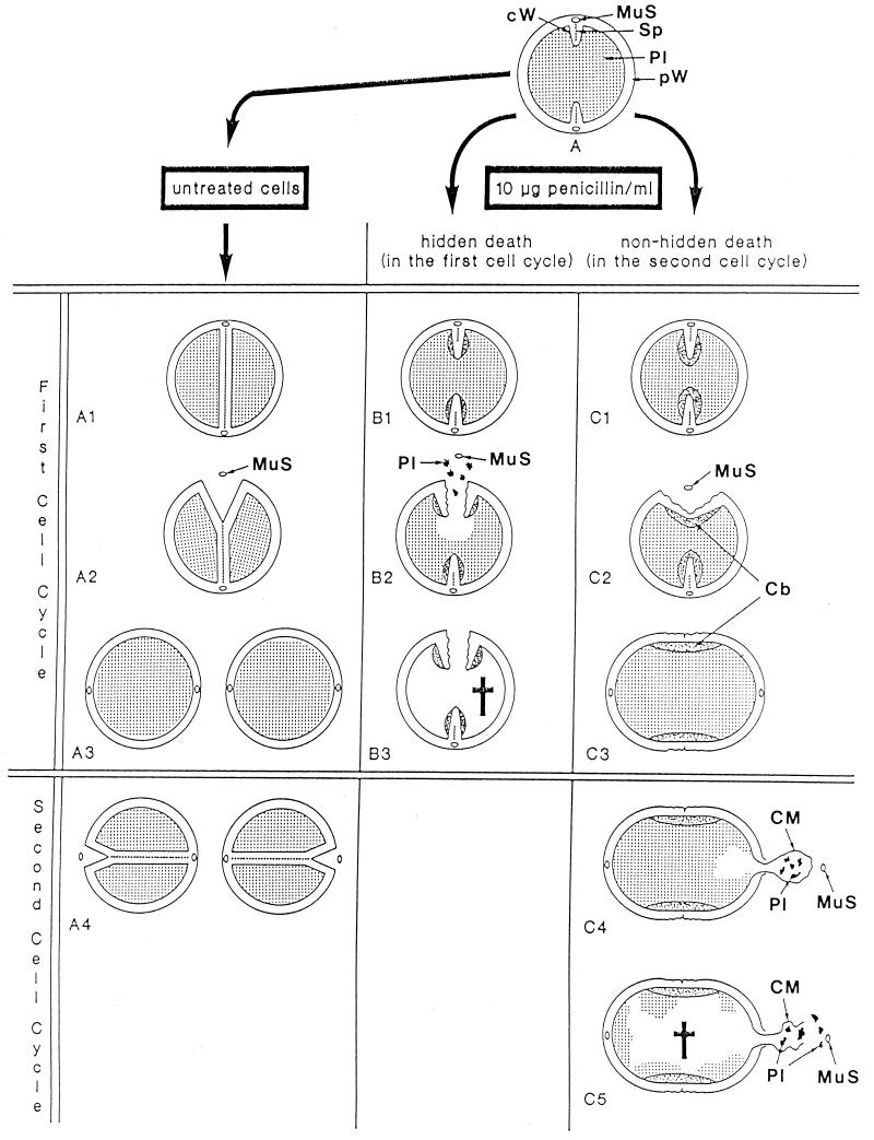
The primary goal of this review is to provide a compilation of the complex architectural features of staphylococcal cell walls and of some of their unusual morphogenetic traits including the utilization of murosomes and two different mechanisms of cell separation. Knowledge of these electron microscopic findings may serve as a prerequisite for a better understanding of the sophisticated events which lead to penicillin-induced death. For more than 50 years there have been controversial disputes about the mechanisms by which penicillin kills bacteria. Many hypotheses have tried to explain this fatal event biochemically and mainly via bacteriolysis. However, indications that penicillin-induced death of staphylococci results from overall biochemical defects or from a fatal attack of bacterial cell walls by bacteriolytic murein hydrolases were not been found. Rather, penicillin, claimed to trigger the activity of murein hydrolases, impaired autolytic wall enzymes of staphylococci. Electron microscopic investigations have meanwhile shown that penicillin-mediated induction of seemingly minute cross wall mistakes is the very reason for this killing. Such "morphogenetic death" taking place at predictable cross wall sites and at a predictable time is based on the initiation of normal cell separations in those staphylococci in which the completion of cross walls had been prevented by local penicillin-mediated impairment of the distribution of newly synthesized peptidoglycan; this death occurs because the high internal pressure of the protoplast abruptly kills such cells via ejection of some cytoplasm during attempted cell separation. An analogous fatal onset of cell partition is considered to take place without involvement of a detectable quantity of autolytic wall enzymes ("mechanical cell separation"). The most prominent feature of penicillin, the disintegration of bacterial cells via bacteriolysis, is shown to represent only a postmortem process resulting from shrinkage of dead cells and perturbation of the cytoplasmic membrane. Several schematic drawings have been included in this review to facilitate an understanding of the complex morphogenetic events.


Figures

FIG. 1
FIG. 1
The structure of peptidoglycan and the sites where peptidoglycan may be attacked by cell wall hydrolases. Three glycan strands of peptidoglycan, consisting of alternating N-acetylmuramic acid and N-acetylglucosamine are depicted. The tetrapeptides (stem peptides), branching from N-acetylmuramic acid, are interconnected by pentaglycine bridges. The sites where cell wall hydrolases may attack peptidoglycan are indicated by arrows, but staphylococci contain only three of these wall hydrolases (amidase, glucosaminidase, and endopeptidase).
FIG. 2
FIG. 2
Scanning electron micrograph (a) and thin sections of staphylococcal cells (b to f). (a) A packet of eight staphylococcal cells was induced by liquoid; this packet is derived from one bacterium by three consecutive cell divisions, each having changed its direction at an angle of 90° to the preceding division plane. The three division planes are indicated by arrows (reproduced with permission from reference 139). (b) Characteristic alternation of consecutive division planes (arrowheads) (reproduced with permission from reference 41). (c) Asymmetrical initiation of cross wall formation (arrowhead), Sp, splitting system (reproduced with permission from reference 41). (d) Centripetal growth of the closing cross wall. The splitting system (Sp) appears as a darker central cross wall layer. (e) At the cell periphery, above the closed cross wall with its splitting system (Sp), there is one of the murosomes (MuS) (reproduced with permission from reference 50). (f) After a 2-h exposure to the antibiotic batumin (1 μg/ml) the peripheral wall appears to be differentiated into an outer layer, the so-called primary wall (prW), and an inner layer, the so-called secondary wall (scW). The dark line between these two layers represents the so-called stripping system (Str) of the staphylococcal cell wall which is involved in cell wall turnover. The remnants of the cutting through of the primary wall, the so-called clefts, are marked by arrows.
FIG. 3
FIG. 3
Nomenclature of the different parts of the staphylococcal cell wall. Schematic overview of the common parts of the cell wall, as seen in the electron microscope by investigating thin sections of fixed staphylococci. (For more details see Fig. 18). (A) Cell wall, splitting system, and murosomes. A highly elastic peripheral cell wall (pW) protects the protoplast against the extremely high turgor of the cytoplasm. The cross wall (cW) contains the splitting system (Sp) consisting of concentrically arranged ring-shaped tubuli. The splitting system is involved in cell separation. Minute, vesicular, extraplasmatic wall organelles, the murosomes (MuS), are located in two circumferential rows above the closed cross wall. They are engaged in lytic processes during cross wall formation and initiation of cell separation. Reference figure, Fig. 2e. (B) Primary and secondary walls. The seemingly homogeneous peripheral cell wall is, in fact, differentiated into an outer layer (the so-called primary wall [prW]) and an inner layer (the so-called secondary wall [scW]). The secondary wall, which continues into the cross wall, is deposited beneath the primary wall in connection with the formation of a new cross wall. The dark line between the primary and the secondary wall represents the so-called stripping system (Str), which is involved in wall turnover. Reference figures, Fig. 2f and Fig. 4a. (C) Clefts. During early stages of cell separation the lytic capacity of the murosomes is activated. The murosomes perforate and, subsequently, cut through the primary wall, sometimes leaving behind characteristic clefts (Cl) on the cell surface. If such clefts are not turned over, they mark the site of cutting through even during later stages in the cell cycle. Reference figures, Fig. 2f and 13b (D) Longitudinal slit of the cross wall. Sandwiched between layers of the cross wall, a preformed, longitudinal slit (Sl) is found in the center of the cross wall, which contains the concentrically arranged tubuli of the splitting system. If the splitting system is removed, the cross wall layers are moved aside, exposing the container-like slit of the cross wall. Reference figure, Fig. 4g.
FIG. 4
FIG. 4
Thin sections of staphylococcal cells. (a) After treatment with penicillin (0.1 μg/ml) the secondary wall (scW) is intensely stained while the primary wall and the central parts of the cross wall (the so-called transitory cross wall material) are hardly stained. A murosome (MuS) is detectable in the secondary wall. (b) This section, running exactly through the middle of the cross wall, reveals the concentrically arranged tubuli of the splitting system. The hexagonally shaped inner edge of the closing cross wall is marked with arrows (reproduced with permission from reference 41). (c) The diameter of the tubuli of the splitting system enlarges continuously during growth in the presence of spermine (arrowheads). (d) Treatment with Triton X-100 likewise resulted in an enlargement of the tubular structures of the staphylococcal splitting system (Sp). (e) By extraction of the LTA, the splitting system disappeared. (f) Isolated cell wall of a staphylococcus after removal of the cytoplasm. The splitting system is still detectable (arrows) (reproduced with permission from reference 78). (g) Extraction of LTA from the isolated cell wall leads not only to the disappearance of the splitting system but also to a premature separation of the central cross wall region (arrows) (reproduced with permission from reference 78).
FIG. 5
FIG. 5
Staphylococcal cells after thin sectioning (a and c to e) or freeze fracturing in the presence of sucrose (b and f) or sodium chloride (g). (a) A pair of murosomes (MuS) is located at the site of a new cross wall initiation. (b) The murosomes (MuS [arrows]) appear to be enclosed by a definite envelope (reproduced with permission from reference 50). (c) Flattened or collapsed vesicular murosomes (arrows) in the peripheral cell wall. (d) For initiation of cross wall formation, the murosomes in both the just-separating daughter cells have been anchored in the secondary wall where they induced centripetal lytic processes which cut the secondary wall. Me, membranous body. (e) Higher magnification of panel d showing details of the new cross wall initiation. At the free ends of the secondary wall created by the lytic activity of the murosomes the first assembling of wall material for the formation of the new cross wall can be seen. A local invagination of the cytoplasmic membrane, the membranous body (Me), consisting of an envelope and a core, is associated with the site of cross wall initiation. (f) A vesicular murosome exhibiting a tubular tail-like extension (arrow) is detectable; this extension connects the extraplasmatic murosome with the surface of the cytoplasm (reproduced with permission from reference 49). (g) The fracturing has exposed an envelope and a vesicular part of the murosome and its tail. At the end of the tail-like extension of the murosome, a ring-like structure is revealed which marks the connection site between the murosome and the cytoplasmic membrane (CM). W, wall.
FIG. 6
FIG. 6
Freeze fractures of staphylococci in the presence of sodium chloride (a and b) or normal freeze fracture (i) and thin sections (c to h) of staphylococcal cells. (a) A row of linearly arranged vesicular murosomes (MuS) on the concave fracture plane (EF). (b) Murosomes (MuS) on the concave (EF) and convex (PF) fracture planes of the cytoplasmic membrane are in different stages of maturation (arrowheads); a ring-like structure is visible in the upper murosomes. (c) In the presence of penicillin (0.1 μg/ml) the underlayered wall material of the secondary wall reveals a higher contrast than the primary wall. Two murosomes (arrows) embedded in the highly contrasted layer are located beneath the primary wall. (d) In the presence of penicillin (0.1 μg/ml) the two murosomes differentiate the nascent cross wall into three parts, a central sector (white arrow) and two lateral ones (black arrows) ★, transparent “lytic” region at the tip of the nascent cross wall. (e) In the presence of penicillin (0.1 μg/ml) the nascent cross wall is divided in three parts by the lytic activity of murosomes (arrows). (f) Even in the presence of trimethoprim (3.13 μg/ml) the murosomes for the second division plane (arrows) are located at a 90° angle to the first division plane. (g) Higher magnification of panel f. The murosomes (arrows) are located outside the protoplast, between the cell wall and an invagination of the cytoplasmic membrane. (h) In the presence of trimethoprim (3.13 μg/ml) the murosomes located between the primary wall and the cytoplasmic membrane (CM) are covered with particles (white arrows) probably derived from the membrane-wall interlayer (MWI) of the cytoplasmic membrane. (i) Hexagonally arranged particles of an isolated cytoplasmic membrane with its membrane-wall interlayer of a control cell (reproduced with permission from reference 43).
FIG. 7
FIG. 7
Cross-wall morphogenesis. This schematic illustration is intended to tentatively represent the involvement of murosomes in morphogenetic processes during neoformation of a staphylococcal cross wall. Becoming acquainted with these morphogenetic steps is an essential prerequisite for understanding the last minutes in the life of a staphylococcus under penicillin (see Fig. 21 and 22). (A) Site for cross-wall neoformation. The murosomes (MuS) are anchored within the secondary wall (scW) directly above the site of the future cross wall initiation. CM, cytoplasmic membrane; MWI, membrane-wall interlayer; prW, primary wall. Reference figures, Fig. 5a and 6c. (B) Initiation of cross wall morphogenesis. For initiation of a new cross wall, the murosomes induce centripetally directed lytic cutting processes considered to separate the secondary wall into three parts: a central sector and two lateral ones. Sometimes the vesicular murosomes show tail-like tubular extensions which, probably, are involved in this cutting process. Reference figure, Fig. 5d to g. (C) Onset of cross wall morphogenesis. The reason for the cutting processes within the secondary wall seems to become evident: the central sector of the sectioned secondary wall starts to form the so-called transitory part of the cross wall, while the two lateral sectors initiate the formation of the so-called permanent parts of the cross wall (see Fig. 12B). Reference figure, Fig. 6d to e. (D) Initiation of the splitting system. No reliable data are available about the genesis of the splitting system (Sp). It is speculated that the splitting system stems from the membrane-wall interlayer of the cytoplasmic membrane. Reference figure, Fig. 2c and d.
FIG. 8
FIG. 8
Formation and positioning of the murosomes. A highly speculative attempt to reconstruct the formation and localization of staphylococcal murosomes, which are derivatives of the cytoplasmic membrane. (A) The primary wall. A part of the primary wall (prW) is depicted. (B-1) Formation of the secondary wall. By apposition growth the secondary wall (scW) is placed beneath the primary wall. The cytoplasmic membrane (CM) is indicated beneath the primary wall. Sandwiched between the wall and the membrane is the so-called membrane-wall interlayer (MWI). (B and B-2) Murosome morphogenesis. The murosomes (MuS) seem to originate from a local invagination of the cytoplasmic membrane followed by evagination of the membrane-wall interlayer (B and B-2). It is not clear, however, whether the secondary wall is formed before genesis of the murosomes (B-1 to B-2) or whether it is synthesized only after the murosomes are formed (B to C). Moreover, it cannot be excluded that murosomes and secondary wall originate synchronously. The surface of the murosomes is covered with particles which seem to originate from the membrane-wall interlayer. Reference figure, Fig. 6g and h. (C) Positioning of the murosomes. At this stage the murosomes are found to be anchored beneath the primary wall within the secondary wall material. The murosomes are always considered to be placed directly above the site where the next cross wall formation will be initiated. If murosome positioning takes place only after formation of the secondary wall (B-1 to B-2) the murosomes must be assumed to be capable of penetrating into the secondary wall (B-2 to C). Reference figure, Fig. 6c.
FIG. 9
FIG. 9
Thin sections of staphylococcal cells. (a) In the presence of sucrose a ring-like structure surrounds the murosome (MuS). (b) In the presence of chloramphenicol (3 μg/ml) the murosome (MuS) appears to be enveloped by a rather thick layer of wall material. (c) In the presence of penicillin (0.1 μg/ml) the murosome is enlarged and its surrounding layer is of rather fibrillar appearance (reproduced with permission from reference 38). (d) During treatment with penicillin (0.05 μg/ml) the compact cross wall has been converted into fibrillar wall material seemingly arranged in an arc-shaped configuration (reproduced with permission from reference 42). (e) Simultaneous treatment of staphylococci with chloramphenicol (20 μg/ml) and penicillin (0.1 μg/ml) has resulted in the formation of an extremely thick cross wall exhibiting a layered architecture. (f) Under penicillin (0.1 μg/ml) cross wall material may be assembled even at an extension of the cytoplasmic membrane (CM), seemingly without any contact with preexisting wall material. (g) Staphylococci having lost their splitting system during penicillin treatment restore this system during growth in drug-free medium (arrows) (reproduced with permission from reference 38).
FIG. 10
FIG. 10
Staphylococcal cells after freeze fracturing (a to c) and thin sectioning (d to g). (a) Cell separation starts with a row of minute wall perforations (pores [arrows]) on the surface of the peripheral wall above the completed cross wall (reproduced with permission from reference 41). (b) After deactivation of autolytic wall hydrolases by chloramphenicol (20 μg/ml) and subsequent reactivation of these enzymes by treatment with lysozyme (10 μg/ml), two parallel rows of pores can be detected on the surface of the peripheral wall (arrowheads) (reproduced with permission from reference 50). (c) After deactivation and subsequent reactivation of autolytic wall enzymes a row of blebs (arrows) is located on the cell surface, indicating the release of murosomes into the medium (reproduced with permission from reference 50). (d) After chloramphenicol-mediated deactivation and lysozyme-induced reactivation of autolytic wall enzymes the release of murosomes (MuS) leaves behind pore-like cavities in the peripheral cell wall (stars) (reproduced with permission from reference 50). (e to g) The peripheral area of the completed cross wall is shown after deactivation and subsequent reactivation of autolytic wall enzymes. An attempt to reconstruct the subsequent steps of murosome-mediated lytic perforation of the peripheral cell wall during which the murosomes seem to disintegrate is shown. (e) A murosome (arrowhead), still consisting of an envelope and a core, appears to be rather well preserved (reproduced with permission from reference 54). (f) The murosome (arrowhead) shows the first signs of swelling and core disintegration (reproduced with permission from reference 54). (g) The murosome (arrowhead), having just perforated the peripheral wall, appears only as an undifferentiated vesicular structure (reproduced with permission from reference 54).
FIG. 11
FIG. 11
Freeze fracturing (a to d) and thin sectioning of staphylococcal cells (e) and B. subtilis (f). (a) After compensating the tension of the elastic cell wall by suspending unfixed cells in 3 M sucrose (a and b), a tripartite architecture of the cross wall was revealed (arrowheads). In the middle of the cross wall the transitory part is located (reproduced with permission from reference 54). (b) During lytic cell separation the central, transitory part of the cross wall is disintegrated (arrowhead) (reproduced with permission from reference 54). (c) After deactivation and subsequent reactivation of autolytic wall enzymes, spoke-like intruding canals or separation scars (stars) appear on the exposed cross wall surface after cell separation. (cW, cross wall, pW, peripheral wall, MuS, murosome) (reproduced with permission from reference 54). (d) A cell recovering from chloramphenicol treatment reveals spoke-like structures (arrows) on the just-exposed surface of the daughter cell during cell separation. (e) After deactivation and subsequent reactivation of autolytic wall enzymes, centripetally directed lytic wall processes (arrowheads) have left behind a lysis-resistant central part of the cross wall (reproduced with permission from reference 54). (f) After deactivation and subsequent reactivation of autolytic wall enzymes, central parts of the cross wall in a B. subtilis cell are disintegrated during cell separation.
FIG. 12
FIG. 12
Components of staphylococcal cross walls. These sketches (modified from reference 54) give preliminary information about the cross wall components involved in cell division and in the different types of cell separation of staphylococci. (A) Situation during initiation of cell separation. Illustrated is a divided staphylococcus with a newly completed cross wall before cell separation liberates the two daughter cells; however, in order to look inside the cross wall with its divers components, the right daughter cell is depicted separately, at some distance from its normal location. Cell separation is just being initiated by the centrifugally directed lytic activity of the murosomes (MuS) which punch two rows of pores (po) into the peripheral cell wall. In slowly growing staphylococci cell separation takes place along the concentrically arranged rings of the splitting system (Sp) which is synthesized during cross wall formation; in rapidly growing staphylococci cell separation takes place along the spoke-shaped canals (spo) which originate only after completion of the cross wall by the centripetally directed lytic activity of the murosomes. The cross wall material located between the two rows of pores including the splitting system is only destined for cell separation and will be disintegrated; this material can only be considered as being transitory parts of the staphylococcal cross wall. Reference figures, Fig. 4b and 11d. (B) Situation after the onset of cell separation. Illustrated are those parts of a completed cross wall which are located directly beneath the peripheral cell wall (pW). In a first lytic step the murosomes (MuS) have punched, via their centrifugal lytic activity, two circumferential rows of pores (po) into the peripheral wall (upward arrow, left side). These pores in the peripheral wall are then torn apart along the perforation line (right row). In a second lytic step the murosomes attack central parts of the cross wall via centripetally directed lytic actions (downward arrow, left side), resulting in the formation of spoke-shaped canals (spo). Between the presumptive cell walls of the future daughter cells (dW) and the peripheral wall of the mother cell (pW) is the location of the so-called stripping system (Str) which is involved in cell wall turnover. Reference figures, Fig. 10a and b and e to g and Fig. 11c to e.
FIG. 13
FIG. 13
Thin sections (a and g) and freeze fractures (b to f) of staphylococcal cells. (a) Cell separation in untreated cells along the small layer of the splitting system without detectable loss of cross wall material. (b) During the separation of untreated cells not even remnants of the concentrically arranged tubuli of the splitting system can be detected on the just-exposed cross wall surfaces of the daughter cells. Only the clefts (arrowheads) are preserved which mark the site of the initial cutting through of the peripheral wall (reproduced with permission from reference 54). (c) After growth in the presence of chloramphenicol (20 μg/ml) and subsequent regeneration in drug-free medium, the concentrically arranged tubuli of the splitting system are preserved on the just-exposed surfaces of the daughter cells (reproduced with permission from reference 44). (d) This untreated cell of S. aureus SA 113 reveals the concentrically arranged tubuli of the splitting system on the cross wall surfaces of both daughter cells during cell separation. (e) After growth in the presence of chloramphenicol (20 μg/ml) and subsequent regeneration in drug-free medium, the next division plane is already initiated beneath the center of the still-preserved concentrically arranged rings of the splitting system (arrowheads) (reproduced with permission from reference 44). (f) In the presence of penicillin (0.1 μg/ml), the murosome-mediated punching of holes into the peripheral wall for cell separation starts in a zigzag-like manner (stars), resulting in the formation of two parallel rows of circumferential pores (reproduced with permission from reference 38). (g) In spite of growth in the presence of penicillin, this murosome just released from the cell appears to be rather well preserved.
FIG. 14
FIG. 14
Thin sections of penicillin-treated staphylococcal cells. (a) Already during growth in the presence of low doses of penicillin (0.01 μg/ml) the formation of the splitting system is blocked and only fibrillar wall material is synthesized instead of a compact cross wall (arrowhead). (b) Pairs of lytic sites are involved in the process of cross wall degradation during cell separation. (c) Under penicillin (0.01 μg/ml), central parts of the cross wall (arrowhead) are lysed without participation of the splitting system (reproduced with permission from reference 54). (d) This section of a penicillin-treated cell (0.1 μg/ml), running parallel to the middle of the cross wall through the intensely stained cross wall fibrils, reveals several murosome-mediated lytic sites located at the cell periphery (arrows). Further presumable lytic sites are indicated by arrowheads (reproduced with permission from reference 50). (e) Section of a penicillin-treated cell (0.1 μg/ml) running parallel to the middle of the cross wall; the lytic sites from the cell periphery have been extended centripetally and form spoke-shaped canals (arrowheads) (reproduced with permission from reference 42). (f) After simultaneous treatment with penicillin (0.1 μg/ml) and liquoid (2 mg/ml) transitory parts of the cross wall disintegrated without, however, affecting the peripheral cell wall above the cross wall, thus more or less inhibiting cell separation.
FIG. 15
FIG. 15
Thin sections (a to c) and a scanning electron micrograph (d) of pseudomulticellular staphylococci. (a) Evans blue-mediated (200 μg/ml) suppression of wall autolysins resulted in the formation of symmetrically arranged pseudomulticellular staphylococci; the two division planes are marked by arrowheads. (b) After simultaneous treatment with penicillin (0.1 μg/ml) and magnesium chloride (0.5%) asymmetrically arranged pseudomulticellular staphylococci were formed due to the inhibition of autolytic wall enzymes. (c) After simultaneous treatment with penicillin (0.05 μg/ml) and trypsin (1%) asymmetrically arranged pseudomulticellular staphylococci were formed via inactivation of autolytic wall enzymes. (d) A femA mutation in S. aureus has induced the formation of pseudomulticellular staphylococci via alteration of the target structure (peptidoglycan) for wall autolysins.
FIG. 16
FIG. 16
Thin sections of staphylococcal cells treated with 20-μg/ml chloramphenicol (a to f) and subsequently transferred into drug-free medium for regeneration (b to f). (a) Considerable wall thickening has taken place (reproduced with permission from reference 40). (b) Wall restoration has started with the formation of a so-called stripping layer (arrowheads) beneath the old thick cell wall; the subsequent synthesis of new wall material has resulted in the formation of a thin peripheral wall underneath the stripping layer (reproduced with permission from reference 44). (c) Removal of superfluous thick wall material starts with the formation of a cutting rim (C1) above the initiation site of the new cross wall. Furthermore, the sequential detachment of the old thick wall material along the stripping layer (St [small arrowheads]) above the underlayered new wall (U [large arrowhead]) is initiated (reproduced with permission from reference 45). (d) The stripping of superfluous thick wall material has proceeded, and the removed wall material is already partially disintegrated above the cutting rim (C1) by a so-called disintegrating system (D). The gap along the stripping layer (St) is enlarged (small arrowheads) (reproduced with permission from reference 45). (e) The continuous disintegration of the thick peripheral wall has already liberated parts of the new peripheral wall from superfluous wall material via sequential activating of the stripping system (St) along the stripping layer (small arrowheads), C2, former cutting rim (reproduced with permission from reference 45). (f) Detachment and disintegration (Di) of thick wall material is sometimes initiated by the formation of periodically arranged holes (arrowheads) (reproduced with permission from reference 44).
FIG. 17
FIG. 17
Wall regeneration sequences after chloramphenicol treatment. Growth-inhibiting bacteriostatic antibiotics, such as chloramphenicol, do not interfere with the synthesis of wall material. Consequently, under such bacteriostatic conditions the slowly growing staphylococci are capable of forming huge masses of wall material, resulting in extremely thick cell walls. After being transferred to normal growth medium, a comprehensive sequence of cutting and separation processes in the thick wall material is initiated to restore the normal width of the cell wall. This simplified schematic drawing gives a survey of these processes. (A to B) Wall thickening. By chloramphenicol-mediated, successive underlayerings of the peripheral cell wall with newly synthesized wall material, the staphylococcal cell wall becomes considerably wider. Reference figure, Fig. 16a. (C) Formation of a stripping layer. After transfer into normal growth medium, staphylococci initiate wall regeneration by placing a so-called stripping layer (Str) beneath the thick peripheral wall. Reference figure, Fig. 16b. (D) Synthesis of a new peripheral wall. Under the stripping layer a completely new peripheral cell wall (pW) is synthesized via inside-to-outside processes of wall assembly. Reference figure, Fig. 16c. (E) Cutting through of the old peripheral wall. Removal of the old and apparently obstructive peripheral wall is initiated by an asymmetrical cutting through of this wall via forming a so-called cutting rim (ri) above the cross wall (cW) which is now synthesized. Reference figure, Fig. 16c. (F) Onset of sequential wall-stripping processes. By activation of autolytic wall hydrolases within the stripping layer (Str), the old superfluous peripheral wall is sequentially peeled off from the underlayered new peripheral wall. Reference figure, Fig. 16d and e. (G) Disintegration of stripped cell walls. The detached old cell wall is disintegrated either already during the stripping of the old peripheral cell wall or after this process. The disintegrating processes start from periodically arranged holes (disintegration system [Di]) located between the stripping layer and the old peripheral wall and result in the liberation of rather large pieces of the old, peripheral cell wall. Reference figure, Fig. 16f. MuS, murosomes. (H and I) Normal growth after detachment of the old cell wall. As soon as the old peripheral wall formed under chloramphenicol is removed, normal growth takes place, starting in staphylococci with completed cross walls, by murosome-mediated punching of pores into the new peripheral wall and subsequent cell separation. Reference figures, Fig. 10a and 13a. Sp, splitting system.
FIG. 18
FIG. 18
Three-dimensional reconstruction of the staphylococcal wall architecture. This sketch depicts all of the essential structural parts of a staphylococcal cell wall which are presently known. CM, cytoplasmic membrane. This membrane, which covers the protoplast, appears in thin sections as a characteristic three-layered structure about 10 nm in width. Reference figure, Fig. 6h. cW, cross wall. The cross wall is differentiated into the central layers comprising about 30% of the cross wall volume including the splitting system (Sp) and the outer layers. The central layers are only destined to be disintegrated during cell separation (transitory cross wall parts [trW]), while the outer layers (dW) represent the presumptive cell walls of the future daughter cells (permanent cross wall parts). Reference figures, Fig. 2d and 11e. Di, disintegrating system. This lytic, wall-disintegrating tool is periodically arranged on the surface of the stripping system (Str) and engaged in cutting through superfluous peripheral cell walls into rather large pieces. Reference figure, Fig. 16f. dW, Presumptive cell wall of the future daughter cell. The outer, permanent layer of the cross wall represents, after completion of the cross wall and subsequent lytic cell separation, the peripheral wall of future daughter cells (see trW below). Reference figure, Fig. 11a, b, and e. MuS, murosome. These minute extraplasmatic wall organelles are found located in two circumferential rows in the peripheral wall above the cross walls, close to the stripping system (Str). They are more or less spherical (diameter about 30 nm) and sometimes equipped with a “tail.” Murosomes contain special autolytic wall enzymes and are involved in three types of lytic disintegration of wall material during wall morphogenesis: (i) initiation of cross wall neoformation, (ii) punching of pores (po) into the peripheral wall, and (iii) attacking transitory cross wall material during cell separation. Reference figures, Fig. 5f and 10e. MuS2, murosomes of the second division plane. They are arranged at a right angle to the first one. Reference figures, Fig. 6a and b. MWI, membrane-wall interlayer. This thin layer sandwiched between the cytoplasmic membrane and the peripheral wall covers the outer surface of the cytoplasmic membrane and, probably, also the murosomes. This layer contains hexagonally arranged particles with center-to-center spacing of about 7 nm. The function of the MWI is unknown. Reference figure, Fig. 6i. po, pore. Pores in the peripheral cell wall are the result of centrifugally directed lytic murosome activities and represent the first step in cell separation. Pore diameters hardly surpass the size of the murosomes. At later stages of cell separation the pores are enlarged and fuse with each other; cutting of circumferential pores marks the beginning of cell separation. Reference figure, Fig. 10a to g. prW, primary wall. Outer layer of the peripheral cell wall. Reference figure, Fig. 2f. pW, peripheral cell wall. The highly elastic peripheral wall determines the bacterial shape and protects the protoplast, having an internal turgor of about 25 atm, against bursting. It functions as an “exoskeleton.” The peripheral wall, about 40 nm wide, is capable of enormous thickening. The seemingly homogeneous peripheral cell wall is, in fact, differentiated into an outer primary wall and an inner secondary wall. Reference figure, Fig. 2f. scW, secondary wall. Inner layer of the peripheral wall which is continued into the cross wall. It is deposited beneath the primary wall in connection with the formation of a new cross wall. The murosomes are positioned in those parts of the secondary wall that are located above the cross wall. Reference figures, Fig. 2f and 6c. Sl, slit. A longitudinal slit in the center of the cross wall which contains the concentrically arranged tubuli of the splitting system. Reference figure, Fig. 4g. Sp, splitting system. The splitting system is located in a container-like longitudinal slit in the center of the cross wall and consists of 14 to 18 concentrically arranged ring-shaped tubuli, each about 7 to 10 nm in diameter. It is involved in mechanical cell separation. Reference figures, Fig. 4b and 13c. spo, spoke-shaped canal. These canals are the result of centripetally directed lytic actions of the murosomes and are formed during lytic cell separation within the transitory cross wall material. Reference figures, Fig. 11c-d and 14e. Str, stripping system. This lytically active system is found sandwiched between the primary wall and the secondary wall and is involved in wall turnover processes. Reference figure, Fig. 16b-c. Str 2, stripping system after removing the cytoplasmic membrane, the membrane-wall interlayer, and the secondary wall, revealing the vast surface of this lytically active system. trW, transitory parts of the cross wall. The transitory parts of the cross wall are only destined to be disintegrated during lytic cell separation (see dW above). Reference figure, Fig. 11a, b, and e.
FIG. 19
FIG. 19
Thin sections of staphylococcal cells grown in the presence of 0.05 (a) or 0.1 (b to f) μg of penicillin/ml. (a) Huge amounts of fibrillar wall material deposited at the cross wall tips have prevented a fatal tearing apart of the nascent cross wall (arrowheads). The premature initiation of cell separation has only resulted in a limited increase of cell size (arc-shaped arrows). (b) A murosome (MuS) is deposited at the initiation site of the second division plane between the cytoplasmic membrane and the peripheral wall. 1, first division plane (reproduced with permission from reference 50). (c) A murosome (MuS) is found within the peripheral wall of the second division plane (2). 1, first division plane (reproduced with permission from reference 50). (d) By attack from the inside, one murosome has disintegrated a sector of some inner layers of the peripheral wall, the so-called secondary wall (arrow), at the initiation site of the second cell division. The outer layers of the peripheral wall, the so-called primary wall, are not yet affected. (e) A pair of murosomes (black stars at the left side) is found deposited within the peripheral wall at the initiation site of the second division plane. The white stars mark the initiation site of the second division plane of the other daughter cell (reproduced with permission from reference 48). (f) By attack from the inside, a pair of murosomes has disintegrated a sector of the secondary wall at the initiation site of the second division plane, leaving behind a rather extended gap in the inner layer of the peripheral wall (arrow) without, however, affecting its outer layers.
FIG. 20
FIG. 20
Thin sectioning (a, b, e), freeze fracturing (c) and scanning electron micrographs (d, f) of staphylococcal cells grown in the presence of 0.1 (a, b, e) or 10 (c, d, f) μg of penicillin/ml. Some cultures were grown in medium supplemented with 3% NaCl and were subsequently transferred to medium with low osmolarity (d and f). (a) The murosome (MuS) has, finally, perforated the outer layers of the peripheral cell wall, the so-called primary wall, at the initiation site of the second division plane. 1, first division plane (reproduced with permission from reference 50). (b) After perforation of the outer, primary wall at the site of the second division plane (2), the murosome (MuS) is ejected together with some plasmatic material (arrowhead). 1, first division plane (reproduced with permission from reference 48). (c) Even in the presence of high concentrations of penicillin the ejection of the murosome can be detected at the initiation site of the second division plane. This ejection starts from a local bulge of the peripheral wall (vo). 1, first division plane. (d) At high concentrations of penicillin the explosion-like liberation of the cytoplasmic membrane (short arrow) has caused a considerable enlargement of the small, murosome-induced wall perforation (E) at the second division plane. 1, first division plane (reproduced with permission from reference 53). (e) A similar explosion-like liberation of the cytoplasmic membrane occurs at low penicillin doses (reproduced with permission from reference 48). (f) At high concentrations of penicillin (10 μg/ml) murosome-induced explosions (stars) can take place simultaneously in the second division planes of both daughter cells, liberating at the same time two cytoplasmic membranes (CM). 1, first division plane.
FIG. 21
FIG. 21
Time course of penicillin-induced death and bacteriolysis. A simplified schematic survey is shown of the events which take place in growing control cells (A) and in staphylococci after the addition of 0.1-μg/ml penicillin (B). The critical first 90 min of treatment with this antibiotic, which include three cell cycles (generation time of staphylococci, 30 min), are illustrated; these events lead to penicillin-induced death and finally to bacteriolysis. First cell cycle. (A1 to A2) In untreated cells a normal, thin, highly organized and complete cross wall will be formed in the first division plane which contains the intact splitting system. Only after this cross wall has been completed do the murosomes of the first cross wall initiate cell separation via perforation of periodically arranged minute pores into the peripheral wall in the first division plane. After such initial step, the subsequent tearing apart of these pores initiates the separation of the two daughter cells. Reference figure, Fig. 10a. (B1 to B2) After the addition of a lethal concentration of penicillin, the staphylococci almost immediately lose the capacity to form a splitting system. Reference figure, Fig. 19a. Furthermore, the cells are now no longer capable of assembling normal, compact cross walls, but they synthesize mainly a network of fine fibrils arranged in rather thick, deformed and often incomplete defective cross walls. Nevertheless, the staphylococci try to start cell separation via murosome-induced perforation of the peripheral wall as if the first cross wall is intact and complete. However, instead of cell separation only rather large, murosome-induced cavities appear in the peripheral wall of the first division plane. Reference figure, Fig. 13f. Second cell cycle. (A3) During separation of the untreated daughter cells the formation of a new cross wall in the second division plane is initiated at a 90° angle with respect to the previous one. Reference figure, Fig. 2b. Cross wall initiation starts with a very localized, murosome-mediated wall lysis which attacks only some inner layers of the peripheral wall; cross wall assembly takes place and proceeds in this small lytic region. Reference figure, Fig. 5e. (B3) In the presence of penicillin, the second division plane is likewise initiated via a very localized, murosome-mediated wall lysis of some inner layers of the peripheral wall. Reference figure, Fig. 19c to f. However, no cross wall assembly takes place here; the cross wall material bound for the second division plane is detoured and deposited further on in the first division plane, so that the deformed, defective first cross wall becomes even thicker. Reference figure, Fig. 19e. (A4) In control cells, only after completion of the cross wall, the next cell separation is initiated via murosome-mediated perforation of the peripheral wall in the second division plane. Reference figure, Fig. 10a. (B4) Like in normal staphylococci, cell separation in the second division plane starts with murosome-mediated punching of pores into the peripheral wall in spite of the fact that in the presence of penicillin no cross wall material has been deposited at this site. Hence, due to their high internal turgor, the cells will burst and eject the murosome and a limited amount of their plasmatic constituents (aneurysm principle). This murosome-induced morphogenetic death takes place about 50 min after addition of the drug. Reference figure, Fig. 20b to f. For further details see Fig. 22. Third cell cycle. (A5) In control cells, already during the course of cell separation in the second division plane, the cross walls for the third division plane are initiated, again at a 90° angle to the previous one. Reference figure, Fig. 2a. (B5) The dead staphylococci, killed in the presence of penicillin during late stages of the second cell cycle, have lost part of their cytoplasm. This loss reduces considerably the tension of the elastic cell wall and results, therefore, in some shrinkage of the cells. The dead staphylococci preserve, however, their seemingly intact cellular integrity and, therefore, hardly differ from bacteria that are still alive. Reference figure, Fig. 23d. (A6) In control cells the cross walls for the third division planes are completed and the next cell separation is initiated (not depicted). (B6) Disintegration of the cell wall and decomposition of the cytoplasm (bacteriolysis) start only about 30 min after penicillin-induced death, leaving behind mainly large pieces of ruptured cell walls, membrane fragments, and plasmatic debris. Reference figure, Fig. 23b. Schematic drawing modified from reference and supplemented and varied. For the sake of simplicity, only one murosome has been depicted at all the murosomal sites.
FIG. 22
FIG. 22
Fatal effects of “normal” lytic wall processes. Two consecutive lytic processes of wall morphogenesis in control cells, induced by the same murosomes in an interval of 10 min, are the key events for understanding penicillin-induced death: (i) initiation of cross wall formation and (ii) initiation of cell separation. In penicillin-treated staphylococci both processes take place during the same phase of wall morphogenesis, at the same site, and in the same way as in control cells. Only some penicillin-induced variations in the distribution of wall material proved to be fatal (for an overview, see Fig. 21). The schematic drawing seeks to provide a visual aid for a better understanding of these crucial processes. The depicted area is the site where both these lytic processes take place during the second cell cycle after the addition of penicillin. (A to D) Initiation of cross wall formation. (A) At the site of the second division plane, murosomes are formed by the cytoplasmic membrane (CM) or its membrane-wall interlayer (MWI) via an evagination process. Pl, cytoplasm; prW, primary wall; scW, secondary wall. Reference figures, Fig. 19b and 6f to h. (B) Immediately before cross wall formation starts, the murosomes (MuS) are found to be located in the lower layer of the peripheral cell wall, the so-called secondary wall. Probably, they have penetrated into this secondary wall or they are formed together with this wall layer. Reference figures, Fig. 19b and c and 6c (see also Fig. 8). (C) Lytic processes of the murosomes, directed to the center of the cell, separate the secondary wall into three parts. Folds of the cytoplasmic membrane indicate the first steps for cross wall formation. Reference figure, Fig. 6d and e. (D) The central part of the secondary cross wall starts the formation of the central, “transitory” layer of the future cross wall while the other parts initiate the “permanent” layers (see Fig. 7). However, while in control cells cross wall formation goes on until it is completed, in the presence of penicillin, cross wall formation at this site ceased because the necessary wall material is deposited at another site; furthermore, lytic processes (lyt 1) within the secondary wall proceed, leaving behind a disintegrated sector in the secondary wall. Reference figure, Fig. 6d and 19c to f. (E and F) Fatal initiation of cell separation. (E) In spite of the fact that in the presence of penicillin there is no cross wall material deposited in the second division plane, staphylococci start normal cell separation with murosome-mediated punching of pores into the primary layer of the peripheral wall via outward directed lytic processes (lyt 2). Reference figure, Fig. 20a. (F) As soon as one of the murosomes (MuS) has succeeded in perforating the outer layer of the peripheral wall and is released into the growth medium, the cell will burst and eject limited amounts of its cytoplasm (Pl), due to its extremely high internal turgor. This death (cross) happens only because a protecting cross wall is missing beneath the single wall perforation. Reference figure, Fig. 20 b to f.
FIG. 23
FIG. 23
Scanning electron micrograph (a) and thin sections (b to d) of staphylococcal cells grown in the presence of penicillin. (a) By varying the osmolarity of the growth medium, several murosomes of the second division plane are ejected simultaneously (arrows); arrowheads mark supposed ejections of murosomes. 1, first division plane (reproduced with permission from reference 50). (b) After a 4-h treatment with 0.1-μg/ml penicillin most cells undergo bacteriolysis and show different degrees of cellular disintegration. (c) Simultaneous treatment with penicillin (0.1 μg/ml) and lysozyme (1 mg/ml) prevents bacteriolysis; the protoplast even remains stabilized in spite of multiple breakages in the peripheral cell wall (arrows) (reproduced with permission from reference 51). (d) After 4 h of simultaneous treatment with 0.1-μg/ml penicillin and 100-μg/ml Evans blue, the staphylococci seem to be intact, although about 99% are already dead.
FIG. 24
FIG. 24
Thin sections (a to c and e to f) and a scanning electron micrograph (d) of staphylococci treated with high doses of penicillin (10 μg/ml). (a) Small amounts of fibrillar wall material are deposited mainly laterally on the nascent cross wall (reproduced with permission from reference 53). (b) In spite of the fact that the cross wall is not yet complete, cell separation has started in the first division plane (1) via ripping up of the cross wall along its splitting system (reproduced with permission from reference 53). (c) Opening of the cell wall via ripping up of the incomplete cross wall along the splitting system has resulted in the eruption of considerable parts of the cytoplasm (Pl) and in cell death (reproduced with permission from reference 53). (d) The upper two dividing staphylococci reveal a certain loss of cytoplasm (Pl) along slit-like openings in the peripheral wall along the first division plane while staphylococci having already completed their cross wall (lower cells) are not affected by the drug. (1, first division plane) (reproduced with permission from reference 53). (e) Deposition of sufficient amounts of newly formed wall fibrils at the tips of the ingrowing cross wall has prevented complete ripping up of the cross wall in spite of the initiation of cell separation (arrows and arrowhead), thus protecting the cells from lysis. (f) The incomplete cross wall of this cell is almost completely ripped up, but the cross wall tips are welded together (asterisks), thus preventing fatal consequences; the paired arrows indicate the region of the peripheral wall which is suggested to be derived from the tearing apart of the cross wall; the elongated cell already shows an initiation site for the formation of the next cross wall (arrow) (reproduced with permission from reference 53).
FIG. 25
FIG. 25
Hidden death at high penicillin concentrations. Schematic drawing illustrating the fate of staphylococcal control cells (A1 to A4) and of cells in the presence of high penicillin concentrations (B1 to B3 and C1 to C5) during the first and the second cell cycles. (A1 to A3) Control cells during the first cell cycle. (A1) Untreated cells divide by completion of their cross wall; (A2) they initiate cell separation via murosome-induced punching of pores into the peripheral wall above the completed cross wall; (A3) two daughter cells have been generated by this first cell separation. MuS, Murosome. (B1 to B3) Hidden death during the first cell cycle. (B1) In the presence of high concentrations of penicillin the formation of the splitting system stops and cell wall synthesis is largely inhibited. Instead of compact, highly organized cross walls, only some loose fibrillar cross wall material is synthesized which is mainly deposited laterally on the ingrowing cross walls, leaving the tips of the cross wall unprotected. Reference figure, Fig. 24a. (B2). Like in untreated cells (A2), cell separation is then initiated in spite of the fact that cross wall formation has not yet been completed. After liberation of the murosomes (MuS), cell separation proceeds along the splitting system synthesized before the action of the drug. Because of the high internal pressure and since the tips of the cross wall are not sufficiently protected by fibrillar cross wall material, the affected cells erupt granular cytoplasm (Pl) through slit-like openings in the peripheral wall. Reference figure, Fig. 24d. (B3) The incomplete cross wall of the first division plane is ripped up along the splitting system, the cell loses considerable parts of its cytoplasm and dies, leaving behind only empty cell walls which still preserve the shape of the primary staphylococcus. Reference figure, Fig. 24c. (C1 to C5) Nonhidden death during the second cell cycle. (C1) For still unknown reasons, a certain percentage of staphylococci treated with high doses of penicillin is still capable of synthesizing considerable amounts of fibrillar cross wall material after blocking the formation of the splitting system. In those cases not only the lateral parts but also the tips of the ingrowing cross walls of the first cell cycle are covered with these fibrils. Reference figure, Fig. 24e. (C2) Since the cross wall fibrils at the tips are capable of welding with each other, initiation of cell separation via murosome-mediated perforations of the peripheral wall (MuS) and ripping up of the cross wall along the primary splitting system will not result in a loss of cytoplasm and in death, and this in spite of the fact that their cross walls are not completed either: a connecting bridge (Cb) formed by the welding of fibrillar cross wall material proves to be tough enough to resist the high internal pressure. Reference figure, Fig. 24e. (C3) Consequently, those cells capable of welding together the individual primary cross walls of the prospective daughter cells can survive at least the first division cycle in the form of more or less elongated cells. Reference figure, Fig. 24f. (C4) Like in normal cells (A4), cell separation during the second cell cycle may be initiated via murosome-mediated formation of peripheral pores in the second division plane (MuS). Since hardly any cross wall material is deposited at this site, the extremely high internal pressure widens one of these pores, and part of the cytoplasmic membrane (CM) together with cytoplasmic constituents (Pl) is thrown out via an explosion-like ejection. Reference figure, Fig. 20d. (C5) Rupture of the thrown-out part of the cytoplasmic membrane (CM) results in the release of much of the granular cytoplasm (Pl), and the cell dies. Reference figure, Fig. 20f. (Modified from reference .)

References

    1. Abraham E P. From penicillins to cephalosporins. In: Kleinkauf H, von Döhren H, editors. 50 years of penicillin application—history and trends. Berlin, Germany: Technische Universität Berlin; 1993. pp. 7–23.
    1. Abraham E P, Fletcher C M, Florey H W, Gardner A D, Heatley N G, Jennings M A. Further observations on penicillin. Lancet. 1941;ii:177–189. - PubMed
    1. Amako K, Umeda A. Scanning electron microscopy of Staphylococcus. J Ultrastruct Res. 1977;58:34–40. - PubMed
    1. Amako K, Umeda A. Concentric circular structure of the cell wall of Staphylococcus epidermidis revealed by scanning electron microscope. J Electron Microsc. 1978;27:147–148. - PubMed
    1. Archibald A R, Coapes H E. Bacteriophage SP50 as a marker for cell wall growth in Bacillus subtilis. J Bacteriol. 1976;125:1195–1206. - PMC - PubMed

MeSH terms

LinkOut - more resources