. 1998 Dec 7;188(11):2091-7.
doi: 10.1084/jem.188.11.2091.

Human toll-like receptor 2 confers responsiveness to bacterial lipopolysaccharide

Affiliations

Human toll-like receptor 2 confers responsiveness to bacterial lipopolysaccharide

C J Kirschning et al. J Exp Med. .

Abstract

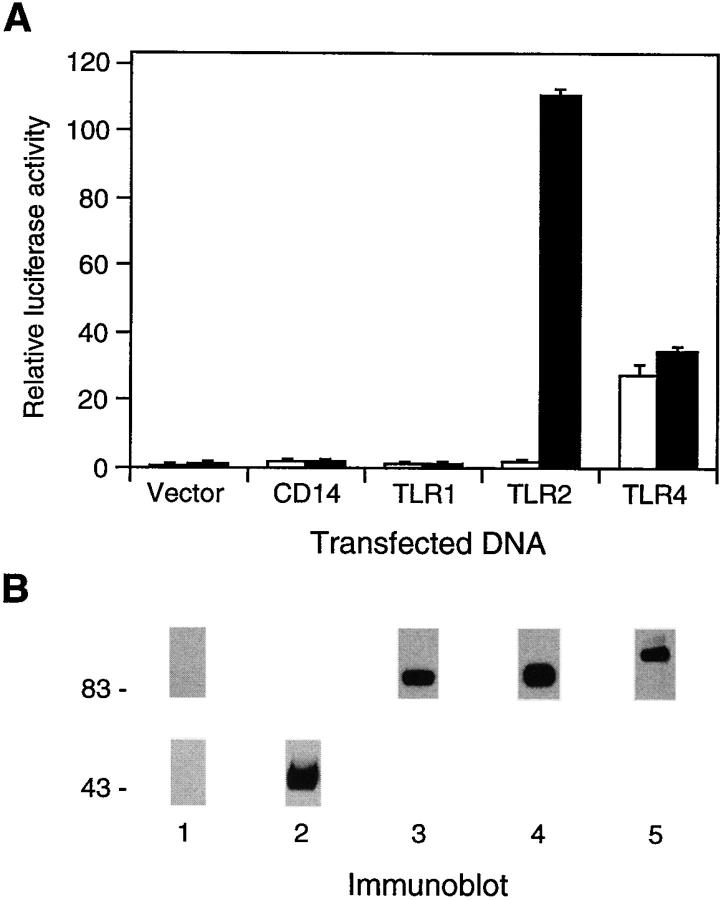
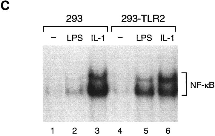
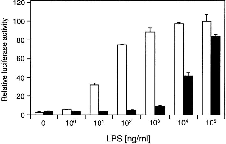
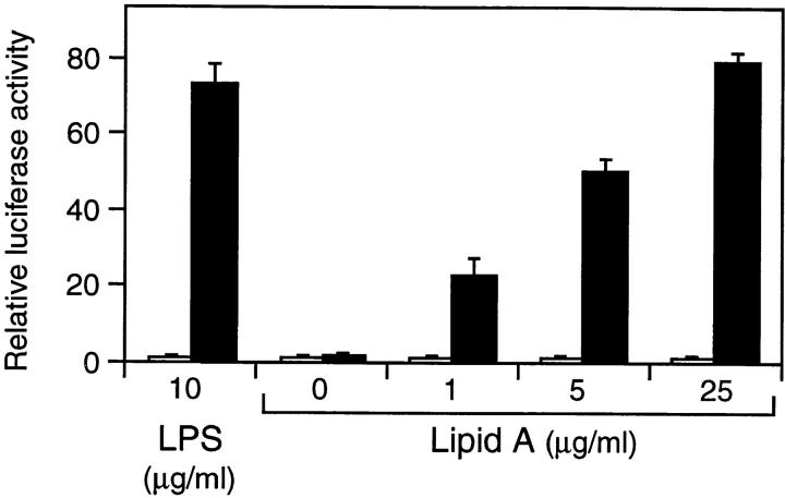
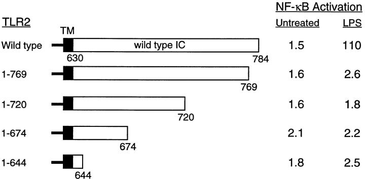
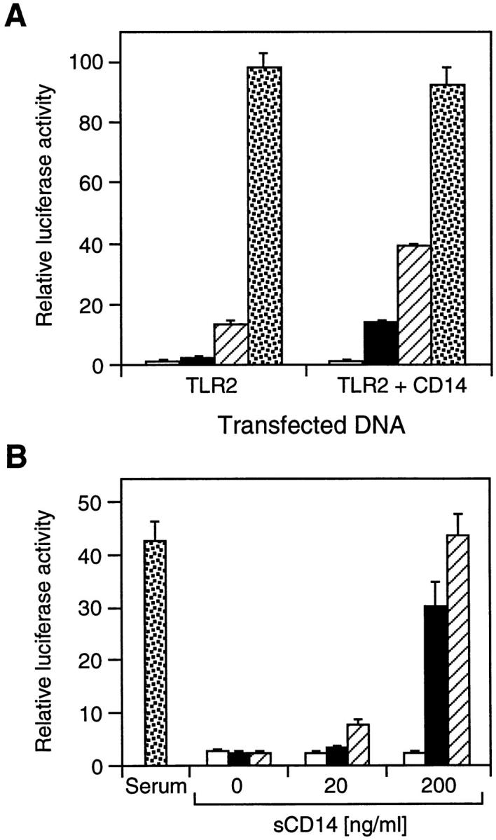
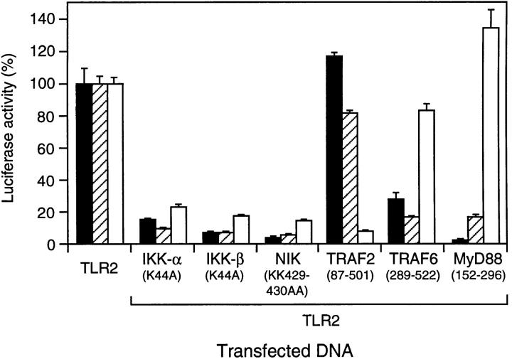
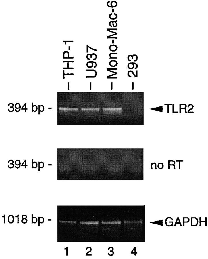
Bacterial lipopolysaccharide (LPS) induces activation of the transcription factor nuclear factor kappaB (NF-kappaB) in host cells upon infection. LPS binds to the glycosylphosphatidylinositol (GPI)- anchored membrane protein CD14, which lacks an intracellular signaling domain. Here we investigated the role of mammalian Toll-like receptors (TLRs) as signal transducers for LPS. Overexpression of TLR2, but not TLR1, TLR4, or CD14 conferred LPS inducibility of NF-kappaB activation in mammalian 293 cells. Mutational analysis demonstrated that this LPS response requires the intracellular domain of TLR2. LPS signaling through TLR2 was dependent on serum which contains soluble CD14 (sCD14). Coexpression of CD14 synergistically enhanced LPS signal transmission through TLR2. In addition, purified recombinant sCD14 could substitute for serum to support LPS-induced TLR2 activation. LPS stimulation of TLR2 initiated an interleukin 1 receptor-like NF-kappaB signaling cascade. These findings suggest that TLR2 may be a signaling component of a cellular receptor for LPS.


Figures

Figure 1
Figure 1
Effect of CD14 and TLRs on LPS-induced NF-κB activation. (A) TLR2-mediated activation of NF-κB by LPS. Luciferase activities were determined in 293 cells transfected with the indicated expression plasmids (1 μg) together with ELAM-1 luciferase reporter plasmid for 24 h, and either left untreated (white bars) or stimulated with LPS (10 μg/ml) for 6 h (black bars) before harvest. (B) Expression of CD14 and TLRs. 293 cells were transiently transfected with aliquots from the same transfection mixtures as in A containing 4 μg of expression plasmid as described in Materials and Methods. Flag epitope–tagged TLRs and CD14 were immunoprecipitated and detected by immunoblotting with anti-Flag antibodies (  lanes 1, 3–5; upper panel  ) and anti-CD14 antibodies (lanes 1 and 2; lower panel), respectively. (C) LPS induction of NF-κB DNA-binding activity by TLR2. EMSA was performed with nuclear extracts from 293 cells and cells stably expressing TLR2 (293-TLR2) that were either left untreated (lanes 1 and 4) or stimulated with LPS (10 μg/ml; lanes 2 and 4) or IL-1 (20 ng/ml; lanes 3 and 6) for 20 min before harvest.
Figure 1
Figure 1
Effect of CD14 and TLRs on LPS-induced NF-κB activation. (A) TLR2-mediated activation of NF-κB by LPS. Luciferase activities were determined in 293 cells transfected with the indicated expression plasmids (1 μg) together with ELAM-1 luciferase reporter plasmid for 24 h, and either left untreated (white bars) or stimulated with LPS (10 μg/ml) for 6 h (black bars) before harvest. (B) Expression of CD14 and TLRs. 293 cells were transiently transfected with aliquots from the same transfection mixtures as in A containing 4 μg of expression plasmid as described in Materials and Methods. Flag epitope–tagged TLRs and CD14 were immunoprecipitated and detected by immunoblotting with anti-Flag antibodies (  lanes 1, 3–5; upper panel  ) and anti-CD14 antibodies (lanes 1 and 2; lower panel), respectively. (C) LPS induction of NF-κB DNA-binding activity by TLR2. EMSA was performed with nuclear extracts from 293 cells and cells stably expressing TLR2 (293-TLR2) that were either left untreated (lanes 1 and 4) or stimulated with LPS (10 μg/ml; lanes 2 and 4) or IL-1 (20 ng/ml; lanes 3 and 6) for 20 min before harvest.
Figure 4
Figure 4
Serum dependence of TLR2-mediated NF-κB activation by LPS. 293 cells were transfected with TLR2 expression plasmid (1 μg) and ELAM-1 luciferase reporter plasmid. After 24 h, cells were stimulated with the indicated concentrations of LPS for 6 h either in the presence (white bars) or absence of 10% fetal bovine serum (black bars) before harvest. Luciferase activities were determined relative to empty vector control.
Figure 2
Figure 2
TLR2-mediated activation of NF-κB by lipid A. 293 cells were transfected with 200 ng empty control vector (white bars) or TLR2 expression plasmid (black bars) and ELAM-1 luciferase reporter plasmid. After 24 h, cells were stimulated with the indicated concentrations of LPS or lipid A for 6 h before determination of luciferase activities.
Figure 3
Figure 3
Analysis of TLR2 deletion mutants. The horizontal bars represent the sequence of TLR2 with transmembrane domain (TM) and intracellular domain (IC) indicated. The amino acids contained in each deletion mutant are stated. 293 cells were transiently cotransfected with ELAM-1 luciferase reporter plasmid and TLR2 expression vectors (1 μg) as indicated. After 24 h, cells were either stimulated with LPS (1 μg/ml) for 6 h or left untreated before harvest. Values shown represent luciferase activities determined relative to empty vector control.
Figure 5
Figure 5
Effect of CD14 on TLR2-mediated NF-κB activation by LPS. (A) Synergistic activation of NF-κB by coexpression of CD14 and TLR2. 293 cells were transfected with ELAM-1 luciferase reporter plasmid (reference 13) and expression vectors for TLR2 (50 ng) and CD14 (10 ng) as indicated. After 24 h, cells were either left untreated (white bars) or stimulated for 6 h with LPS at 1 ng/ml (black bars), 10 ng/ml (hatched bars), and 10 μg/ml (stippled bars) before harvest. Luciferase activities were determined relative to empty vector control. (B) Effect of purified recombinant sCD14 on LPS-induced NF-κB activation by TLR2. 293 cells were transfected with 200 ng TLR2 expression plasmid and ELAM-1 luciferase reporter plasmid. After 24 h, cells were left untreated (white bars) or stimulated for 6 h with LPS at 10 ng/ml (black bars), 100 ng/ml (hatched bars), or 1 μg/ml (stippled bars) in medium containing either serum or purified recombinant sCD14 as indicated. Luciferase activities were determined relative to empty vector control.
Figure 6
Figure 6
Involvement of IL-1 and TNF signaling molecules in TLR2-mediated NF-κB activation by LPS. 293–IL-1RI cells (reference 14) were transfected with ELAM-1 luciferase reporter plasmid and expression vectors (0.3 μg) for TLR2 and dominant-negative mutants of components of the IL-1– and TNF-induced NF-κB pathways as indicated. After 24 h, cells were stimulated for 6 h with LPS (10 μg/ml; black bars), IL-1β (20 ng/ml; hatched bars), or TNF-α (100 ng/ml; white bars) before harvest. Luciferase values were determined and are expressed as percent activation for each stimulus relative to cells transfected with TLR2 alone. LPS, IL-1, and TNF induced 117-, 436-, and 28-fold activation of the reporter gene, respectively, compared with untreated cells.
Figure 7
Figure 7
TLR2 expression in tissue culture cells. Expression of TLR2 mRNA in THP-1 (lane 1), U937 (lane 2), Mono-Mac-6 (lane 3), and 293 cells was analyzed by PCR after RT (top) or without RT (middle). RT-PCR analysis of GAPDH expression was used as control (bottom).

References

    1. Schletter J, Heine H, Ulmer AJ, Rietschel ET. Molecular mechanisms of endotoxin activity. Arch Microbiol. 1995;164:383–389. - PubMed
    1. Ulevitch RJ, Tobias PS. Receptor-dependent mechanisms of cell stimulation by bacterial endotoxin. Annu Rev Immunol. 1995;13:437–457. - PubMed
    1. Belvin MP, Anderson KV. A conserved signaling pathway: the Drosophilatoll-dorsal pathway. Annu Rev Cell Dev Biol. 1996;12:393–416. - PubMed
    1. Hoffmann JA, Reichhart J-M. Drosophilaimmunity. Trends Cell Biol. 1997;7:309–316. - PubMed
    1. Mitcham JL, Partnet P, Bonnert TP, Garka KE, Gerhart MJ, Slack JL, Gayle MA, Dower SK, Sims JE. T1/ST2 signaling establishes it as a member of an expanding interleukin-1 receptor family. J Biol Chem. 1996;271:5777–5783. - PubMed

MeSH terms