. 1998 Dec 7;188(11):2127-37.
doi: 10.1084/jem.188.11.2127.

Yersinia-induced apoptosis in vivo aids in the establishment of a systemic infection of mice

Affiliations

Yersinia-induced apoptosis in vivo aids in the establishment of a systemic infection of mice

D M Monack et al. J Exp Med. .

Abstract

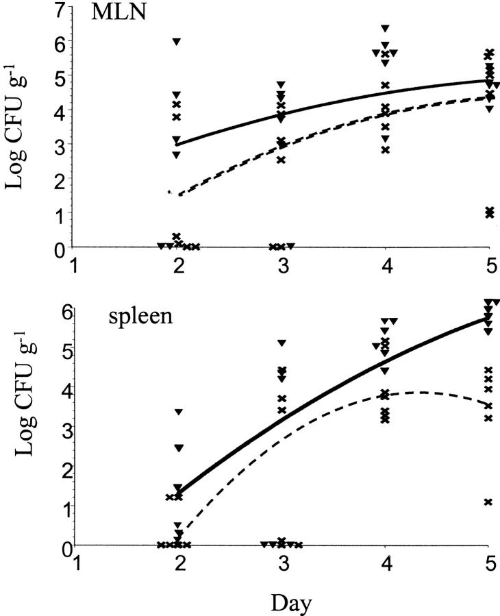
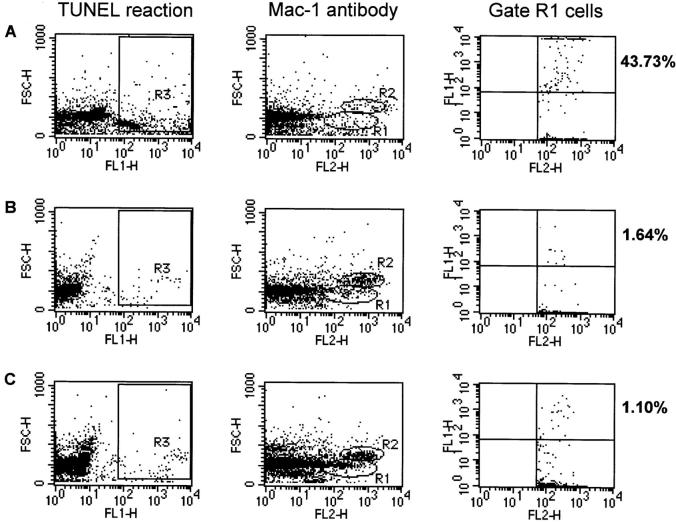
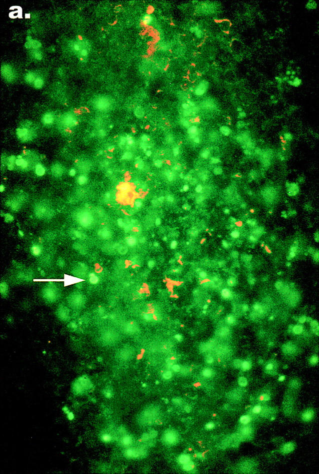
Pathogenic Yersinia cause a systemic infection in mice that is dependent on the presence of a large plasmid encoding a number of secreted virulence proteins called Yops. We previously demonstrated that a plasmid-encoded Yop, YopJ, was essential for inducing apoptosis in cultured macrophages. Here we report that YopJ is a virulence factor in mice and is important for the establishment of a systemic infection. The oral LD50 for a yopJ mutant Yersinia pseudotuberculosis increases 64-fold compared with wild-type. Although the yopJ mutant strain is able to reach the spleen of infected mice, the mutant strain seldom reaches the same high bacterial load that is seen with wild-type Yersinia strain and begins to be cleared from infected spleens on day 4 after infection. Furthermore, when in competition with wild-type Yersinia in a mixed infection, the yopJ mutant strain is deficient for spread from the Peyer's patches to other lymphoid tissue. We also show that wild-type Yersinia induces apoptosis in vivo of Mac-1(+) cells from infected mesenteric lymph nodes or spleens, as measured by quantitative flow cytometry of TUNEL (Tdt-mediated dUTP-biotin nick-end labeling)-positive cells. The levels of Mac-1(+), TUNEL+ cells from tissue infected with the yopJ mutant strain were equivalent to the levels detected in cells from uninfected tissue. YopJ is necessary for the suppression of TNF-alpha production seen in macrophages infected with wild-type Yersinia, based on previous in vitro studies (Palmer, L.E., S. Hobbie, J.E. Galan, and J.B. Bliska. 1998. Mol. Microbiol. 27:953-965). We conclude here that YopJ plays a role in the establishment of a systemic infection by inducing apoptosis and that this is consistent with the ability to suppress the production of the proinflammatory cytokine tumor necrosis factor alpha.


Figures

Figure 1
Figure 1
The yopJ mutant does not colonize spleens as well as wild-type Y. pseudotuberculosis. Colonization kinetics from mice infected orally with 109 YPIIIpYV or 109 YPIIIpyopJ. Log CFU g−1 MLNs or spleen for YPIIIpYV, and YPIIIpyopJ, on days 2, 3, 4, and 5 after inoculation are shown (n = 6 mice/strain for each time point). Curve for YPIIIpYV colonization is a solid line. Curve for YPIIIpyopJ colonization is a dashed line.
Figure 2
Figure 2
An increase in the number of TUNEL-positive cells in infected tissues correlates with YopJ production. Flow cytometry analysis of single cell suspensions prepared from spleens infected for 4 d with (A) YPIIIpYV (n for R1 = 1006) or (B) YPIIIpyopJ (n for R1 = 1546), or (C) from uninfected mice (n for R1 = 2255). TUNEL-positive cells were detected in FL1 and cells immunostained with Mac-1 antibody were detected in FL2. Gates were drawn around three populations of cells. R1 represents a population of cells that were stained with Mac-1 antibody and had a mean forward scatter of 170. R2 represents a population of cells that also stained with Mac-1 antibody and had a mean forward scatter of 350. R3 represents a population of cells that were specifically labeled in the TUNEL reaction and had a positive signal in FL1 relative to negative controls. Percentages indicate the percentage of cells within the gate R1 cell population that were TUNEL positive.
Figure 3
Figure 3
Histology shows cells with condensed nuclei in MLNs infected with wild-type Yersinia. Hematoxylin and eosin staining of fixed MLNs infected with 5 × 10⁹ YPIIIpYV or YPIIIpyopJ for 3 days. (a) This photomicrograph shows an abscess typical of the inflammation seen in mice infected with wild-type Yersinia. (b) At higher magnification note the predominance of condensed nuclei and pyknotic cell debris, (arrowheads) typical of apoptotic cells. (c) Typical lymph node abscess seen in mice infected with the yopJ mutant strain which at higher magnification (d) shows numerous viable inflammatory cells (arrows) that maintain their nuclear morphology. Original magnifications: a and c, ×100; b and d, ×250 oil.
Figure 4
Figure 4
TUNEL reactions show an increase in apoptosis in MLNs infected with wild-type Y. pseudotuberculosis. Sections from the same fixed tissues used for histology were processed as described in Materials and Methods, permeabilized, and subjected to the TUNEL reaction. Nuclei of apoptotic cells in tissue infected with either (a) wild type Yersinia or (b) yopJ mutant Yersinia, were detected with an FITC filter on an epifluorescent microscope (arrows). The presence of TUNEL-positive nuclei in MLNs from the mouse infected with the yopJ mutant was similar to the background level of apoptosis seen in MLNs from uninfected mice. Bacteria were detected with an anti–Y. pseudotuberculosis antibody and a secondary TRITC-conjugated antibody with a rhodamine filter. Original magnification: ×640.
Figure 4
Figure 4
TUNEL reactions show an increase in apoptosis in MLNs infected with wild-type Y. pseudotuberculosis. Sections from the same fixed tissues used for histology were processed as described in Materials and Methods, permeabilized, and subjected to the TUNEL reaction. Nuclei of apoptotic cells in tissue infected with either (a) wild type Yersinia or (b) yopJ mutant Yersinia, were detected with an FITC filter on an epifluorescent microscope (arrows). The presence of TUNEL-positive nuclei in MLNs from the mouse infected with the yopJ mutant was similar to the background level of apoptosis seen in MLNs from uninfected mice. Bacteria were detected with an anti–Y. pseudotuberculosis antibody and a secondary TRITC-conjugated antibody with a rhodamine filter. Original magnification: ×640.

References

    1. Carter PB. Animal model of human disease. Yersinia enteritis. Animal model: oral Yersinia enterocoliticainfection of mice. Am J Pathol. 1975;81:703–706. - PMC - PubMed
    1. Cornelis G, Laroche Y, Balligand G, Sory MP, Wauters G. Yersinia enterocolitica, a primary model for bacterial invasiveness. Rev Infect Dis. 1987;9:64–87. - PubMed
    1. Bottone EJ. Yersinia enterocolitica: a panoramic view of a charismatic microorganism. CRC Crit Rev Microbiol. 1977;5:211–241. - PubMed
    1. Ahvonen P, Sievers K, Aho K. Arthritis associated with Yersinia enterocoliticainfection. Acta Rheumatol Scand. 1969;15:232–253. - PubMed
    1. Bouza E, Dominguez A, Meseguer M, Buzon L, Boixeda D, Revillo MJ, de Rafael L, Martinez-Beltran J. Yersinia enterocoliticaSepticemia. Am J Clin Pathol. 1980;74:404–409. - PubMed

Publication types