. 1998 Dec 7;188(11):2175-80.
doi: 10.1084/jem.188.11.2175.

Dendritic cell survival and maturation are regulated by different signaling pathways

Affiliations

Dendritic cell survival and maturation are regulated by different signaling pathways

M Rescigno et al. J Exp Med. .

Abstract

Although dendritic cell (DC) activation is a critical event for the induction of immune responses, the signaling pathways involved in this process have not been characterized. In this report, we show that DC activation induced by lipopolysaccharide (LPS) can be separated into two distinct processes: first, maturation, leading to upregulation of MHC and costimulatory molecules, and second, rescue from immediate apoptosis after withdrawal of growth factors (survival). Using a DC culture system that allowed us to propagate immature growth factor-dependent DCs, we have investigated the signaling pathways activated by LPS. We found that LPS induced nuclear translocation of the nuclear factor (NF)-kappaB transcription factor. Inhibition of NF-kappaB activation blocked maturation of DCs in terms of upregulation of major histocompatibility complex and costimulatory molecules. In addition, we found that LPS activated the extracellular signal-regulated kinase (ERK), and that specific inhibition of MEK1, the kinase which activates ERK, abrogated the ability of LPS to prevent apoptosis but did not inhibit DC maturation or NF-kappaB nuclear translocation. These results indicate that ERK and NF-kappaB regulate different aspects of LPS-induced DC activation: ERK regulates DC survival whereas NF-kappaB is responsible for DC maturation.


Figures

Figure 1
Figure 1
LPS induces growth arrest and survival of D1 cells after CM deprivation. Cells were incubated either in medium or CM with or without 10 μg/ml of LPS for the indicated times. The number of viable cells remaining at the different time points is shown as a percentage of the cells seeded at time 0.
Figure 2
Figure 2
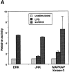
Activation of MAP kinases by LPS. D1 cells were incubated with or without 10 μg/ ml LPS for 15 min. As a positive control for activation of the stress-activated MAP kinase pathways (the JNK and p38/MAPKAP kinase-2 pathways), D1 cells were exposed to high osmolarity conditions by treating them with 0.6 M sorbitol for 15 min. In vitro kinase assays were performed using MBP as a substrate for ERK, GST–c-Jun as a substrate for JNK, and hsp25 as a substrate for MAPKAP kinase-2. (A) MAP kinase activities relative to those in unstimulated cells (defined as 1.0). The transfer of 32P to the various substrates was quantitated with a PhosphorImager. The data represent the average and range of two independent experiments. (B) Representative autoradiographs of the in vitro kinase assays for ERK, JNK, and MAPKAP kinase-2. Lane 1, unstimulated cells; lane 2, LPS-stimulated cells; lane 3, sorbitol-treated cells.
Figure 2
Figure 2
Activation of MAP kinases by LPS. D1 cells were incubated with or without 10 μg/ ml LPS for 15 min. As a positive control for activation of the stress-activated MAP kinase pathways (the JNK and p38/MAPKAP kinase-2 pathways), D1 cells were exposed to high osmolarity conditions by treating them with 0.6 M sorbitol for 15 min. In vitro kinase assays were performed using MBP as a substrate for ERK, GST–c-Jun as a substrate for JNK, and hsp25 as a substrate for MAPKAP kinase-2. (A) MAP kinase activities relative to those in unstimulated cells (defined as 1.0). The transfer of 32P to the various substrates was quantitated with a PhosphorImager. The data represent the average and range of two independent experiments. (B) Representative autoradiographs of the in vitro kinase assays for ERK, JNK, and MAPKAP kinase-2. Lane 1, unstimulated cells; lane 2, LPS-stimulated cells; lane 3, sorbitol-treated cells.
Figure 3
Figure 3
MEK inhibitor does not influence maturation but induces apoptosis of DCs. (A) Flow cytometric analysis of B7.2 and MHC class II in D1 cells. Cells were preincubated or not with 50 μM PD98059 for 30 min and then treated with 10 μg/ml of LPS for 24 h in medium alone (IMDM + 10% FCS). Histograms: black, treated cells; white, untreated cells; broken outline, isotype control. (B) DCs were incubated with 10 μg/ml LPS in the presence or absence of 50 μM PD98059 (I) in medium alone or with or without 50 μM PD98059 in CM. Cells were double stained with annexin V-FITC and propidium iodide and analysed by flow cytometry. Percentage of single positive cells for annexin V-FITC has been plotted. In the inset the number of viable cells remaining at the different time points is shown as a percentage of the cells seeded at time 0. (C) TNF-α production induced by LPS is inhibited by PD98059. Cells were preincubated with 50 or 100 μM inhibitor (I) and then treated for 24 h with 10 μg/ml LPS. Culture supernatants were tested by ELISA for the presence of TNF-α.
Figure 4
Figure 4
(A–C) Nuclear translocation of NF-κB following LPS activation. Cells were stained with a rabbit polyclonal antibody to p65 (Rel A) protein and with anti-rabbit Cy3 conjugated antibody. A, untreated cells; B, cells treated for 30 min with 10 μg/ml LPS; C, cells preincubated for 30 min with 50 μM PD98059 and then treated with 10 μg/ml LPS for an additional 30 min. (D) Flow cytometry of LPS activated D1 cells. D1 cells were preincubated for 90 min with 15 μM TPCK and then treated for 18 h with 10 μg/ml LPS.

References

    1. Banchereau J, Steinman RM. Dendritic cells and the control of immunity. Nature. 1998;392:245–251. - PubMed
    1. Winzler C, Rovere P, Rescigno M, Granucci F, Penna G, Adorini L, Zimmermann VS, Davoust J, Ricciardi-Castagnoli P. Maturation stages of mouse dendritic cells in growth factor-dependent long-term cultures. J Exp Med. 1997;185:317–328. - PMC - PubMed
    1. De Smedt T, Pajak B, Muraille E, Lespagnard L, Heinen E, De Baetselier P, Urbain J, Leo O, Moser M. Regulation of dendritic cell numbers and maturation by lipopolysaccharide in vivo. J Exp Med. 1996;184:1413–1424. - PMC - PubMed
    1. Cella M, Hengering A, Pinet V, Pieters J, Lanzavecchia A. Inflammatory stimuli induces accumulation of MHC class II complexes on dendritic cells. Nature. 1997;388:782–787. - PubMed
    1. May MJ, Ghosh S. Signal transduction through NF-kappa B. Immunol Today. 1998;19:80–88. - PubMed

Publication types