. 1998 Dec;9(12):3367-82.
doi: 10.1091/mbc.9.12.3367.

Nck-2, a novel Src homology2/3-containing adaptor protein that interacts with the LIM-only protein PINCH and components of growth factor receptor kinase-signaling pathways

Y Tu  1 F LiC Wu
Affiliations
Free PMC article

Nck-2, a novel Src homology2/3-containing adaptor protein that interacts with the LIM-only protein PINCH and components of growth factor receptor kinase-signaling pathways

Y Tu et al. Mol Biol Cell. 1998 Dec.
Free PMC article

Abstract

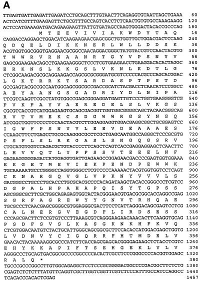
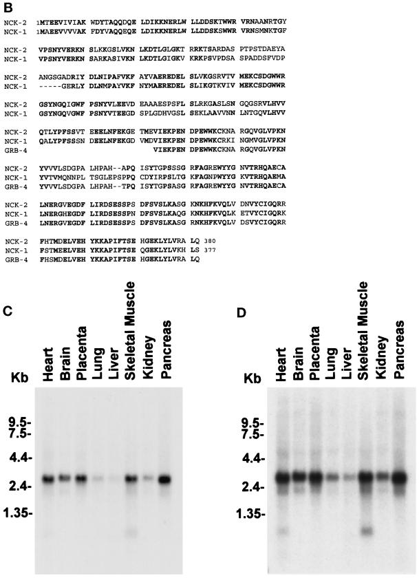
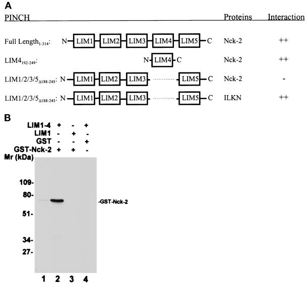
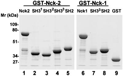
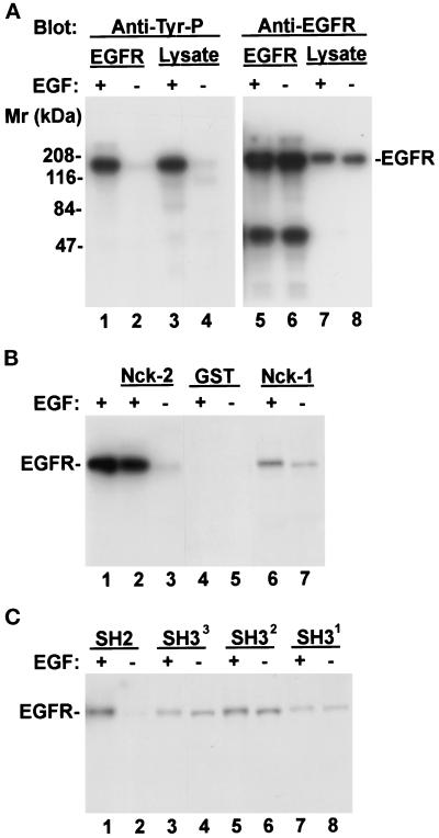
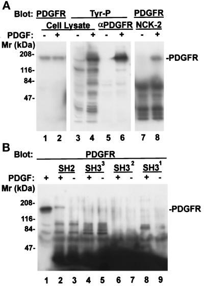
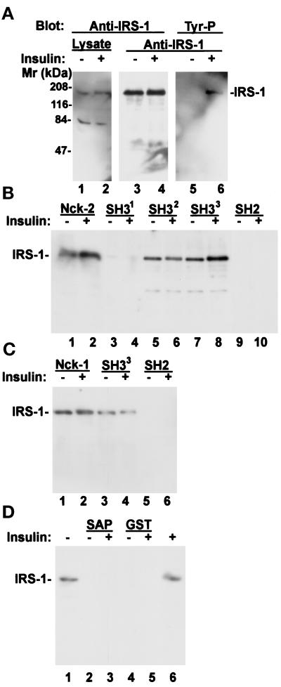
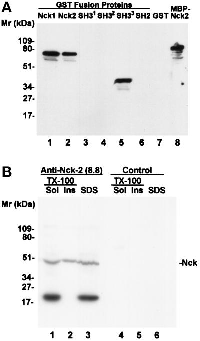
Many of the protein-protein interactions that are essential for eukaryotic intracellular signal transduction are mediated by protein binding modules including SH2, SH3, and LIM domains. Nck is a SH3- and SH2-containing adaptor protein implicated in coordinating various signaling pathways, including those of growth factor receptors and cell adhesion receptors. We report here the identification, cloning, and characterization of a widely expressed, Nck-related adaptor protein termed Nck-2. Nck-2 comprises primarily three N-terminal SH3 domains and one C-terminal SH2 domain. We show that Nck-2 interacts with PINCH, a LIM-only protein implicated in integrin-linked kinase signaling. The PINCH-Nck-2 interaction is mediated by the fourth LIM domain of PINCH and the third SH3 domain of Nck-2. Furthermore, we show that Nck-2 is capable of recognizing several key components of growth factor receptor kinase-signaling pathways including EGF receptors, PDGF receptor-beta, and IRS-1. The association of Nck-2 with EGF receptors was regulated by EGF stimulation and involved largely the SH2 domain of Nck-2, although the SH3 domains of Nck-2 also contributed to the complex formation. The association of Nck-2 with PDGF receptor-beta was dependent on PDGF activation and was mediated solely by the SH2 domain of Nck-2. Additionally, we have detected a stable association between Nck-2 and IRS-1 that was mediated primarily via the second and third SH3 domain of Nck-2. Thus, Nck-2 associates with PINCH and components of different growth factor receptor-signaling pathways via distinct mechanisms. Finally, we provide evidence indicating that a fraction of the Nck-2 and/or Nck-1 proteins are associated with the cytoskeleton. These results identify a novel Nck-related SH2- and SH3-domain-containing protein and suggest that it may function as an adaptor protein connecting the growth factor receptor-signaling pathways with the integrin-signaling pathways.


Figures

Figure 1
Figure 1
Primary structure and tissue distribution of human Nck-2. (A) Nucleotide and deduced amino acid sequences of human Nck-2. The Nck-2 amino acid sequence is shown below the nucleotide sequence, and the stop codon is indicated by a star. The sequence data reported here have been deposited in the GenBank (accession number AF047487). (B) Human Nck-2 protein sequence is aligned with protein sequences of human Nck-1 and mouse GRB-4, and the identical residues are in bold. (C and D) Northern blot analysis of Nck-2 mRNA in human tissues. Two micrograms of polyA+ RNA from human tissues as indicated in the figure were hybridized with a 32P-labeled Nck-2 cDNA probe, and the hybridized mRNA bands were visualized by autoradiography after exposure for 3 (C) or 13 (D) d.
Figure 1
Figure 1
Primary structure and tissue distribution of human Nck-2. (A) Nucleotide and deduced amino acid sequences of human Nck-2. The Nck-2 amino acid sequence is shown below the nucleotide sequence, and the stop codon is indicated by a star. The sequence data reported here have been deposited in the GenBank (accession number AF047487). (B) Human Nck-2 protein sequence is aligned with protein sequences of human Nck-1 and mouse GRB-4, and the identical residues are in bold. (C and D) Northern blot analysis of Nck-2 mRNA in human tissues. Two micrograms of polyA+ RNA from human tissues as indicated in the figure were hybridized with a 32P-labeled Nck-2 cDNA probe, and the hybridized mRNA bands were visualized by autoradiography after exposure for 3 (C) or 13 (D) d.
Figure 2
Figure 2
Analyses of the PINCH-Nck-2 interaction. (A) Yeast two-hybrid binding assays. The cDNAs encoding PINCH sequences were inserted into the pB42AD vector. The cDNAs encoding Nck-2 and ILK sequences were inserted into the pLexA vector. The protein–protein interactions were analyzed by yeast two-hybrid assays as described in MATERIALS AND METHODS. ++, Growth of blue colonies on the leucine-deficient selection medium containing 80 μg/ml X-gal was detected within 1 d; −, no blue colony was detected after 5 d. LIM1/2/3/5▵188–245, PINCH mutant in which the LIM4 domain (residues 188–245) was deleted; LIM4192–249, PINCH mutant containing the LIM4 domain (residues 192–249); ILKN, the N-terminal domain of ILK (residues 1–163). (B) Coprecipitation assays. Affinity-purified GST-Nck-2 (lanes 2 and 3), or GST as a control (lane 4), was mixed with His-tagged PINCH LIM1–4 (residues 1–249)(lanes 2 and 4) or His-tagged PINCH LIM1 (residues 1–70)(lane 3). The GST-Nck-2 was coprecipitated with the His-tagged LIM1–4 and detected by immunoblotting with a polyclonal rabbit anti-GST-Nck-2 antibody as described in MATERIALS AND METHODS. Lane 1 was loaded with 10 ng of affinity- purified GST-Nck-2 fusion protein.
Figure 3
Figure 3
Mapping of the PINCH binding site on Nck-2. The cDNA sequences encoding the full-length or various domains of Nck-2 and Nck-1 were inserted into the pB42AD vector. The cDNAs encoding the full-length PINCH or its LIM4 domain were inserted into the pLexA vector. (A) PINCH binding activity. The numbers in subscript indicate amino acid residues of Nck-2, Nck-1, and PINCH encoded by each construct. W234→K, the conserved W (amino acid residue 234) in the third SH3 domain of Nck-2 was changed to K. The PINCH binding activity was determined based on the interaction of each of the Nck proteins with the LIM4 domain and/or the full length PINCH in yeast two-hybrid assays as described in MATERIALS AND METHODS. ++, Growth of blue colonies on the leucine-deficient selection medium containing 80 μg/ml X-gal was detected within 1 d; +, growth of blue colonies on the leucine-deficient selection medium containing 80 μg/ml X-gal was detected within 2 or 3 d; −, no blue colony was detected after 5 d. ND, not determined. (B) Expression of B42AD fusion proteins containing various Nck-2 and Nck-1 sequences. The expression of the Nck fusion proteins in yeast cells harboring the pLexA-LIM4 and the pB42AD that contains the first SH3 domain of Nck-2 (residues 1–76)(lanes 1 and 9), the second SH3 domain of Nck-2 (residues 115–190)(lane 2), the third SH3 domain of Nck-2 (long form, residues 176–274)(lane 3), the third SH3 domain of Nck-2 (short form, residues 195–274)(lane 4), the SH2 domain of Nck-2 (residues 267–380)(lane 5), the full- length Nck-1 (lane 6), or the full-length Nck-2 (lane 8) were determined by immunoblotting. Yeast cells from 3-ml cultures were extracted with 200 μl of urea/SDS protein extraction buffer (40 mM Tris-HCl, pH 6.8, containing 8 M urea, 5% (wt/vol) SDS, 0.1 mM EDTA, and 0.4 mg/ml) and lanes 1–6, 8, and 9 were loaded with 20 μl of the yeast extracts per lane. Lane 7 was loaded with 1 ng MBP fusion protein containing the full-length Nck-2. Lanes 1–7 were probed with a rabbit anti-Nck antibody raised against a GST fusion protein containing the two C-terminal SH3 domains and the SH2 domain of Nck-2 (residues 115–380), and lanes 8 and 9 were probed with a rabbit anti-HA antibody (Zymed Laboratories; 1 μg/ml).
Figure 4
Figure 4
Expression of recombinant Nck-2 fusion proteins. The GST fusion proteins containing various Nck-2 and Nck-1 sequences were expressed in E. coli cells as described in MATERIALS AND METHODS. The fusion proteins were isolated with glutathione-Sepharose 4B beads, separated on 10% SDS-PAGE (reduced), and detected by staining with Coomassie Brilliant Blue R-250. Lanes were loaded with GST fusion proteins containing the full- length Nck-2 (residues 1–380) (lane 1), the first SH3 domain of Nck-2 (residues 1–76) (lane 2), the second SH3 domain of Nck-2 (residues 115–190) (lane 3), the third SH3 domain of Nck-2 (residues 176–274) (lane 4), the Nck-2 SH2 domain (residues 267–380) (lane 5), the full-length Nck-1 (residues 1–377)(lane 6), the third SH3 domain of Nck-1 (residues 171–270) (lane 7), the Nck-1 SH2 domain (residues 264–377), and GST (lane 9) (10 μg protein/lane).
Figure 5
Figure 5
Association of Nck-2 with EGF receptors. Human A431 epidermoid carcinoma cell monolayers (∼80% confluent) were starved in DMEM medium containing 0.1% FBS for 18 h. At the end of serum starvation, the cells were either harvested (−EGF), or stimulated with 250 ng/ml EGF (+EGF) for 5 min and then lysed. (A) Tyrosine phosphorylation of EGF receptors. The cell lysates (1 ml of 2.8 mg protein/ml) were incubated with 1 μg of rabbit polyclonal anti-EGF receptor IgG (1005, Santa Cruz Biotechnology), and the EGF receptor immune complex was precipitated with 20 μl protein G-Sepharose 4B beads. The EGF receptor immunoprecipitates (EGFR, lanes 1, 2, 5, and 6, ¼ of the immunoprecipitates/lane) and cell lysates (Lysate, lanes 3 and 4, 21 μg protein/lane; lanes 7 and 8, 3 μg protein/lane) were analyzed by immunoblotting with mouse monoclonal anti-phosphotyrosine antibody (PY20, 0.2 μg/ml) (lanes 1–4) or rabbit anti-EGF receptor antibody (1005, 2 μg/ml) (lanes 5–8), appropriate horseradish peroxidase-conjugated secondary antibodies and the SuperSignal chemiluminescent substrate(Pierce Chemical). (B and C) Association of Nck-2 with EGF receptors. The cell lysates (0.9 mg) were incubated with equal amount (10 μg) of GST fusion proteins containing the full-length Nck-2 (panel B, lanes 2 and 3), the full-length Nck-1 (panel B, lanes 6 and 7), each of the four SH domains of Nck-2 (panel C), or GST (panel B, lanes 4 and 5) as indicated in the figure (final volume = 500 μl). The GST fusion proteins and associated proteins were precipitated with 23 μl glutathione-Sepharose 4B beads. The EGF receptors associated with the Nck-2 or Nck-1 fusion proteins were detected with the anti-EGF receptor (1005, 2 μg/ml) by immunoblotting. The exposure times for the x-ray films shown in panels B and C were identical. Lane 1 in panel B was loaded with 4 μg of the EGF-stimulated cell lysate.
Figure 6
Figure 6
Association of Nck-2 with PDGF receptor-β. NIH3T3 cell monolayers (∼80% confluent) were starved in DMEM containing 0.2% FBS for 18 h. At the end of serum starvation, the cells were either harvested (−PDGF), or stimulated with 50 ng/ml PDGF (+PDGF) for 5 min and then lysed. The PDGF receptor-β was precipitated from the cell lysates (1 ml of 2.8 mg protein/ml) with 1 μg of rabbit polyclonal anti-PDGF receptor-β antibody (P-20, Santa Cruz Biotechnology) and protein G-Sepharose 4B beads (panel A, lanes 5 and 6, one half of the immunoprecipitates/lane). The protein tyrosine phosphorylation was determined by immunoblotting with a mouse monoclonal anti-phosphotyrosine antibody (PY20, 0.2 μg/ml) (panel A, lanes 3–6). For GST fusion protein pull-down experiments, the cell lysates (0.37 mg protein) were incubated with 10 μg of the GST fusion protein containing the full-length Nck-2 (panel A, lanes 7 and 8) or its individual SH domains (panel B, lanes 2–9) as indicated in the figure (final volume = 220 μl). The GST fusion proteins were precipitated with glutathione-Sepharose beads. The PDGF receptor-β that was associated with the Nck-2 proteins (panel A, lanes 7 and 8; panel B, lanes 1–9) or those in the total cell lysates (panel A, lanes 1 and 2) were detected with a rabbit anti-PDGF receptor-β antibody (P-20, 1 μg/ml). Lanes 1–4 in panel A and lane 1 in panel B were loaded with cell lysates (21 μg protein/lane).
Figure 7
Figure 7
Association of Nck-2 with IRS-1. Human 293 cell monolayers (∼70% confluent) were starved in serum-free medium (Eagle’s MEM) for 20 h. At the end of serum starvation, the cells were either harvested (−insulin) or stimulated with 6 μg/ml insulin (+insulin) for 5 min and then lysed. (A) Tyrosine phosphorylation of IRS-1. The cell lysates (0.8 mg) were incubated with 1 μg of rabbit polyclonal anti-IRS-1 receptor IgG (C-20, Santa Cruz Biotechnology), and the IRS-1 immune complex was precipitated with protein G-Sepharose 4B beads. The cell lysates (Lysate, lanes 1 and 2; 2 μg protein/lane) and the IRS-1 immunoprecipitates (anti-IRS-1, lanes 3–6, ¼ of the immunoprecipitates/lane) were analyzed by immunoblotting with rabbit anti-IRS-1 antibody (C-20, 1 μg/ml) (lanes 1–4) or mouse monoclonal anti-phosphotyrosine antibody (PY20, 0.2 μg/ml) (lanes 5 and 6), appropriate horseradish peroxidase-conjugated secondary antibodies and the SuperSignal chemiluminescent substrate (Pierce Chemical). (B and C) Association of Nck-2 with IRS-1. The cell lysates (100 μg) were incubated with equalamounts (10 μg) of GST fusion proteins containing the full-length Nck-2 or its individual SH domains (panel B), the full-length Nck-1 or its SH domains (panel C), the SH3 domain of SAP97 (panel D, lanes 2 and 3), or GST alone (panel D, lanes 4 and 5) as indicated in the figure (final volume = 170 μl). The GST fusion proteins and associated proteins were precipitated with glutathione-Sepharose 4B beads. The IRS-1 associated with the Nck-2 or Nck-1 fusion proteins were detected with the anti-IRS-1 antibody (C-20, 1 μg/ml) by immunoblotting. The exposure time for the x-ray films shown in panels B–D was identical. Lanes 1 and 6 in panel D were loaded with cell lysates (6 μg protein/lane).
Figure 8
Figure 8
Immunoblot detection of Nck proteins with monoclonal antibody 8.8. (A) Lanes 1–8 were loaded with GST fusion proteins containing the full-length Nck-1 (lane 1), the full-length Nck-2 (lane 2), the first SH3 domain of Nck-2 (residues 1–76) (lane 3), the second SH3 domain of Nck-2 (residues 115–190) (lane 4), the third SH3 domain of Nck-2 (residues 176–274) (lane 5), the Nck-2 SH2 domain (residues 267–380) (lane 6), GST (lane 7), or an MBP fusion protein containing the full-length Nck-2 (lane 8)(5 ng protein/lane). (B) Equal amount of human 293 cells was extracted with 0.4 ml of SDS sample buffer (2% [wt/vol] SDS, 5% [vol/vol] 2-mercaptoethanol, 10% [vol/vol] glycerol, 0.05% [wt/vol] bromophenol blue in 62.5 mM Tris-HCl, pH 6.8) (lanes 3 and 6), or 0.2 ml of 0.5% Triton X-100 in 10 mM Tris-HCl buffer, pH 7.1, containing 50 mM NaCl, 30 mM sodium pyrophosphate, 50 mM sodium fluoride, 200 μm sodium orthovanadate, 1 mg/ml BSA, 0.2 mM 4-(2-aminoethyl)benzenesulfonylfluoride, HCl, 10 μg/ml aprotinin, 1 μg/ml pepstatin A and 5 μg/ml leupeptin. The Triton X-100 insoluble fraction was pelleted by centrifugation, and then mixed with 0.4 ml (insoluble fraction) of the SDS sample buffer (lanes 2 and 5). The Triton X-100 soluble fraction (total volume = 0.2 ml) was mixed with 0.2 ml of the SDS sample buffer (lanes 1 and 4). Each lane was loaded with 20 μl of the samples as indicated in the figure. The membranes were probed with monoclonal antibody 8.8-conditioned culture supernatant (lanes 1–3) and the unconditioned plain medium (lanes 4–6), respectively.

References

    1. Anton IM, Lu W, Mayer BJ, Ramesh N, Geha RS. The Wiskott-Aldrich Syndrome Protein-interacting Protein (WIP) binds to the adaptor protein Nck. J Biol Chem. 1998;273:20992–20995. - PubMed
    1. Bartfeld NS, Pasquale EB, Geltosky JE, Languino LR. The alpha v beta 3 integrin associates with a 190-kDa protein that is phosphorylated on tyrosine in response to platelet-derived growth factor. J Biol Chem. 1993;268:17270–17276. - PubMed
    1. Birge RB, Knudsen BS, Besser D, Hanafusa H. SH2 and SH3-containing adaptor proteins: redundant or independent mediators of intracellular signal transduction. Genes Cells. 1996;1:595–613. - PubMed
    1. Bokoch GM, Wang Y, Bohl BP, Sells MA, Quilliam LA, Knaus UG. Interaction of the Nck adapter protein with p21-activated kinase (PAK1) J Biol Chem. 1996;271:25746–25749. - PubMed
    1. Chou MM, Fajardo JE, Hanafusa H. The SH2- and SH3-containing Nck protein transforms mammalian fibroblasts in the absence of elevated phosphotyrosine levels. Mol Cell Biol. 1992;12:5834–5842. - PMC - PubMed

Publication types

MeSH terms

Substances

Associated data

LinkOut - more resources