. 1998 Dec;9(12):3399-415.
doi: 10.1091/mbc.9.12.3399.

The cdr2(+) gene encodes a regulator of G2/M progression and cytokinesis in Schizosaccharomyces pombe

Affiliations
Free PMC article

The cdr2(+) gene encodes a regulator of G2/M progression and cytokinesis in Schizosaccharomyces pombe

C S Breeding et al. Mol Biol Cell. 1998 Dec.
Free PMC article

Abstract

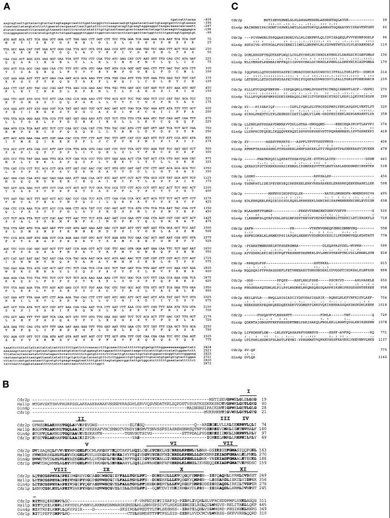
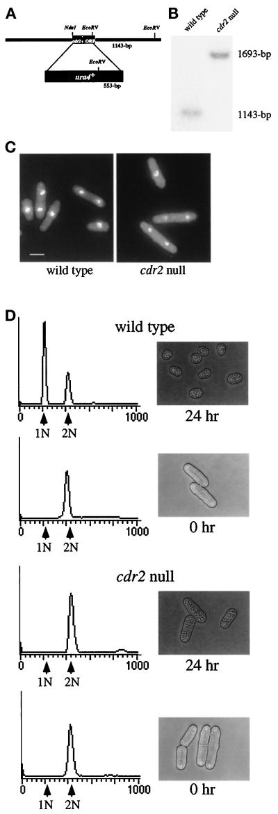
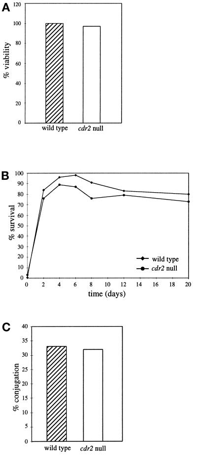
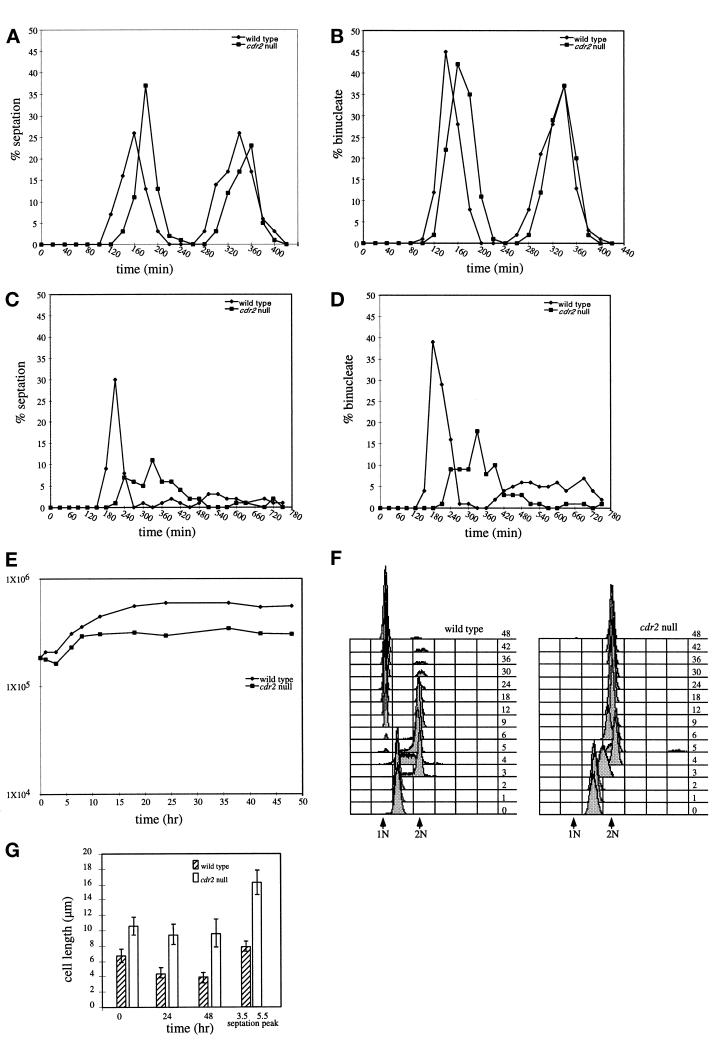
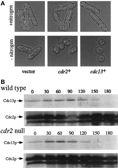
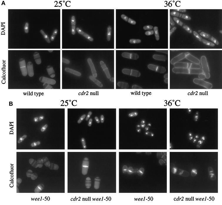
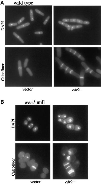
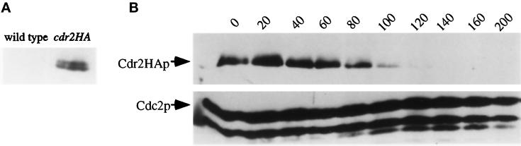
Schizosaccharomyces pombe cells respond to nutrient deprivation by altering G2/M cell size control. The G2/M transition is controlled by activation of the cyclin-dependent kinase Cdc2p. Cdc2p activation is regulated both positively and negatively. cdr2(+) was identified in a screen for regulators of mitotic control during nutrient deprivation. We have cloned cdr2(+) and have found that it encodes a putative serine-threonine protein kinase that is related to Saccharomyces cerevisiae Gin4p and S. pombe Cdr1p/Nim1p. cdr2(+) is not essential for viability, but cells lacking cdr2(+) are elongated relative to wild-type cells, spending a longer period of time in G2. Because of this property, upon nitrogen deprivation cdr2(+) mutants do not arrest in G1, but rather undergo another round of S phase and arrest in G2 from which they are able to enter a state of quiescence. Genetic evidence suggests that cdr2(+) acts as a mitotic inducer, functioning through wee1(+), and is also important for the completion of cytokinesis at 36 degrees C. Defects in cytokinesis are also generated by the overproduction of Cdr2p, but these defects are independent of wee1(+), suggesting that cdr2(+) encodes a second activity involved in cytokinesis.


Figures

Figure 1
Figure 1
Sequence analysis of cdr2+. (A) Nucleotide sequence and predicted amino acid sequence of cdr2+. Sequencing of the cdr2+ complementing DNA generated bases −409 to +2247 that did not include a stop codon. Comparison of this sequence to the S. pombe sequence database revealed that it was identical to a ORF on cosmid c57A10 from chromosome I. Bases +2248 to +2872 were obtained solely from the sequence of cosmid c57A10. The GenBank accession number for the cdr2+ sequence is AF092508. (B) Alignment of the Cdr2p putative catalytic domain with the Snf1p subfamily members S. cerevisiae proteins Hsl1p and Gin4p and S. pombe Cdr1p/Nim1p. Subdomains conserved among protein kinases I–XI are indicated by black lines (Hanks et al., 1988). Amino acid residues conserved in three of four sequences are highlighted in black. (C) Amino acid sequence alignment of Cdr2p and Gin4p. Identical residues are indicated by colons (:). Similar residues are indicated by a period (.).
Figure 2
Figure 2
Figure 2. Deletion of cdr2+. (A) Diagram of the cdr2 deletion construct. (B) Southern blot of a wild-type and a cdr2::ura4+ strain (KGY246 and KGY1519). The 1143-bp band represents the wild- type cdr2+ locus while the 1693-bp band represents the disrupted cdr2::ura4+ locus. (C) Phenotype of the cdr2 null cells. Wild-type and cdr2::ura4+ strains (KGY69 and KGY1475) were grown at 30°C to midlog in YE, fixed with formaldehyde, and stained with DAPI to visualize DNA. Scale bar, 5 μm. (D) Response of cdr2 null and wild-type cells to nitrogen deprivation. Wild-type (upper panels) and cdr2 null (lower panels) strains (KGY69 and KGY1475) were grown at 30°C to midlog, harvested by filtration, washed, and released into minimal medium lacking nitrogen. Samples were collected at 0 and 24 h for DNA content (left-hand panels) and to visualize the phenotype of the cells by light microscopy (right-hand panels).
Figure 3
Figure 3
Cells lacking cdr2+ are able to enter quiescence upon nitrogen deprivation. Wild-type and cdr2 null strains (KGY69 and KGY1475) were grown at 30°C to midlog, filtered, washed, and released into minimal medium lacking nitrogen. Cells were incubated for a total of 20 d with samples collected periodically to determine total viability (A) and heat shock resistance (B). (C) Conjugation assay. Homothallic wild- type and cdr2 null strains were plated onto glutamate agar and incubated at 25°C for 48 h.
Figure 4
Figure 4
Mitotic response to nitrogen deprivation. Wild-type and cdr2 null strains (KGY69 and KGY1475) were grown to midlog phase in minimal medium, and then separated on the basis of size by centrifugal elutriation. The smallest newborn cells were collected and split between two cultures, and each half was filtered, washed, and released into minimal medium (A and B) or minimal medium lacking nitrogen (C, D, E, F, and G). Samples were collected initially every 20 min for 6 h (A and B) or every 30 min for 12 h (C and D), and the percentage of binucleate (B and D) and cells containing a septum (A and C) were determined microscopically. From 12–48 h, samples were collected less frequently, and total cell number (E) and DNA content (F) were determined by Coulter counting and flow cytometry, respectively. Samples of cells grown in medium lacking nitrogen were also collected to measure average cell length at 0, 24, and 48 h and cell length at the first peak of septation (3.3 h for wild type and 5.5 h for cdr2 null) (G).
Figure 5
Figure 5
Cdc13p levels in the cdr2 null. (A) Overexpression of cdc13+ in the cdr2 null. A cdr2 null leu1-32 strain was transformed with either empty pREP1 vector (left panel), pREP1cdr2+ (middle panel), or pREP1cdc13+ (right panel). Transformants were selected and grown to midlog in selective medium. Transformants were then collected, filtered, washed, and released into selective medium lacking nitrogen for 48 h. Micrographs were taken just before nitrogen deprivation (top panels; +nitrogen) and after 48 h (bottom panels; −nitrogen). (B) Detection of endogenous Cdc13p during nitrogen deprivation. Cell samples acquired from the experiment described in Figure 4 were analyzed for Cdc13p levels at 30-min intervals for 3 h. Cdc2p served as a loading control.
Figure 6
Figure 6
cdr2 null and cdr2 null wee1-50 strains at 36°C. (A) cdr2 null at 36°C. Wild-type and cdr2 null strains (KGY69 and KGY1475) were grown to midlog at 25°C (left panels) and to midlog at 36°C (right panels). Cells were fixed in methanol and then stained with DAPI to visualize the DNA (top panels) or with Calcofluor to visualize cell walls and septa (bottom panels). (B) wee1-50 and cdr2 null wee1-50 strains at 36°C. wee1-50 and cdr2 null wee1-50 strains (KGY460 and KGY1636) were grown to midlog at 25°C (left panels) and then shifted to 36°C for 6 h (right panels). Cells were fixed in methanol and then stained with DAPI to visualize the DNA (top panels) or with Calcofluor to visualize cell walls and septa (bottom panels).
Figure 7
Figure 7
Overexpression of cdr2+ in wild-type and wee1 null cells. A leu1-32 strain (KGY246; panel A) and a wee1 null leu 1-32 strain (KGY4; panel B) were transformed with either empty pREP1 vector (left panels) or pREP1cdr2+ (right panels). Transformants were selected and grown to midlog in selective medium containing thiamine. Cells were collected, washed, and released into selective medium lacking thiamine for 18 h. Samples were fixed with methanol and stained with DAPI (top panels) to visualize the DNA or Calcofluor (bottom panels) to visualize cell walls and septa.
Figure 8
Figure 8
Regulation of Cdr2p levels. (A) Detection of Cdr2p-HA. Protein generated from a HA epitope-tagged cdr2+ strain (cdr2HA+; KGY1628) and a wild-type strain (KGY68) was probed with the HA antibody (12CA5) to detect Cdr2p-HA. (B) Detection of Cdr2p-HA levels during nitrogen deprivation. The cdr2HA+ strain (KGY1628) was grown to midlog in minimal medium, and then separated on the basis of size by centrifugal elutriation. Early G2 cells were collected, filtered, washed, and released into minimal medium lacking nitrogen. Time points were taken every 20 min for 3 h to analyze Cdr2p-HA levels. Cdc2p served as a loading control.

References

    1. Aligue R, Wu L, Russell P. Regulation of Schizosaccharomyces pombe Wee1 tyrosine kinase. J Biol Chem. 1997;272:13320–13325. - PubMed
    1. Atman R, Kellogg D. Control of mitotic events by Nap1 and the Gin4 kinase. J Cell Biol. 1997;138:119–130. - PMC - PubMed
    1. Bähler J, Wu J, Longtine MS, Shah NS, McKenzie A, III, Steever AB, Wach A, Philippsen P, Pringle JR. Heterologous molecules for efficient and versatile PCR-based gene targeting in Schizosaccharomyces pombe. Yeast. 1998;14:943–951. - PubMed
    1. Balasubramanian MK, McCollum D, Gould KL. Cytokinesis in the fission yeast Schizosaccharomyes pombe. Methods Enzymol. 1997;283:494–506. - PubMed
    1. Belenguer P, Pelloquin L, Oustrin ML, Ducommun B. Role of the fission yeast nim1 protein kinase in the cell cycle response to nutritional signals. Biochem Biophys Res Commun. 1997;232:204–208. - PubMed

Publication types

MeSH terms