

Semirational design of active tumor suppressor p53 DNA binding domain with enhanced stability
- PMID: 9843948
- PMCID: PMC24508
- DOI: 10.1073/pnas.95.25.14675
Semirational design of active tumor suppressor p53 DNA binding domain with enhanced stability
Abstract
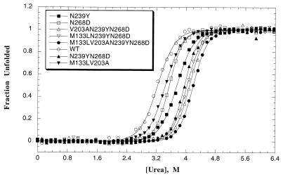
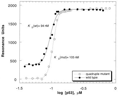
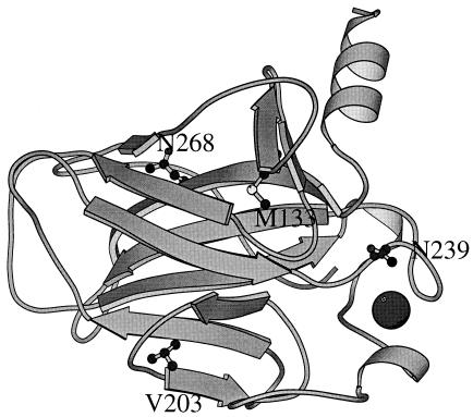
We have designed a p53 DNA binding domain that has virtually the same binding affinity for the gadd45 promoter as does wild-type protein but is considerably more stable. The design strategy was based on molecular evolution of the protein domain. Naturally occurring amino acid substitutions were identified by comparing the sequences of p53 homologues from 23 species, introducing them into wild-type human p53, and measuring the changes in stability. The most stable substitutions were combined in a multiple mutant. The advantage of this strategy is that, by substituting with naturally occurring residues, the function is likely to be unimpaired. All point mutants bind the consensus DNA sequence. The changes in stability ranged from +1.27 (less stable Q165K) to -1.49 (more stable N239Y) kcal mol-1, respectively. The changes in free energy of unfolding on mutation are additive. Of interest, the two most stable mutants (N239Y and N268D) have been known to act as suppressors and restored the activity of two of the most common tumorigenic mutants. Of the 20 single mutants, 10 are cancer-associated, though their frequency of occurrence is extremely low: A129D, Q165K, Q167E, and D148E are less stable and M133L, V203A and N239Y are more stable whereas the rest are neutral. The quadruple mutant (M133LV203AN239YN268D), which is stabilized by 2.65 kcal mol-1 and Tm raised by 5.6 degreesC is of potential interest for trials in vivo.
Figures
References
Publication types
MeSH terms
Substances
LinkOut - more resources
Full Text Sources
Other Literature Sources
Research Materials
Miscellaneous
