. 1998 Dec 8;95(25):14733-8.
doi: 10.1073/pnas.95.25.14733.

Apoprotein B100 has a prolonged interaction with the translocon during which its lipidation and translocation change from dependence on the microsomal triglyceride transfer protein to independence

Affiliations

Apoprotein B100 has a prolonged interaction with the translocon during which its lipidation and translocation change from dependence on the microsomal triglyceride transfer protein to independence

D M Mitchell et al. Proc Natl Acad Sci U S A. .

Abstract

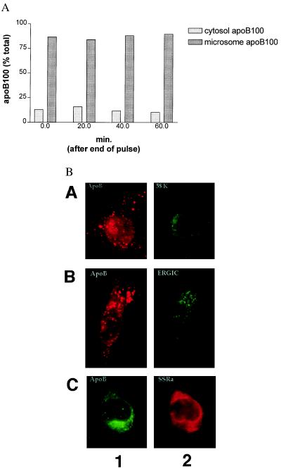
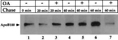
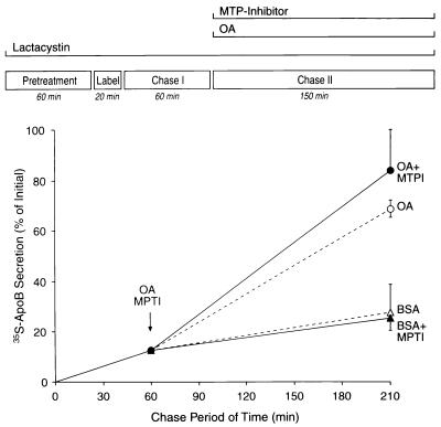
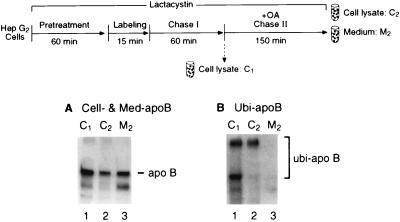
When lipid synthesis is limited in HepG2 cells, apoprotein B100 (apoB100) is not secreted but rapidly degraded by the ubiquitin-proteasome pathway. To investigate apoB100 biosynthesis and secretion further, the physical and functional states of apoB100 destined for either degradation or lipoprotein assembly were studied under conditions in which lipid synthesis, proteasomal activity, and microsomal triglyceride transfer protein (MTP) lipid-transfer activity were varied. Cells were pretreated with a proteasomal inhibitor (which remained with the cells throughout the experiment) and radiolabeled for 15 min. During the chase period, labeled apoB100 remained associated with the microsomes. Furthermore, by crosslinking sec61beta to apoB100, we showed that apoB100 remained close to the translocon at the same time apoB100-ubiquitin conjugates could be detected. When lipid synthesis and lipoprotein assembly/secretion were stimulated by adding oleic acid (OA) to the chase medium, apoB100 was deubiquitinated, and its interaction with sec61beta was disrupted, signifying completion of translocation concomitant with the formation of lipoprotein particles. MTP participates in apoB100 translocation and lipoprotein assembly. In the presence of OA, when MTP lipid-transfer activity was inhibited at the end of pulse labeling, apoB100 secretion was abolished. In contrast, when the labeled apoB100 was allowed to accumulate in the cell for 60 min before adding OA and the inhibitor, apoB100 lipidation and secretion were no longer impaired. Overall, the data imply that during most of its association with the endoplasmic reticulum, apoB100 is close to or within the translocon and is accessible to both the ubiquitin-proteasome and lipoprotein-assembly pathways. Furthermore, MTP lipid-transfer activity seems to be necessary only for early translocation and lipidation events.


Figures

Figure 1
Figure 1
ApoB100 protected from degradation by the proteasome is associated stably with microsomes. (A) HepG2 cells were treated with lactacystin starting 30 min before pulse labeling with [35S]methionine/cysteine. At the end of the 15-min labeling period, the medium was changed to one that was radioisotope-free. At the indicated times in the chase period, cell lysates were collected and separated into supernatant (cytosol) and pellet (microsomes) fractions by ultracentrifugation. Labeled apoB100 in each fraction was measured by immunoprecipitation-SDS/PAGE analysis, and the percentage of distribution was calculated. The results shown are the averages of two separate experiments. (B) HepG2 cells were treated with lactacystin for 2 h and then subjected to double-label immunofluorescence analysis by using antisera to apoB100 and markers for the Golgi apparatus (58-kDa protein; row A), the intermediate compartment (ERGIC; row B), or the ER (SSRα, indicated by SSRa; row C).
Figure 2
Figure 2
ER-associated apoB100 is in close proximity to the translocon protein sec61β before and during lipoprotein assembly. HepG2 cells were treated with lactacystin or MG132 starting 30 min before pulse labeling with [35S]methionine/cysteine. At the end of the 15-min labeling period, the medium was changed to one that was isotope-free and contained either OA complexed to BSA (+) or BSA alone (−). At the indicated times in the chase period, cells were subjected to dithiobis(succinimidyl propionate)-crosslinking and sequential immunoprecipitation analysis with sec61β antiserum followed by apoB100 antiserum to isolate the labeled apoB100 that was in close proximity to the translocon. The immunoprecipitates were resolved by SDS/PAGE, and the labeled apoB100 bands were visualized by fluorography.
Figure 3
Figure 3
Recruitment of proteasome-protected apoB100 to lipoprotein assembly and the secretory pathway and the effects of MTP inhibition. HepG2 cells were treated as summarized at the top of the figure. At the end of the labeling period, samples from three wells were measured to determine the initial cellular pool of labeled apoB100 by immunoprecipitation-SDS/PAGE analysis of lysates. At the ends of the chase I and II periods, conditioned media samples from three wells were taken to measure the cumulative secretion of labeled apoB100. Some wells of HepG2 cells were similarly treated and analyzed, except that an inhibitor of MTP lipid-transfer activity was added at the end of chase I (+MTPI). The results are expressed as the mean percentage (± standard error) of the initial cellular pool.
Figure 4
Figure 4
Recruitment into the secretory pathway of apoB100 protected from degradation by the proteasome involves deubiquitination. HepG2 cells were treated as summarized at the top of the figure. At the ends of chase I (C1) and chase II (C2), cells were harvested to measure labeled apoB100 (A) or to identify cellular apoB100–ubiquitin conjugates (B) by immunoprecipitation-SDS/PAGE analysis of lysates. In addition, at the end of chase II, conditioned medium (M2) was taken for a similar analysis of the content of labeled apoB100 (A) or of ubiquitin–apoB100 conjugates (B). The lower bands in A represent a reported (24) crossreacting nonspecific protein.

References

    1. Borén J, Graham L, Wettesten M, Scott J, White A, Olofsson S. J Biol Chem. 1992;267:9858–9867. - PubMed
    1. Wu X, Zhou M, Huang L-S, Wetterau J, Ginsberg H N. J Biol Chem. 1996;271:10277–10281. - PubMed
    1. Patel S B, Grundy S M. J Biol Chem. 1996;271:18686–18694. - PubMed
    1. Hussain M M, Bakillah A, Jamil H. Biochemistry. 1997;21:13060–13067. - PubMed
    1. Vance J E, Vance D E. Annu Rev Nutr. 1990;10:337–356. - PubMed

Publication types

LinkOut - more resources