. 1998 Dec 8;95(25):14805-10.
doi: 10.1073/pnas.95.25.14805.

The increased level of beta1,4-galactosyltransferase required for lactose biosynthesis is achieved in part by translational control

Affiliations

The increased level of beta1,4-galactosyltransferase required for lactose biosynthesis is achieved in part by translational control

M Charron et al. Proc Natl Acad Sci U S A. .

Abstract

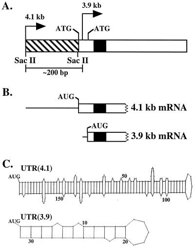
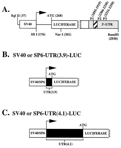
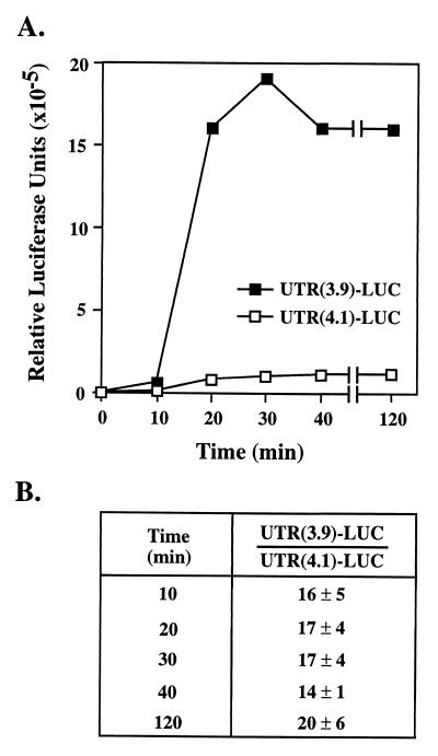
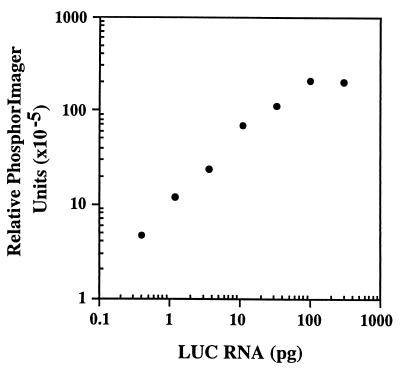
beta1,4-Galactosyltransferase (beta4GalT-I) participates in both glycoconjugate biosynthesis (ubiquitous activity) and lactose biosynthesis (mammary gland-specific activity). In somatic tissues, transcription of the mammalian beta4GalT-I gene results in a 4.1-kb mRNA and a 3.9-kb mRNA as a consequence of initiation at two start sites separated by approximately 200 bp. In the mammary gland, coincident with the increased beta4GalT-I enzyme level ( approximately 50-fold) required for lactose biosynthesis, there is a switch from the 4.1-kb start site to the preferential use of the 3.9-kb start site, which is governed by a stronger tissue-restricted promoter. The use of the 3.9-kb start site results in a beta4GalT-I transcript in which the 5'- untranslated region (UTR) has been truncated from approximately 175 nt to approximately 28 nt. The 5'-UTR of the 4.1-kb transcript [UTR(4.1)] is predicted to contain extensive secondary structure, a feature previously shown to reduce translational efficiency of an mRNA. In contrast, the 5'-UTR of the 3.9-kb mRNA [UTR(3.9)] lacks extensive secondary structure; thus, this transcript is predicted to be more efficiently translated relative to the 4.1-kb mRNA. To test this prediction, constructs were assembled in which the respective 5'-UTRs were fused to the luciferase-coding sequence and enzyme levels were determined after translation in vitro and in vivo. The luciferase mRNA containing the truncated UTR(3.9) was translated more efficiently both in vitro (approximately 14-fold) and in vivo (3- to 5-fold) relative to the luciferase mRNA containing the UTR(4.1). Consequently, in addition to control at the transcriptional level, beta4GalT-I enzyme levels are further augmented in the lactating mammary gland as a result of translational control.


Figures

Figure 1
Figure 1
(A) Schematic representation of exon one of the murine β4GalT-I gene. The bent arrows denote the position of the 4.1-kb transcriptional start site (4.1 kb) and the 3.9-kb transcriptional start site (3.9 kb) relative to the two in-frame ATGs that are separated by 39 bp. The 3.9-kb start site is positioned between the two in-frame ATGs, ≈200 bp downstream of the 4.1-kb start site. The hatched and open rectangles represent the 5′-UTR and coding sequence of the 4.1-kb mRNA, respectively. Note the UTR(3.9) is embedded within the coding sequence of the 4.1 kb-mRNA. The black box indicates the position of the transmembrane domain. (B) The 5′ end of the 4.1- and 3.9-kb β4GalT-I mRNA is shown, where the thin line and open rectangle represent the 5′-UTR and part of the coding sequence, respectively. Translation of the 4.1- and 3.9-kb β4GalT-I mRNA results in two catalytically identical, trans-Golgi resident protein isoforms with NH2-terminal cytoplasmic domains of 24 and 11 aa, respectively. (C) Potential secondary structure of the 5′-UTR of the 4.1- and 3.9-kb β4GalT-I transcript. The long UTR(4.1) has a calculated ΔG of −76 kcal/mol, whereas the short UTR(3.9) has a calculated ΔG of only −7 kcal/mol (13).
Figure 2
Figure 2
(A) Schematic representation of the pGL2-promoter vector. The horizontal arrow indicates the SV40 transcriptional start site, the open box designated SV40 indicates the SV40 promoter, the open box designated LUC indicates the LUC coding sequence, the shaded box indicates the SV40 3′-UTR, and the hatched box indicates the position of the intron. P1 (forward), P2 (reverse), and P3 (reverse) designate the position of the primers used for the RT-PCR experiments. Numbers in parentheses indicate relevant nucleotide positions discussed in the text. (B) Schematic representation of the LUC construct containing either the SV40 or SP6 promoter plus UTR(3.9). (C) Schematic representation of the LUC construct containing either the SV40 or SP6 promoter plus UTR(4.1).
Figure 3
Figure 3
mRNA containing the UTR(3.9) is translated in vitro ≈14-fold more efficiently relative to mRNA containing the UTR(4.1). (A) The two SP6 constructs were linearized with BamHI and transcribed in vitro by using SP6 polymerase. An equal amount of each RNA (0.5 μg) was translated by using the rabbit reticulocyte lysate system as described in Materials and Methods. Aliquots (5 μl) from each reaction were assayed for LUC activity at the indicated times. A representative time course is shown. (B) The ratio between the LUC activity generated by the translation of the UTR(3.9)–LUC mRNA relative to that of the UTR(4.1)–LUC mRNA is shown at different time points. Data were averaged from three independent sets of in vitro-transcribed RNA.
Figure 4
Figure 4
Standard curve for quantitation of LUC RNA. Samples representing 3-fold serial dilutions of in vitro-transcribed LUC RNA (300–0.4 pg) were mixed with 1 μg of total RNA from nontransfected COS-1 cells. RNA was reverse-transcribed by using primer P3, and PCR was performed by using primers P1 and P2 in the presence of [32P]dCTP as described in Materials and Methods. The PCR products were electrophoresed on an agarose gel and transferred to Nytran. The relative intensities of the bands were measured by using the PhosphorImager. The background signal obtained by using 1 μg of total RNA from nontransfected COS-1 cells was subtracted from the signal of each sample.

Similar articles

Cited by

References

    1. Beyer T A, Hill R L. In: The Glycoconjugates. Horowitz M, editor. New York: Academic; 1982. pp. 25–45.
    1. Brodbeck U, Denton W L, Tanahashi N, Ebner K E. J Biol Chem. 1967;242:1391–1397. - PubMed
    1. Brew K, Vanaman T C, Hill R L. Proc Natl Acad Sci USA. 1968;59:491–497. - PMC - PubMed
    1. Hill R L, Brew K, Vanaman T C, Trayer I P, Mattock P. Brookhaven Symp Biol. 1968;21:139–154. - PubMed
    1. Palmiter R D. Biochem J. 1969;113:409–417. - PMC - PubMed

Publication types

MeSH terms

Substances

LinkOut - more resources