

Phosphorylation of the rRNA transcription factor upstream binding factor promotes its association with TATA binding protein
- PMID: 9843972
- PMCID: PMC24532
- DOI: 10.1073/pnas.95.25.14816
Phosphorylation of the rRNA transcription factor upstream binding factor promotes its association with TATA binding protein
Abstract
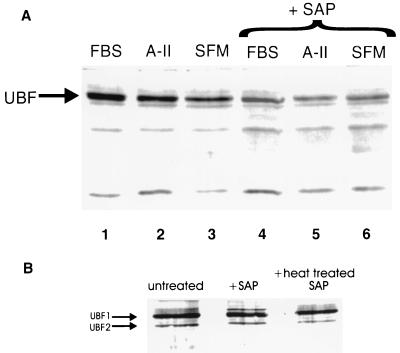
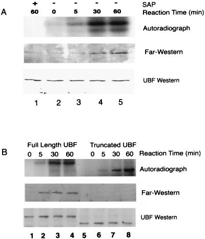
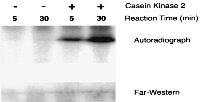
rRNA synthesis by RNA polymerase I requires both the promoter selectivity factor 1, which is composed of TATA binding protein (TBP) and three TBP-associated factors, and the activator upstream binding factor (UBF). Whereas there is strong evidence implicating a role for phosphorylation of UBF in the control of growth-induced increases in rRNA transcription, the mechanism of this effect is not known. Results of immunoprecipitation studies with TBP antibodies showed increased recovery of phosphorylated UBF from growth-stimulated smooth muscle cells. Moreover, using an immobilized protein-binding assay, we found that phosphorylation of UBF in vivo in response to stimulation with different growth factors or in vitro with smooth muscle cell nuclear extract increased its binding to TBP. Finally, we demonstrated that UBF-TBP binding depended on the C-terminal 'acidic tail' of UBF that was hyperphosphorylated at multiple serine sites after growth factor stimulation. Results of these studies suggest that phosphorylation of UBF and subsequent binding to TBP represent a key regulatory step in control of growth-induced increases in rRNA synthesis.
Figures


Similar articles
-
Recruitment of TATA-binding protein-TAFI complex SL1 to the human ribosomal DNA promoter is mediated by the carboxy-terminal activation domain of upstream binding factor (UBF) and is regulated by UBF phosphorylation.Mol Cell Biol. 1999 Apr;19(4):2872-9. doi: 10.1128/MCB.19.4.2872. Mol Cell Biol. 1999. PMID: 10082553 Free PMC article.
-
In vivo evidence that TATA-binding protein/SL1 colocalizes with UBF and RNA polymerase I when rRNA synthesis is either active or inactive.J Cell Biol. 1996 Apr;133(2):225-34. doi: 10.1083/jcb.133.2.225. J Cell Biol. 1996. PMID: 8609157 Free PMC article.
-
A kinase activity associated with simian virus 40 large T antigen phosphorylates upstream binding factor (UBF) and promotes formation of a stable initiation complex between UBF and SL1.Mol Cell Biol. 1999 Apr;19(4):2791-802. doi: 10.1128/MCB.19.4.2791. Mol Cell Biol. 1999. PMID: 10082545 Free PMC article.
-
Mass spectrometric identification of phosphorylation sites of rRNA transcription factor upstream binding factor.Am J Physiol Cell Physiol. 2007 May;292(5):C1617-24. doi: 10.1152/ajpcell.00176.2006. Epub 2006 Dec 20. Am J Physiol Cell Physiol. 2007. PMID: 17182730
-
HMG-boxes, ribosomopathies and neurodegenerative disease.Front Genet. 2023 Aug 3;14:1225832. doi: 10.3389/fgene.2023.1225832. eCollection 2023. Front Genet. 2023. PMID: 37600660 Free PMC article. Review.
Cited by
-
Targeting of Hmo1 to subcompartments of the budding yeast nucleolus.Mol Biol Cell. 2023 Mar 1;34(3):ar22. doi: 10.1091/mbc.E22-07-0261. Epub 2023 Jan 25. Mol Biol Cell. 2023. PMID: 36696177 Free PMC article.
-
The Treacher Collins syndrome (TCOF1) gene product is involved in ribosomal DNA gene transcription by interacting with upstream binding factor.Proc Natl Acad Sci U S A. 2004 Jul 20;101(29):10709-14. doi: 10.1073/pnas.0402492101. Epub 2004 Jul 12. Proc Natl Acad Sci U S A. 2004. PMID: 15249688 Free PMC article.
-
The Ribosomal Gene Loci-The Power behind the Throne.Genes (Basel). 2021 May 18;12(5):763. doi: 10.3390/genes12050763. Genes (Basel). 2021. PMID: 34069807 Free PMC article. Review.
-
Signaling mechanisms in the regulation of renal matrix metabolism in diabetes.Exp Diabetes Res. 2012;2012:749812. doi: 10.1155/2012/749812. Epub 2012 Feb 19. Exp Diabetes Res. 2012. PMID: 22454628 Free PMC article. Review.
-
Transcription factors that influence RNA polymerases I and II: To what extent is mechanism of action conserved?Biochim Biophys Acta Gene Regul Mech. 2017 Feb;1860(2):246-255. doi: 10.1016/j.bbagrm.2016.10.010. Epub 2016 Oct 27. Biochim Biophys Acta Gene Regul Mech. 2017. PMID: 27989933 Free PMC article. Review.
References
Publication types
MeSH terms
Substances
Grants and funding
LinkOut - more resources
Full Text Sources
