

Mitogen-activated protein kinase and neural specification in Xenopus
- PMID: 9843975
- PMCID: PMC24535
- DOI: 10.1073/pnas.95.25.14833
Mitogen-activated protein kinase and neural specification in Xenopus
Abstract
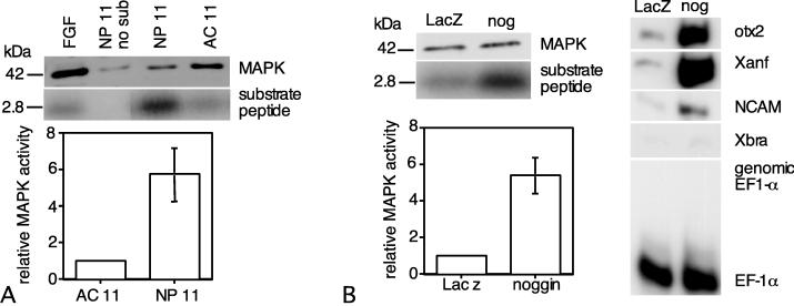
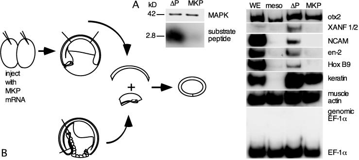
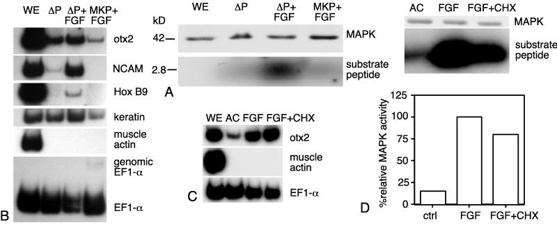
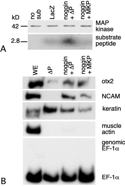
We have investigated the activity and function of mitogen-activated protein kinase (MAPK) during neural specification in Xenopus. Ectodermal MAPK activity increased between late blastula and midgastrula stages. At midgastrula, MAPK activity in both newly induced neural ectoderm and ectoderm overexpressing the anterior neural inducer noggin was 5-fold higher than in uninduced ectoderm. Overexpression of MAPK phosphatase-1 (MKP-1) in ectoderm inhibited MAPK activity and prevented neurectoderm-specific gene expression when the ectoderm was recombined with dorsal mesoderm or treated with fibroblast growth factor (FGF). Neurectoderm-specific gene expression was observed, however, in ectoderm overexpressing both noggin and MKP-1. To evaluate the role of MAPK in posterior regionalization, ectodermal isolates were treated with increasing concentrations of FGF and assayed for MAPK activity and neurectoderm-specific gene expression. Although induction of posterior neural ectoderm by FGF was accompanied by an elevation of MAPK activity, relative MAPK activity associated with posterior neural fate was no higher than that of ectoderm specified to adopt an anterior neural fate. Thus, increasingly posterior neural fates are not correlated with quantitative increases in MAPK activity. Because MAPK has been shown to down-regulate Smad1, MAPK may disrupt bone morphogenetic protein 4 (BMP-4) signaling during neural specification. Our results suggest that MAPK plays an essential role in the establishment of neural fate in vivo.
Figures


Similar articles
-
Regulation of MAP kinase by the BMP-4/TAK1 pathway in Xenopus ectoderm.Dev Biol. 2001 Aug 15;236(2):259-70. doi: 10.1006/dbio.2001.0338. Dev Biol. 2001. PMID: 11476570
-
Evidence for antagonism of BMP-4 signals by MAP kinase during Xenopus axis determination and neural specification.Differentiation. 2003 Sep;71(7):434-44. doi: 10.1046/j.1432-0436.2003.7107006.x. Differentiation. 2003. PMID: 12969336
-
Fibroblast growth factor is a direct neural inducer, which combined with noggin generates anterior-posterior neural pattern.Development. 1995 Nov;121(11):3627-36. doi: 10.1242/dev.121.11.3627. Development. 1995. PMID: 8582276
-
Ectodermal patterning in vertebrate embryos.Dev Biol. 1997 Feb 1;182(1):5-20. doi: 10.1006/dbio.1996.8445. Dev Biol. 1997. PMID: 9073437 Review.
-
Calcium transients and calcium signalling during early neurogenesis in the amphibian embryo Xenopus laevis.Biochim Biophys Acta. 2006 Nov;1763(11):1184-91. doi: 10.1016/j.bbamcr.2006.08.005. Epub 2006 Aug 10. Biochim Biophys Acta. 2006. PMID: 16987559 Review.
Cited by
-
BMP antagonists and FGF signaling contribute to different domains of the neural plate in Xenopus.Dev Biol. 2010 Jan 15;337(2):335-50. doi: 10.1016/j.ydbio.2009.11.008. Epub 2009 Nov 10. Dev Biol. 2010. PMID: 19913009 Free PMC article.
-
Posttranscriptional control of the stem cell and neurogenic programs by the nonsense-mediated RNA decay pathway.Cell Rep. 2014 Feb 27;6(4):748-64. doi: 10.1016/j.celrep.2014.01.028. Epub 2014 Feb 13. Cell Rep. 2014. PMID: 24529710 Free PMC article.
-
Neural induction in the absence of organizer in salamanders is mediated by MAPK.Dev Biol. 2007 Jul 15;307(2):282-9. doi: 10.1016/j.ydbio.2007.04.049. Epub 2007 May 6. Dev Biol. 2007. PMID: 17540356 Free PMC article.
-
Interplay between electrical activity and bone morphogenetic protein signaling regulates spinal neuron differentiation.Proc Natl Acad Sci U S A. 2012 Oct 2;109(40):16336-41. doi: 10.1073/pnas.1202818109. Epub 2012 Sep 18. Proc Natl Acad Sci U S A. 2012. PMID: 22991474 Free PMC article.
-
Transcriptome Regulation by Oncogenic ALK Pathway in Mammalian Cortical Development Revealed by Single-Cell RNA Sequencing.Cereb Cortex. 2021 Jul 5;31(8):3911-3924. doi: 10.1093/cercor/bhab058. Cereb Cortex. 2021. PMID: 33791755 Free PMC article.
References
-
- Harland R, Gerhart J. Annu Rev Cell Biol. 1997;11:611–667. - PubMed
-
- Wilson P A, Hemmati-Brivanlou A. Neuron. 1997;18:699–710. - PubMed
-
- Sasai Y, DeRobertis E M. Dev Biol. 1997;182:5–20. - PubMed
-
- Sato S, M, Sargent T D. Dev Biol. 1989;134:263–266. - PubMed
-
- Godsave S F, Slack J M W. Dev Biol. 1989;134:486–490. - PubMed
Publication types
MeSH terms
Substances
LinkOut - more resources
Full Text Sources
Miscellaneous
