. 1998 Dec 8;95(25):14932-7.
doi: 10.1073/pnas.95.25.14932.

Improved cervical smear assessment using antibodies against proteins that regulate DNA replication

Affiliations

Improved cervical smear assessment using antibodies against proteins that regulate DNA replication

G H Williams et al. Proc Natl Acad Sci U S A. .

Abstract

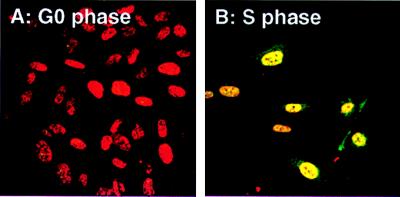
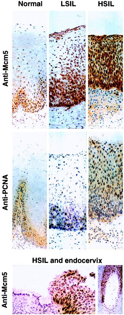
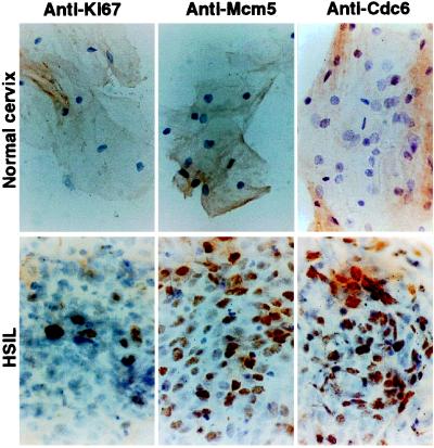
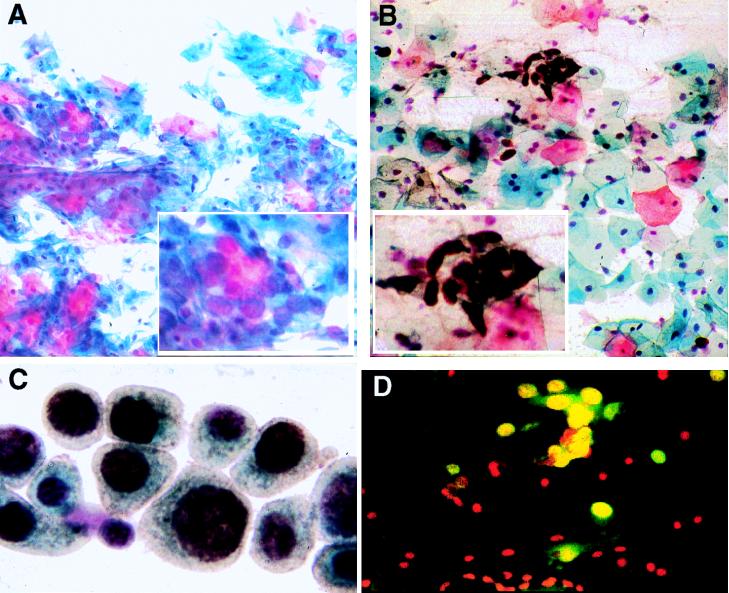
Carcinoma of the cervix is one of the most common malignancies. Papanicolaou (Pap) smear tests have reduced mortality by up to 70%. Nevertheless their interpretation is notoriously difficult with high false-negative rates and frequently fatal consequences. We have addressed this problem by using affinity-purified antibodies against human proteins that regulate DNA replication, namely Cdc6 and Mcm5. These antibodies were applied to sections and smears of normal and diseased uterine cervix by using immunoperoxidase or immunofluorescence to detect abnormal precursor malignant cells. Antibodies against Cdc6 and Mcm5 stain abnormal cells in cervical smears and sections with remarkably high specificity and sensitivity. Proliferation markers Ki-67 and proliferating cell nuclear antigen are much less effective. The majority of abnormal precursor malignant cells are stained in both low-grade and high-grade squamous intraepithelial lesions. Immunostaining of cervical smears can be combined with the conventional Pap stain so that all the morphological information from the conventional method is conserved. Thus antibodies against proteins that regulate DNA replication can reduce the high false-negative rate of the Pap smear test and may facilitate mass automated screening.


Figures

Figure 1
Figure 1
Cdc6 is expressed in cycling but not quiescent human cells. (A) Contact inhibited, quiescent, or (B) synchronized S phase newborn human fibroblasts were grown on coverslips and stained with propidium iodide to reveal DNA (red) and with anti-Cdc6 antibody followed by secondary fluorescein isothiocyanate-conjugated antibody (green: note that green + red = yellow). Magnification: ×130.
Figure 2
Figure 2
Immunoperoxidase staining of frozen sections of normal cervix, LSILs, and HSILs with antibodies against PCNA and Mcm5. Frozen sections of normal cervix, LSILs, and HSILs were immunostained for Mcm5 or PCNA (as a conventional proliferation marker) by the immunoperoxidase method. In normal cervix, surface cells likely to be sampled by cervical smear examination are negative with both antibodies. In LSIL with associated koilocytosis (cells showing HPV cytopathic effect), anti-PCNA antibodies stain basal and some parabasal nuclei, but surface koilocytes are negative. In contrast, anti-Mcm5 antibodies stain nuclei in superficial as well as basal epithelial layers including most koilocytes. In HSIL, anti-PCNA antibodies show focal staining of 10% of nuclei in the surface layers, whereas anti-Mcm5 antibodies stain virtually all nuclei, including those at the epithelial surface. Antibodies against Ki-67 (another conventional proliferation marker), show essentially similar results to anti-PCNA. In contrast, antibodies against Cdc6 stain all cells in HSIL as seen with anti-Mcm5 (data not shown; Top and Middle; magnification ×108). Although HSIL cells show strong nuclear immunostaining with anti-Mcm5, endocervical surface, and glandular cells are negative (Bottom Left, magnification ×225; Bottom Right, magnification ×87).
Figure 3
Figure 3
Immunoperoxidase staining of cells obtained by cervical smear examination from surface of normal ectocervix or HSIL with antibodies against Ki-67, Mcm5, or Cdc6. Superficial squamous cells from normal ectocervix show no nuclear staining with any of the antibodies tested. In HSIL, anti-Ki-67 antibodies (and anti-PCNA; data not shown) show nuclear staining of only a minority population of the abnormal cells in the exfoliated sheets. In contrast, anti-Mcm5 and anti-Cdc6 antibodies stain most HSIL cells. Magnification: ×240.
Figure 4
Figure 4
Staining of HSIL cells in cervical smears by Pap stain alone (A) and with anti-Mcm5 antibody using a combination of immunoperoxidase and Pap staining (B and C) or immunofluorescence (D). (A and B) Cells are viewed at the scanning magnification used in the standard cervical screening test. The field in A stained with Pap stain alone contains several foci of HSIL cells (one is shown in detail, Inset), although they are difficult to recognize. The field in B stained with Pap stain and anti-Mcm5 antibodies contains immature-type HSIL cells, which are readily identified by the anti-Mcm5 antibody. As the Pap stain also has been performed on this latter sample, no information derived from the Pap stain has been lost (magnification: ×130). The field in C contains metaplastic type HSIL cells. Superimposition of the immunoperoxidase method on the Pap smear still allows detailed examination of the cellular and nuclear morphology of positively immunostained cells. The irregular nuclear outlines, prominent nucleoli, and abnormal chromatin patterns are still clearly visible (magnification: ×400). (D) Predominant nuclear immunofluorescence of Mcm5 (green), with a low level of cytoplasmic staining. DNA is counterstained with propidium iodide (red) so that immunoreactive nuclei appear yellow. HSIL cells stain positively, whereas the surrounding squamous and metaplastic cells are negative (magnification: ×243).

References

    1. Cancer Research Campaign. Cancer of the Cervix Uteri, Fact Sheet 12. London: CRC; 1994.
    1. Cancer Research Campaign. Cancer: World Perspectives, Fact Sheet 22. London: CRC; 1995.
    1. Park, T. W., Fujiwara, H. & Wright, T. C. (1995) Cancer 76, Suppl., 1902–1913. - PubMed
    1. Wright T C, Ferenczy A F, Kurman R J. In: Blaustein’s Pathology of the Female Genital Tract. Kurman R J, editor. New York: Springer; 1994. pp. 229–278.
    1. Wright T C, Kurman R J. In: Papillomavirus Reviews: Current Research on Papillomaviruses. Lacey C, editor. Leeds: Leeds Univ. Press; 1996. pp. 215–225.

Publication types

MeSH terms