. 1998 Dec 8;95(25):14979-84.
doi: 10.1073/pnas.95.25.14979.

Bacteroides fragilis enterotoxin cleaves the zonula adherens protein, E-cadherin

Affiliations

Bacteroides fragilis enterotoxin cleaves the zonula adherens protein, E-cadherin

S Wu et al. Proc Natl Acad Sci U S A. .

Abstract

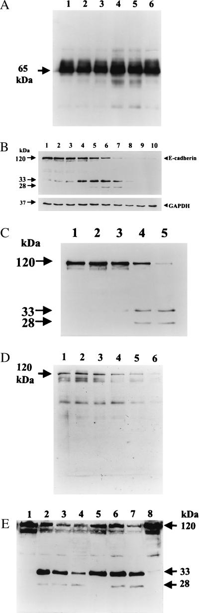
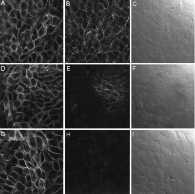
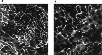
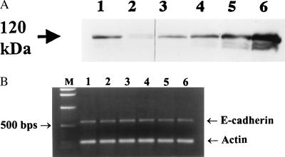
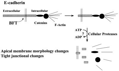
Strains of Bacteroides fragilis associated with diarrheal disease (enterotoxigenic B. fragilis) produce a 20-kDa zinc-dependent metalloprotease toxin (B. fragilis enterotoxin; BFT) that reversibly stimulates chloride secretion and alters tight junctional function in polarized intestinal epithelial cells. BFT alters cellular morphology and physiology most potently and rapidly when placed on the basolateral membrane of epithelial cells, suggesting that the cellular substrate for BFT may be present on this membrane. Herein, we demonstrate that BFT specifically cleaves within 1 min the extracellular domain of the zonula adherens protein, E-cadherin. Cleavage of E-cadherin by BFT is ATP-independent and essential to the morphologic and physiologic activity of BFT. However, the morphologic changes occurring in response to BFT are dependent on target-cell ATP. E-cadherin is shown here to be a cellular substrate for a bacterial toxin and represents the identification of a mechanism of action, cell-surface proteolytic activity, for a bacterial toxin.


Figures

Figure 1
Figure 1
Cleavage of E-cadherin, but not occludin, by BFT. (A) Occludin immunoblot of HT29/C1 cells. HT29/C1 cells were treated with BFT (5 nM) for various times, lysed, and examined by Western blotting using a polyclonal anti-human occludin antibody. Occludin is a 65-kDa protein (arrow). Lane 1, untreated HT29/C1 cells; lanes 2–5, BFT for 30 min, 60 min, 2 hr, 3 hr, respectively; lane 6, untreated HT29/C1 cells. (B) Time course of BFT proteolysis of E-cadherin. HT29/C1 cells were treated for various times with BFT (5 nM), lysed in 1× SDS/gel loading buffer, and examined by Western blot using the E2 antibody (34). Cell-shape changes were observed beginning 10 min after treatment with BFT, with 100% of cells affected by 30 min. Arrows indicate intact E-cadherin (120 kDa) and E-cadherin fragments (33 and 28 kDa) observed after BFT treatment. Lane 1, untreated HT29/C1 cells; lanes 2–10, BFT for 1 min, 3 min, 5 min, 10 min, 15 min, 30 min, 1 hr, 2 h, 3 hr, respectively. Immunostaining of the housekeeping protein GAPDH showed no change over time. (C) Concentration dependency of proteolysis of E-cadherin by BFT. HT29/C1 cells were treated with 0.005–5 nM BFT for 30 min. The immunoblot was processed and probed with E2 antibody as in 1B. Lane 1, untreated HT29/C1 cells; lane 2, 0.005 nM BFT; lane 3, 0.05 nM BFT; lane 4, 0.5 nM BFT; lane 5, 5 nM BFT. (D) Time course of BFT proteolysis of E-cadherin expressed by LE cells. LE cells (E-cadherin-transfected L cells) were induced overnight with 1 μM dexamethasone followed by treatment for various times with BFT (5 nM). The immunoblot was processed and probed with the E2 antibody as in Fig. 1B. Lane 1, control LE cells; lanes 2–6, BFT for 5 min, 10 min, 15 min, 30 min, 1 hr, respectively. (E) Effect of CCCP on BFT-stimulated E-cadherin proteolysis. HT29/C1 cells were treated with CCCP as described in Materials and Methods. CCCP-treated HT29/C1 cells were compared with HT29/C1 cells treated with BFT (5 nM) in standard HT29/C1 medium. Lanes 1–4, HT29/C1 cells without CCCP treatment. Lane 1, control HT29/C1 cells; lanes 2–4, BFT for 10 min, 30 min, 60 min, respectively. Lanes 5–8, HT29/C1 cells treated with CCCP. Lane 5, BFT for 10 min, 30 min, 60 min, control HT29/C1 cells, respectively.
Figure 2
Figure 2
Immunofluorescent confocal microscopy of BFT-treated HT29/C1 cells. (A) Control β1-integrin. (B) Control E-cadherin. (C) Control Nomarski optics. Edges of individual control (untreated) cells are indistinct by Nomarski optics. (D) Thirty minutes after BFT-β1-integrin treatment. (E) Thirty minutes after BFT-E-cadherin treatment. (F) Thirty-minute BFT-Nomarski optics. Note the dramatic loss of E-cadherin immunofluorescence without a change in β1-integrin immunofluorescence. By Nomarski optics, HT29/C1 cellular borders are more distinct in areas of loss of E-cadherin immunofluorescence but remain indistinct where E-cadherin immunofluorescence remains intact. (G) Sixty minutes after BFT- β1-integrin treatment. (H) Sixty minutes after BFT-E-cadherin treatment. (I) Sixty-minute BFT-Nomarski optics. By Nomarski optics, individual HT29/C1 cell borders are visible. (Magnification, ×1,200.)
Figure 3
Figure 3
ZO1 redistribution in HT29/C1 cells after BFT treatment. Polarized HT29/C1 cells were treated with BFT (5 nM) for 1 hr and assessed by confocal immunofluorescent microscopy. (A) Control cells reveal the even membrane distribution of ZO1. (B) ZO1 immunofluorescence is more diffuse and punctate after BFT treatment. (Magnification, ×1,000.)
Figure 4
Figure 4
E-cadherin expression in recovering BFT-treated HT29/C1 cells. HT29/C1 cells were treated with BFT (5 nM) for 3 hr, washed, placed in normal medium, and assessed at various times. (A) Western blot of E-cadherin changes over time after BFT treatment. After 3 hr of BFT treatment, all cells exhibited altered morphology. Cellular morphology gradually returned to normal by 45 hr after washing to remove BFT from the cells. At each time point, cell lysates were examined by Western blot using the E2 antibody as described in Fig. 1B. Lane 1, 3-hr control HT29/C1 cells; lane 2, BFT for 3 hr; lane 3, 19 hr after removal of BFT; lane 4, 33 hr after removal of BFT; lane 5, 45 hr after removal of BFT; lane 6, 48-hr control HT29/C1 cells. (B) Analysis of E-cadherin expression. Lane 1, untreated HT29/C1 cells; lanes 2–6, BFT for 1 hr, 3 hr, 6 hr, 10 hr, and 24 hr, respectively.
Figure 5
Figure 5
Model of BFT action. BFT cleaves the extracellular domain of the zonula adherens protein, E-cadherin, in an ATP-independent manner. Removal of the extracellular domain of E-cadherin results in the ATP-dependent proteolysis of the intracellular domain of E-cadherin, most likely by cellular proteases. Loss of intact E-cadherin disrupts its linkages with β-catenin and secondarily α-catenin and actin, leading to the characteristic disruption of the apical cytoskeleton of polarized epithelial cells as previously reported (16).

References

    1. Polk F B, Kasper D L. Ann Intern Med. 1996;86:569–571. - PubMed
    1. Tzianabos A O, Onderdonk A B, Rosner B, Cisneros R L, Kasper D L. Science. 1993;262:416–419. - PubMed
    1. Mundy L M, Sears C L. Clin Infect Dis. 1996;23:269–276. - PubMed
    1. Sears, C. L., Myers, L. L., Lazenby, A. & Van Tassell, R. L. (1995) Clin. Infect. Dis. 20, Suppl. 2, S142–S148. - PubMed
    1. Myers L L, Shoop D S, Stackhouse L L, Newman F S, Flaherty R J, Letson G W, Sack R B. J Clin Microbiol. 1987;25:2330–2333. - PMC - PubMed

Publication types

LinkOut - more resources