. 1998 Dec 8;95(25):15043-8.
doi: 10.1073/pnas.95.25.15043.

The neuropeptide Y/agouti gene-related protein (AGRP) brain circuitry in normal, anorectic, and monosodium glutamate-treated mice

Affiliations

The neuropeptide Y/agouti gene-related protein (AGRP) brain circuitry in normal, anorectic, and monosodium glutamate-treated mice

C Broberger et al. Proc Natl Acad Sci U S A. .

Abstract

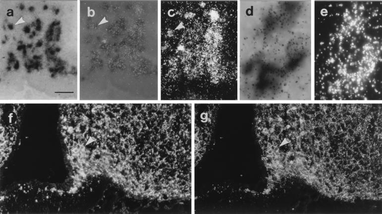
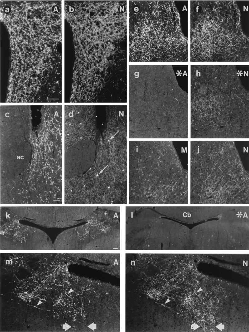
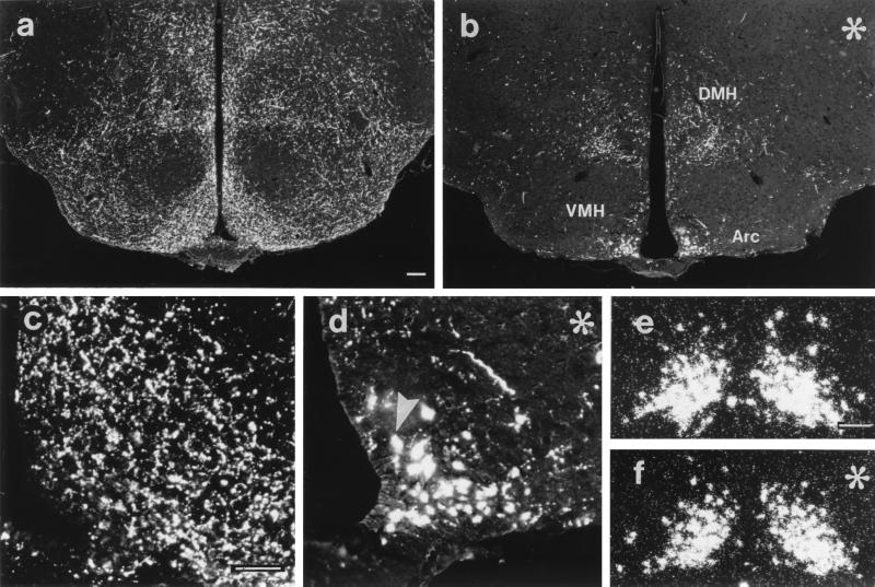
Neuropeptide Y (NPY) and the endogenous melanocortin receptor antagonist, agouti gene-related protein (AGRP), coexist in the arcuate nucleus, and both exert orexigenic effects. The present study aimed primarily at determining the brain distribution of AGRP. AGRP mRNA-expressing cells were limited to the arcuate nucleus, representing a major subpopulation (95%) of the NPY neurons, which also was confirmed with immunohistochemistry. AGRP-immunoreactive (-ir) terminals all contained NPY and were observed in many brain regions extending from the rostral telencephalon to the pons, including the parabrachial nucleus. NPY-positive, AGRP-negative terminals were observed in many areas. AGRP-ir terminals were reduced dramatically in all brain regions of mice treated neonatally with monosodium glutamate as well as of mice homozygous for the anorexia mutation. Terminals immunoreactive for the melanocortin peptide alpha-melanocyte-stimulating hormone formed a population separate from, but parallel to, the AGRP-ir terminals. Our results show that arcuate NPY neurons, identified by the presence of AGRP, project more extensively in the brain than previously known and indicate that the feeding regulatory actions of NPY may extend beyond the hypothalamus.


Figures

Figure 1
Figure 1
Micrographs from mouse arcuate nucleus sections processed for in situ hybridization (ae) or immunohistochemistry (f and g) double-labeling for AGRP or NPY mRNA (ae) or peptide (f and g), respectively. Micrographs taken with bright-field (a and d), bright-field with epiillumination (b), dark-field (c and e), or fluorescence illumination (f and g). NPY mRNA visualized as dark precipitate (a, b, and d) and AGRP mRNA detected as silver grains (b, c, and e) are shown. Note coexistence of AGRP and NPY mRNA/peptide in cell bodies (arrowheads). [Bar = 50 μm (ac, f, and g) and 250 μm (d and e).]
Figure 2
Figure 2
Micrographs from sections of mouse brain processed for double-labeling immunohistochemistry for AGRP (A; a, c, e, g, k, l, and m), NPY (N; b, d, f, h, j, and n), or αMSH (M; i) showing the PVH (a and b), bed nucleus of the stria terminalis (BST; c and d), periaqueductal gray (ej), and PBN (kn) from normal (af, ik, m, and n) or MSG-treated (asterisk; g, h, and l) mice. Note colocalization of AGRP and NPY in several fibers (arrowheads in m and n), but not in cell bodies of the BST (thin arrows in d), or in NPY fibers of the central gray of the pons (thick arrows in m and n). MSG treatment virtually abolishes AGRP (g) and decreases NPY staining (h). ac, anterior commissure; Cb, cerebellum. [Bar in a = 50 μm (a, b, ej, m, and n); in c = 50 μm (c and d); and in k = 100 μm (k and l).]
Figure 3
Figure 3
Fluorescence (ad) and dark-field (e and f) micrographs from arcuate nucleus sections processed for immunohistochemistry (ad) or in situ hybridization (e and f) showing AGRP peptide (ad) or mRNA (e and f), respectively, from anx/anx (asterisk; b, d, and f) and control (a, c, and e) mice. AGRP staining in anx/anx mice is decreased in hypothalamic terminals (cf. a with b), increased in arcuate cell bodies (arrowhead; cf. c with d), whereas AGRP mRNA is unchanged (cf. e with f). Arc, arcuate; DMH, dorsomedial nucleus; VMH, ventromedial nucleus. [Bar in a = 100 μm (a and b), in c = 50 μm (c and d), and in e = 50 μm (e and f).]
Figure 4
Figure 4
Quantification of line crossings (see Materials and Methods) of AGRP fibers in the dorsomedial hypothalamic nucleus of anx/anx mice (n = 10) and control littermates (n = 7). The density of AGRP terminals is decreased significantly in anx/anx mice. ∗∗∗, P < 0.001.

References

    1. Tatemoto K, Carlquist M, Mutt V. Nature (London) 1982;296:659–660. - PubMed
    1. Chronwall B M, DiMaggio D A, Massari V J, Pickel V M, Ruggiero D A, O’Donohue T L. Neuroscience. 1985;15:1159–1181. - PubMed
    1. de Quidt M E, Emson P C. Neuroscience. 1986;18:545–618. - PubMed
    1. Clark J T, Kalra P S, Crowley W R, Kalra S P. Endocrinology. 1984;115:427–429. - PubMed
    1. Stanley B G, Leibowitz S F. Proc Natl Acad Sci USA. 1985;82:3940–3943. - PMC - PubMed

Publication types

LinkOut - more resources