. 1998 Dec 8;95(25):15078-83.
doi: 10.1073/pnas.95.25.15078.

Metabotropic glutamate receptor-initiated translocation of protein kinase p90rsk to polyribosomes: a possible factor regulating synaptic protein synthesis

Affiliations

Metabotropic glutamate receptor-initiated translocation of protein kinase p90rsk to polyribosomes: a possible factor regulating synaptic protein synthesis

F Angenstein et al. Proc Natl Acad Sci U S A. .

Abstract

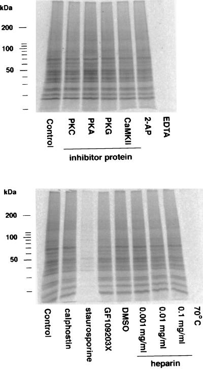
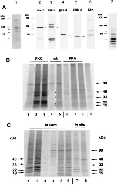
Maintenance of lasting synaptic efficacy changes requires protein synthesis. We report here a mechanism that might influence translation control at the level of the single synapse. Stimulation of metabotropic glutamate receptors in hippocampal slices induces a rapid protein kinase C-dependent translocation of multifunction kinase p90rsk to polyribosomes; concomitantly, there is enhanced phosphorylation of at least six polyribosome binding proteins. Among the polyribosome bound proteins are the p90rsk-activating kinase ERK-2 and a known p90rsk substrate, glycogen synthase kinase 3beta, which regulates translation efficiency via eukaryotic initiation factor 2B. Thus metabotropic glutamate receptor stimulation could induce synaptic activity-dependent translation via translocation of p90rsk to ribosomes.


Figures

Figure 1
Figure 1
PhosphorImager record of in situ phosphorylation of PRBPs isolated from hippocampal slices of 12-day-old rats, labeled with 33P. (A) Stimulation with 0.1 mM DHPG for 10 min increased endogenous phosphorylation of proteins with molecular masses of 90, 85, 48, 33, 22, and 19 kDa (arrows). A comparable effect could be observed after addition of a PKC activator, 1 μM PDBu, but not after stimulation with 50 μM forskolin. This enhanced phosphorylation elicited by DHPG was not detectable if 10 μM calphostin C, an inhibitor of PKC, or 10 μM GF109203X, an inhibitor of both PKC and p90rsk, was present during stimulation. (B) Scanning profiles obtained from the shown autoradiographs. Black line: unstimulated samples; gray line: stimulated samples (DHPG, PDBu, and forskolin). Arrows indicate peaks corresponding to the indicated bands in the autoradiography.
Figure 2
Figure 2
Detection of polyribosome-bound kinase activity. In vitro phosphorylation assay of PRBP (10 μg per lane) prepared from cerebral cortical synaptoneurosomes from 12-day-old rats (24). This fraction includes ribosome-bound kinases as well as substrates. Incorporated 33P was detected by PhosphorImaging. The activity of the polyribosome-bound kinases was not blocked by addition of specific inhibitor peptides against PKC (fragment 19–36, Sigma), PKA (PKI, Sigma), PKG (Arg-Lys-Arg-Ala-Arg-Lys-Glu, Sigma), or CaMKII (fragment 290–309, Sigma), each 2 μg per assay, or inhibitors against protein kinase R (100 mM 2-aminopurine = 2-AP), casein kinase II (increasing concentrations of heparin), PKC (10 μM calphostin C). GF109203X (10 μM), an inhibitor of PKC and p90rsk, had a small inhibitory effect. The kinase activities could be blocked completely by capturing divalent cations (Mg2+, Ca2+) with 50 mM EDTA or heating the fraction to 70°C for 5 min and inhibited almost completely by 10 μM staurosporine, a nonspecific inhibitor of Ser/Thr kinases.
Figure 3
Figure 3
A group of kinases is bound to polyribosomal complexes prepared from synaptoneurosomes. (A) Detection of autophosphorylation activity in kinases from the 0.5 M K+ fraction. Lane 1, PRBPs (25 μg) were prepared as described (15, 16) and after electrophoretic separation blotted to a poly(vinylidene difluoride) membrane. The proteins were renatured, and the presence of kinases was detected by autophosphorylation in the presence of [γ-33P]-ATP according to the method of Ferrell (27). This procedure revealed five kinases with molecular masses of about 65, 85, 90, 95, and 180 kDa. Identification of different kinases among PRBP by Western blot assay. Lane 2, p90rsk-1. Lane 3, p90rsk-2, the antiserum to p90rsk-2 crossreacts with two unknown additional proteins with molecular masses of about 100 and 115 kDa. Lane 4, gsk-3β. Lane 5, ribosomal S6 kinase p70S6K. Lane 6, ERK-2 (p42MAPK). Lane 7, an amido black protein staining of PRBPs on nitrocellulose. (B) In vitro phosphorylation of PRBPs by added kinases after blocking the activity of all endogenous kinases by heating the fraction to 70°C for 5 min. Lane 1, autophosphorylated form of PKC alone; lane 2, PRBP and PKC without activators; lane 3, PRBP and PKC with activators; lane 4, autophosphorylated p90rsk-2; lane 5, PRBP and p90rsk-2 (* indicates the position of proteins that are also phosphorylated in situ); lane 6, autophosphorylated PKA; lane 7, PRBP and PKA; lane 8, PRBP, PKA, and cAMP; lane 9, PRBP. Note that the specific activities of the kinases are not equal, because p90rsk-2 is only partially purified. (C) Phosphorylation pattern of PRBP under different conditions. Lanes 1–2, all polyribosome-bound kinases (lane 1 with 10 μM GF109203X and lane 2 without inhibitor). Lanes 3–6, effect of p90rsk-2 (lane 3, heated PRBP; lane 4, p90rsk-2 alone, lane 5 and 6 p90rsk-2 with heated PRBP). Lanes 7–8, phosphorylation pattern of PRBP under in situ conditions. All endogenous phosphorylated proteins (arrows on right) are in vitro substrates for p90rsk-2. The 85- and 90-kDa protein phosphorylated under in situ conditions might be p90rsk-1 and p90rsk-2 itself.
Figure 4
Figure 4
mGluR induced translocation of p90rsk-1 to polyribosomes. (A, Left) Stimulation of hippocampal slices with 0.1 mM DHPG for 10 min induced an increase in the amount of polyribosome-bound p90rsk-1. This increase is mediated by an enhanced binding of p90rsk-1 to polyribosomes as revealed by Western blot assay. (Center) PRBPs were prepared from cerebral cortical synaptoneurosomes that were untreated (C), stimulated with 0.1 mM DHPG for 1.5 min (DHPG), or stimulated with 0.1 mM DHPG in presence of 10 μM calphostin C (DHPG/calph.). Identical amounts of proteins were loaded on each lane. P90rsk-1 is indicated by an arrow. (Right) Quantification of nine independent experiments; C = control 100%; *P < 0.05%, Wilcoxon pair test. (B) Blots were washed and restained with antibody to gsk-3β; no significant changes in the amount of polyribosome-bound gsk-3β were found (n = 4).

References

    1. Bailey C H, Bartsch D, Kandel E R. Proc Natl Acad Sci USA. 1996;93:13445–13452. - PMC - PubMed
    1. Frey U, Morris R G M. Nature (London) 1997;385:533–536. - PubMed
    1. Leski M L, Steward O. Neurochem Res. 1996;21:681–690. - PubMed
    1. Torre E R, Steward O. J Neurosci. 1996;16:5967–5978. - PMC - PubMed
    1. Feig S, Lipton P. J Neurosci. 1993;13:1010–1021. - PMC - PubMed

Publication types

Substances

LinkOut - more resources