. 1998 Dec;4(12):1401-8.
doi: 10.1038/3992.

Vpx is required for dissemination and pathogenesis of SIV(SM) PBj: evidence of macrophage-dependent viral amplification

Affiliations

Vpx is required for dissemination and pathogenesis of SIV(SM) PBj: evidence of macrophage-dependent viral amplification

V M Hirsch et al. Nat Med. 1998 Dec.

Abstract

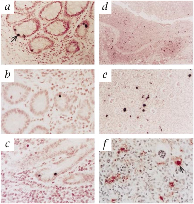
The viral accessory protein Vpx is required for productive in vitro infection of macrophages by simian immunodeficiency virus from sooty mangabey monkeys (SIV(SM)). To evaluate the roles of Vpx and macrophage infection in vivo, we inoculated pigtailed macaques intravenously or intrarectally with the molecularly cloned, macrophage tropic, acutely pathogenic virus SIV(SM) PBj 6.6, or accessory gene deletion mutants (deltaVpr or deltaVpx) of this virus. Both wild-type and SIV(SM) PBj deltaVpx viruses were readily transmitted across the rectal mucosa. A subsequent 'stepwise' process of local amplification of infection and dissemination was observed for wild-type virus, but not for SIV(SM) PBj deltaVpx, which also showed considerable impairment of the overall kinetics and extent of its replication. In animals co-inoculated with equivalent amounts of wild-type and SIV(SM) Pbj deltaVpx intravenously or intrarectally, the deltaVpx mutant was at a strong competitive disadvantage. Vpx-dependent viral amplification at local sites of initial infection, perhaps through a macrophage-dependent mechanism, may be a prerequisite for efficient dissemination of infection and pathogenic consequences after exposure through either mucosal or intravenous routes.


Figures

Fig. 1
Fig. 1
Kinetics of plasma viremia and lymphopenia in macaques inoculated intrarectally with ΔVpr (left graphs: □, 541; ○, 547) or ΔVpx mutants (right graphs: □, 607; ○, 608) or with WT SIVSM PBj (■, 526; ●, 539). Values shown are averages for duplicate determinations on RNA extracted from individual plasma samples.
Fig. 2
Fig. 2
Kinetics of plasma viremia and lymphopenia in macaques inoculated intravenously with ΔVpr (left graphs: □, 437; ○, 453) or ΔVpx mutants (right graphs: □, 537; ○, 549) or with WT SIVSM PBj (■, 123; ●, 389). Values shown are averages for duplicate determinations on RNA extracted from individual plasma samples.
Fig. 3
Fig. 3
Representative SIV-specific in situ hybridization and immunohistochemistry for Ham-56 (macrophage marker) in tissues of macaques inoculated intrarectally with either WT SIVSM PBj (a,dg) or the ΔVpx mutant virus (b and c). a, Intraepithelial lymphocyte (IEL) expressing SIV RNA (arrow) in the rectum of macaque PT550 at 4 days after inoculation with WT virus. b, SIV+ IEL in the rectum of macaque PT612 at 7 days after inoculation with ΔVpx virus. c, SIV+ IEL in the ileum of macaque PT612 at 7 days after inoculation with ΔVpx virus. d, Low magnification in situ hybridization of a characteristic local lesion in the rectum of macaque PT523 at 7 days after inoculation with WT virus, showing local infiltration of lymphocytes and macrophages with many SIV-expressing cells. e, Higher magnification of the localized rectal lesion shown in d. f, High magnification of immunohistochemical staining for HAM-56 expression in rectal lesion in macaque PT523, inoculated with WT virus, showing infiltration of macrophages (arrow) into the lesion.
Fig. 4
Fig. 4
Double labeling for SIV expression and cell surface expression of CD3 or Ham56 (macrophage) in rectal sections collected at peak viremia, demonstrating both SIV-infected T lymphocytes (top row, ac) and macrophages (middle row, df). a and d, cell surface marker (green fluorescence); b and e, SIV expression (green); c and f double exposure or the same field showing double-labeled cells (yellow). Approximately 90% of SIV-expressing cells at this time point were identified as CD3+ T cells. Bottom row (gi), SIV-infected T lymphocytes on the same field of a rectal sample on day 4 after intrarectal exposure. g, CD3+ cells (green); h, SIV-infected cells (red); i, SIV-infected T lymphocytes (yellow).
Fig. 5
Fig. 5
Replication and proportional representation of SIVSM PBj WT, ΔVpr and ΔVpx mutant viruses after intravenous or intrarectal co-inoculation. a, Identification of WT, Δvpr and Δvpx genotypes by differential oligonucleotide hybridization. Bacterial colonies transformed with WT, ΔVpr and ΔVpx SIVSM PBj molecular clones (genotype) were hybridized with probes recognizing WT, Δvpr or Δvpx sequences or with a common probe that hybridizes to both WT and mutant genomes. Hybridization conditions were adjusted to promote specific binding of each probe to its cognate sequence. b and c, Proportional representations of WT, ΔVpr and ΔVpx variants of SIVSM PBj in plasma after intravenous (b) or intrarectal (c) co-inoculation. The relative proportions of WT and mutant genomes were determined by differential oligonucleotide probe hybridization and are shown as mutant genotype fraction. Absolute plasma RNA copy numbers for WT and mutant genomes were calculated on the basis of the measured total (WT and mutant) plasma SIV RNA copy number and the percentages of WT and mutant genotype determined by differential probe hybridization at each indicated time point.

Comment in

References

    1. Wolfs TFW, Zwart G, Bakker M & Goudsmit J HIV-1 genomic RNA diversification following sexual and parenteral virus transmission. Virology 189, 103–110 (1992). - PubMed
    1. Keet IPM et al. Predictors of rapid progression to AIDS in HIV-1 seroconverters. AIDS 7, 51–57 (1993). - PubMed
    1. Zhu T et al. Genotypic and phenotypic characterization of HIV-1 in patients with primary infection. Science 261, 1179–1181 (1993). - PubMed
    1. Roos MTL et al. Viral phenotype and immune response in primary human immunodeficiency virus type 1 infection. J. Infect. Dis 165, 427–432 (1992). - PubMed
    1. Nielsen C, Pedersen C, Lundgren JD & Gerstoft J Biological properties of HIV isolates in primary HIV infection: consequences for the subsequent course of infection. AIDS 7, 1035–1040 (1993). - PubMed

Publication types

MeSH terms

Substances