

Expression status of p16 protein is associated with human papillomavirus oncogenic potential in cervical and genital lesions
- PMID: 9846965
- PMCID: PMC1866324
- DOI: 10.1016/S0002-9440(10)65689-1
Expression status of p16 protein is associated with human papillomavirus oncogenic potential in cervical and genital lesions
Abstract
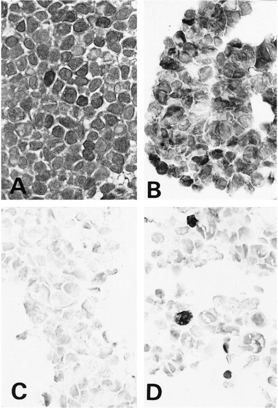
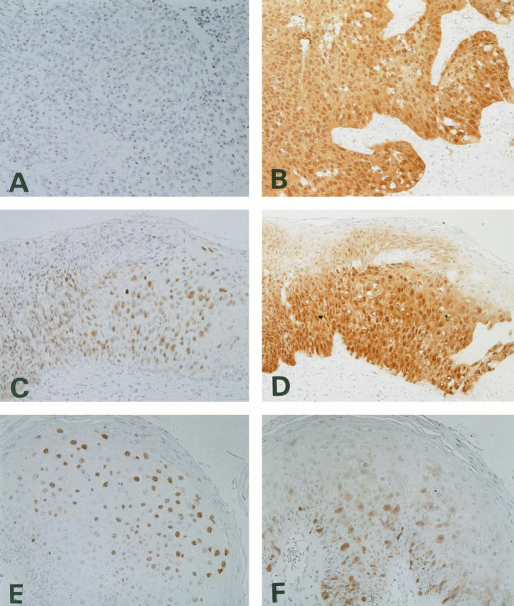
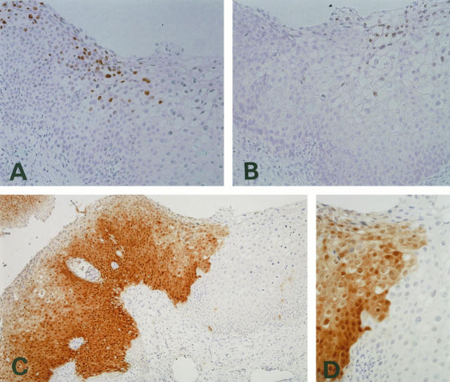
The p16 protein (p16) is a cyclin-dependent kinase (CDK) inhibitor that decelerates the cell cycle by inactivating the CDKs that phosphorylate retinoblastoma (Rb) protein. Recent biological studies have revealed that p16 expression is markedly influenced by the status of Rb expression, and p16 overexpression has been demonstrated in cervical cancers because of functional inactivation of Rb by human papillomavirus (HPV) E7 protein. To clarify the relationship between p16 overexpression and HPV infection in cervical carcinogenesis, immunohistochemical analysis of p16 and detection of HPV by in situ hybridization and polymerase chain reaction were performed on 139 formalin-fixed and paraffin-embedded samples of cervical and genital condylomatous and neoplastic lesions. Marked overexpression of p16 protein, ie, diffuse and strong immunostaining, was observed in all cervical cancers and preneoplastic lesions with infection by high- and intermediate-risk HPVs, ie, subtypes 16, 18, 31, 33, 52, and 58. Condylomata acuminata and low-grade squamous intraepithelial lesions with infection by low-risk HPV such as HPV-6/11 showed focal and weak immunohistochemical staining for p16. Our results clearly showed that the mode of p16 expression in lesions with high- and intermediate-risk HPVs differed from its expression in lesions with low-risk HPVs and thus might be attributable to differences in functional inactivation of Rb protein by different HPVs.
Figures
References
-
- zur Hausen H: Papillomavirus infections: a major cause of human cancers. Biochim Biophys Acta 1996, 1288:F55-F78 - PubMed
-
- Lorincz AT, Reid R, Bennett Jenson A, Greenberg MD, Lancaster W, Kurman RJ: Human papillomavirus infection of the cervix: relative risk associations of 15 common anogenital types. Obstet Gynecol 1992, 79:328-337 - PubMed
-
- Matsukura T, Sugase M: Identification of genital human papillomaviruses in cervical biopsy specimens: segregation of specific virus types in specific clinicopathologic lesions. Int J Cancer 1995, 61:13-22 - PubMed
MeSH terms
Substances
LinkOut - more resources
Full Text Sources
Other Literature Sources
Medical
