. 1998 Dec;153(6):1775-85.
doi: 10.1016/S0002-9440(10)65692-1.

Arsenic enhancement of skin neoplasia by chronic stimulation of growth factors

Affiliations

Arsenic enhancement of skin neoplasia by chronic stimulation of growth factors

D R Germolec et al. Am J Pathol. 1998 Dec.

Abstract

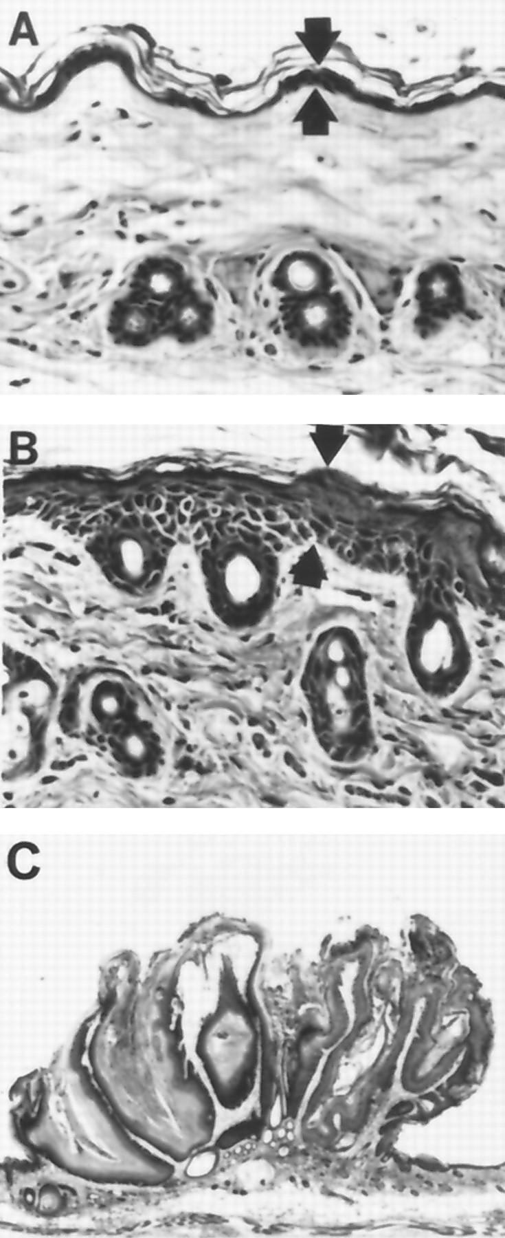
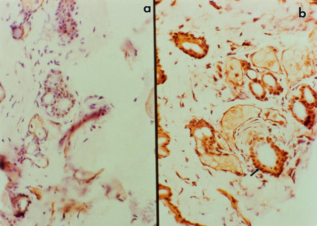
Although numerous epidemiological studies have shown that inorganic arsenicals cause skin cancers and hyperkeratoses in humans, there are currently no established mechanisms for their action or animal models. Previous studies in our laboratory using primary human keratinocyte cultures demonstrated that micromolar concentrations of inorganic arsenite increased cell proliferation via the production of keratinocyte-derived growth factors. As recent reports demonstrate that overexpression of keratinocyte-derived growth factors, such as transforming growth factor (TGF)-alpha, promote the formation of skin tumors, we hypothesized that similar events may be responsible for those associated with arsenic skin diseases. Thus, the influence of arsenic in humans with arsenic skin disease and on mouse skin tumor development in transgenic mice was studied. After low-dose application of tetradecanoyl phorbol acetate (TPA), a marked increase in the number of skin papillomas occurred in Tg.AC mice, which carry the v-Ha-ras oncogene, that received arsenic in the drinking water as compared with control drinking water, whereas no papillomas developed in arsenic-treated transgenic mice that did not receive TPA or arsenic/TPA-treated wild-type FVB/N mice. Consistent with earlier in vitro findings, increases in granulocyte/macrophage colony-stimulating factor (GM-CSF) and TGF-alpha mRNA transcripts were found in the epidermis at clinically normal sites within 10 weeks after arsenic treatment. Immunohistochemical staining localized TGF-alpha overexpression to the hair follicles. Injection of neutralizing antibodies to GM-CSF after TPA application reduced the number of papillomas in Tg.AC mice. Analysis of gene expression in samples of skin lesions obtained from humans chronically exposed to arsenic via their drinking water also showed similar alterations in growth factor expression. Although confirmation will be required in nontransgenic mice, these results suggest that arsenic enhances development of skin neoplasias via the chronic stimulation of keratinocyte-derived growth factors and may be a rare example of a chemical carcinogen that acts as a co-promoter.


Figures

Figure 1.
Figure 1.
BrdU staining of Tg.AC mouse skin after exposure to 0.02% sodium arsenite in the drinking water. Magnification, ×340. Tg.AC mice were injected with BrdU 30 minutes before sacrifice, and tissues and slides were prepared and stained as described in Materials and Methods. A: In normal skin, the epithelium is thin with a moderate amount of keratin on the surface of the skin. B: Mouse skin after 10 weeks of sodium arsenite is moderately thickened (hyperkeratosis) with the stratum corneum containing a moderate increase in the cornified cell layer.
Figure 2.
Figure 2.
Kinetics of BrdU expression in Tg.AC mice receiving control (□) and 0.02% sodium-arsenite-treated ([circo) drinking water. *Significantly different from controls at P < 0.05.
Figure 3.
Figure 3.
Kinetics of mRNA expression in Tg.AC mouse skin over time. Total RNA was isolated from dorsal skin of Tg.AC transgenic mice receiving sodium arsenite in the drinking water. A: RT-PCR was performed as described in Materials and Methods and used to determine relative differences between skin from control (c) or arsenic-exposed animals at 1, 4, and 10 weeks. B: Image analysis of ethidium-bromide-stained PCR products. Values represent means ± SEM peak intensities for the samples shown in A after normalization for G3PDH. *Significantly different from controls at P < 0.05. +, positive control; MW, molecular weight standard.
Figure 4.
Figure 4.
Histopathology of mouse skin from animals either vehicle treated, sodium arsenite exposed (0.02% drinking water), TPA treated (four doses of 2.5 μg of TPA twice a week for 2 weeks) or combined treated. Full-thickness skin sections were cut parallel to the dorsal midline, and 6-μm sections were prepared and stained with H&E. The magnification is ×200 for all panels, and the hair cycle is in the resting stage (telogen) for all samples shown. A: A section of normal skin from an untreated animal. Note the presence of the fatty layer of tissue below the dermal mesenchyme. B: A section of skin taken from an animal exposed to 0.02% sodium arsenite for 6 weeks. Although the epidermal layer of cells does not appear to be more hyperplastic than that observed in the control section, hyperkeratosis is present, as indicated by the increased layers of stratum corneum (arrows). Note the absence of fat cells below the dermal mesenchyme. C: A section of skin from a mouse that had received control drinking water taken 24 hours after the last of four treatments of 2.5 μg of TPA. There is a continuum of the hyperplasia in the outer root sheath epithelium and of the interfollicular epidermis (arrows). The epithelial hyperplasia is accompanied by an increase in hyperkeratosis. D: A section of skin from a mouse exposed to 0.02% sodium arsenite for 6 weeks and taken 24 hours after the last of four treatments with 2.5 μg of TPA. In contrast to C, there is a pronounced hyperplasia of the outer root sheath and interfollicular epithelia (arrows), which is accompanied by a marked increase in hyperkeratosis.
Figure 5.
Figure 5.
Histology (H&E) of murine skin from control and treated animals taken from full-thickness skin sections cut parallel to the dorsal midline. Tg.AC transgenic mice were provided 0.02% sodium arsenite in the drinking water starting at 12 weeks of age. A: In mice receiving control drinking water, the epithelium (arrows) is thin with a moderate amount of keratin on the surface of the skin. Magnification, ×340. B: After 4 weeks of sodium arsenite exposure, the epithelium (arrows) is minimally thickened with a moderate amount of keratin on the surface of the skin. Magnification, ×340. C: Squamous cell papilloma from the back of a mouse administered TPA and receiving arsenic for 12 weeks. Magnification, ×27.
Figure 6.
Figure 6.
Papilloma incidence in Tg.AC transgenic mice provided 0.02% sodium arsenite in the drinking water starting at 12 weeks of age. A: Mice were pretreated with arsenite or water for 4 weeks and sub-grouped, and 1.25 (non-inducing; NITPA) or 2.5 μg of TPA were applied twice per week for 2 consecutive weeks. ×, control water; □, TPA plus water; ▪, TPA plus arsenic; ○, NITPA plus water; •, NITPA plus arsenic; n = 20 mice per treatment group. B: Same as A, using 2.5 μg of TPA, plus additional groups were treated with monoclonal antibodies to mouse GM-CSF 2 weeks before TPA treatment, 2 hours after each application of TPA, and 2 weeks after TPA treatment. ×, control water; □, TPA plus water; ▪, TPA plus As; ▵, TPA plus water plus anti-GM-CSF antibodies; ▴, TPA plus As plus anti-GM-CSF antibodies; n = 13 to 14 mice per treatment group. Papillomas were not observed in non-TPA-promoted, arsenic-treated transgenic mice or TPA-promoted, arsenic-treated wild-type FVB/N mice (data not shown).
Figure 7.
Figure 7.
v-Ha-ras transgene mRNA expression in Tg.AC mouse skin. Total RNA was isolated and RT-PCR performed on dorsal skin of Tg.AC transgenic mice as previously described. Transgene expression was evaluated at 2, 4, 6, or 8 weeks in mice receiving control (C) or 0.02% sodium-arsenite-treated drinking water (A). MW, molecular weight standard; +, positive control.
Figure 8.
Figure 8.
Immunohistochemical localization of TGF in the skin of Tg.AC transgenic mice. Magnification, ×360. Samples of skin from control and arsenic-treated mice were quick-frozen, and 6-μm sections were prepared for immunohistochemistry as described in Materials and Methods. Shown are representative samples from Tg.AC transgenic mice after 14 weeks of exposure to either control (a) or arsenite-treated (b) drinking water. Note intense staining in follicular region of skin from arsenite-treated mice (arrow).
Figure 9.
Figure 9.
TGF-α, GM-CSF, TNF-α, and G3PDH mRNA expression in lesioned skin from Bowen’s disease patients drinking arsenic-contaminated drinking water from the Pa Chang Valley in Taiwan. Control skin samples were from individuals in the same village who had visible skin lesions. Poly(A+) mRNA was isolated from skin biopsies as described in Materials and Methods. A: RT-PCR was performed as described in Materials and Methods to determine relative differences between samples of lesioned and nonlesioned skin. Lanes 1 to 4, nonlesioned skin; lanes 5 to 10, lesioned skin. B: Image analysis of ethidium-bromide-stained PCR products after normalization relative to the density of each corresponding band for G3PDH. Values represent means ± SEM for the samples shown in A. *Significantly different from controls at P < 0.05.

References

    1. US Environmental Protection Agency: Special Report on Ingested Inorganic Arsenic: Skin Cancer and Nutritional Essentiality. Risk Assessment Forum. Washington, DC, US Environmental Protection Agency, 1987
    1. Nriagu JO: Arsenic in the Environment. II. Human Health and Ecosystem Effects. 1994. Wiley and Sons, New York
    1. Smith AH, Hopenhayn-Rich C, Bates MN, Goeden HM, Hertz-Picciotto P, Duggan HM, Wood R, Kornett MJ, Smith MT: Cancer risks from arsenic in drinking water. Environ Health Perspect 1992, 97:259-267 - PMC - PubMed
    1. Chiou HY, Hsueh YM, Liaw KF, Horng SF, Chiang MH, Pu YS, Lin JS, Huang CH, Chen CJ: Incidence of internal cancers and ingested inorganic arsenic: a 7-year follow-up study in Taiwan. Cancer Res 1995, 55:1296-1300 - PubMed
    1. Maloney ME: Arsenic in dermatology. Dermatol Surg 1996, 22:301-304 - PubMed

MeSH terms