. 1998 Dec;153(6):1873-84.
doi: 10.1016/S0002-9440(10)65701-X.

Acute effects of inhaled urban particles and ozone: lung morphology, macrophage activity, and plasma endothelin-1

Affiliations

Acute effects of inhaled urban particles and ozone: lung morphology, macrophage activity, and plasma endothelin-1

L Bouthillier et al. Am J Pathol. 1998 Dec.

Abstract

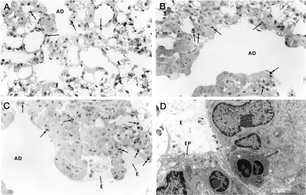
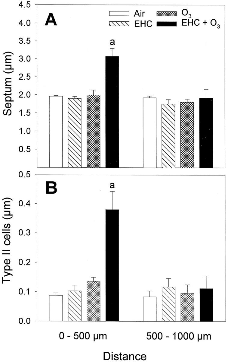
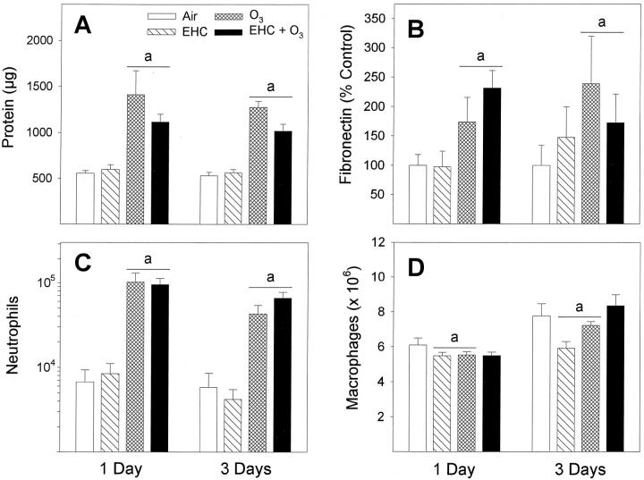
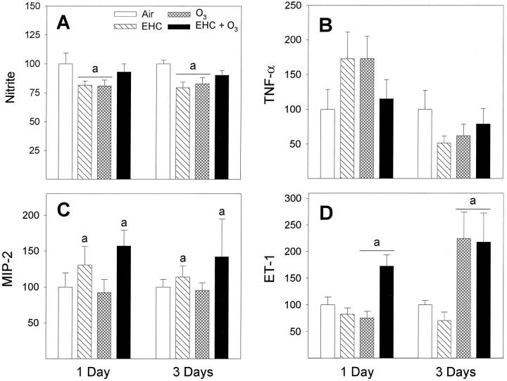
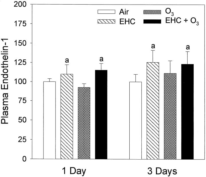
We studied acute responses of rat lungs to inhalation of urban particulate matter and ozone. Exposure to particles (40 mg/m3 for 4 hours; mass median aerodynamic diameter, 4 to 5 microm; Ottawa urban dust, EHC-93), followed by 20 hours in clean air, did not result in acute lung injury. Nevertheless, inhalation of particles resulted in decreased production of nitric oxide (nitrite) and elevated secretion of macrophage inflammatory protein-2 from lung lavage cells. Inhalation of ozone (0.8 parts per million for 4 hours) resulted in increased neutrophils and protein in lung lavage fluid. Ozone alone also decreased phagocytosis and nitric oxide production and stimulated endothelin-1 secretion by lung lavage cells but did not modify secretion of macrophage inflammatory protein-2. Co-exposure to particles potentiated the ozone-induced septal cellularity in the central acinus but without measurable exacerbation of the ozone-related alveolar neutrophilia and permeability to protein detected by lung lavage. The enhanced septal thickening was associated with elevated production of both macrophage inflammatory protein-2 and endothelin-1 by lung lavage cells. Interestingly, inhalation of urban particulate matter increased the plasma levels of endothelin-1, but this response was not influenced by the synergistic effects of ozone and particles on centriacinar septal tissue changes. This suggests an impact of the distally distributed particulate dose on capillary endothelial production or filtration of the vasoconstrictor. Overall, equivalent patterns of effects were observed after a single exposure or three consecutive daily exposures to the pollutants. The experimental data are consistent with epidemiological evidence for acute pulmonary effects of ozone and respirable particulate matter and suggest a possible mechanism whereby cardiovascular effects may be induced by particle exposure. In a broad sense, acute biological effects of respirable particulate matter from ambient air appear related to paracrine/endocrine disruption mechanisms.


Figures

Figure 1.
Figure 1.
Lung histology assessed after a single, 4-hour inhalation exposure to the pollutants, followed by 20-hour recovery in clean air. The lungs were collapsed and fixed by vascular perfusion to minimize displacement of focal intra-alveolar changes. A: Alveolar duct region after exposure to EHC-93. A few neutrophils (arrows) can be seen in tissue closest to the duct, but the lung structure is otherwise normal. GMA section; toluidine blue; magnification, ×540. B: Alveolar duct region after ozone exposure. There is some increase in cellularity, and several neutrophils are found in the lung tissue. GMA section; toluidine blue; magnification, ×560. C: Alveolar duct region after combined dust plus ozone exposure. Septal cellularity is much increased, and a few neutrophils and macrophages are seen with edema fluid in the alveolar space. Several neutrophils are seen in the septum. GMA section; toluidine blue; magnification, ×560. D: Electron micrograph of alveolar duct wall after combined dust plus ozone exposure. Cell debris and edema are seen in the lumen at an area of epithelial cell injury. In the interstitium, neutrophils are observed. Magnification, ×5400. AD, alveolar duct; E, edema; EP, epithelial cell; P, neutrophil.
Figure 2.
Figure 2.
Lung morphometry after a single, 4-hour inhalation exposure to the pollutants, followed by 32-hour recovery in clean air. Lungs were fixed by intratracheal inflation. Results are shown as normalized volume density (volume to surface ratio, in microns) of septum and type II cells of septal edges in alveolar duct paths 0 to 500 μm (proximal) and 500 to 1000 μm (distal), relative to terminal bronchiole. Bars are mean ± SE; n = 6 animals. Repeated measures analyses of variance indicated significant EHC × OZONE × DISTANCE factor interactions for septum (P = 0.008) and type II cells (P = 0.002). By Student t-test, proximal overall was different from distal (α = 0.05). Two-way analyses of variance and multiple comparisons for the proximal (0 to 500 μm) measurements are summarized below. A: Septum thickness, excluding capillary lumen. aEHC × OZONE factor interaction, P < 0.001. Tukey, in the 0- to 500-μm alveolar duct segment, 0 mg/m3 versus 48 mg/m EHC-93 within 0.8 ppm O3 and 0 ppm versus 0.8 ppm O3 within 48 mg/m EHC-93 (α = 0.05). B: Type II alveolar epithelial cell hyperplasia/hypertrophy. aEHC × OZONE factor interaction, P < 0.001. Tukey, in the 0- to 500-μm alveolar duct segment, 0 mg/m3versus 48 mg/m EHC-93 within 0.8 ppm O3 and 0 ppm versus 0.8 ppm O3 within 48 mg/m EHC-93 (α = 0.05).
Figure 3.
Figure 3.
Assessment of lung injury by bronchoalveolar lavage after inhalation of urban dust and ozone for 4 hours, followed by 20-hour recovery in clean air. A: Soluble proteins were analyzed by Coomassie blue dye-binding assay. aOZONE main effect, P < 0.001. B: Fibronectin was measured by Western blotting and imaging densitometry and shown as a percentage of control. aOZONE main effect, P < 0.001. C: Number of neutrophils. aOZONE main effect, P < 0.001. D: Number of macrophages. aEHC × OZONE factor interaction, P < 0.001. Tukey, 0 mg/m3versus 40 mg/m EHC-93 within 0 ppm O3 and within 0.8 ppm O3 (α = 0.05). The significant DAYS main effect, P < 0.001, indicated overall higher recoveries of macrophages for the 3-day versus 1-day exposure groups (not associated with treatments). All results are expressed as mean ± SE; n = 8 to 12 animals.
Figure 4.
Figure 4.
Integrity of macrophages after inhalation exposure of animals to particles and ozone. A: Viability was measured by reduction of alamarBlue and shown as a percentage of control. Results are expressed as mean ± SE; n = 12 animals. There were no effects of treatments. B: Phagocytosis of fluorescein-labeled E. coli by freshly isolated macrophages (2-hour incubation), shown as a percentage of control. Results are expressed as mean ± SE; n = 12 to 18 animals. aOZONE main effect, P < 0.004.
Figure 5.
Figure 5.
Functional assays of macrophages after inhalation exposure of animals to particles and ozone. All data are presented as percentage of control. A: Nitric oxide production as determined by the assay of nitrite in 24-hour culture supernatants of macrophages stimulated with LPS. Results are expressed as mean ± SE; n = 12 to 18 animals. aEHC × OZONE factor interaction, P < 0.001. Tukey, 0 ppm versus 0.8 ppm O3 within 0 mg/m EHC-93 and 0 mg/m3 versus 40 mg/m EHC-93 within 0 ppm O3 (α = 0.05). B: TNF-α measured in 24-hour culture supernatants by ELISA. Results are expressed as mean ± SE; n = 5 to 12 animals. The DAYS main effect, P < 0.001, indicated overall higher amounts secreted for 1-day versus 3-day exposure groups. The data suggest a temporal pattern in the TNF-α response. C: MIP-2 was measured in 24-hour culture supernatants by ELISA. Results are expressed as mean ± SE; n = 9 to 10 animals. aEHC main effect, P = 0.035. D: ET-1 was measured in 24-hour cell culture supernatants by ELISA. Results are expressed as mean ± SE; n = 6 to 10 animals. aOZONE main effect, P < 0.001.
Figure 6.
Figure 6.
Alteration of levels of immunoreactive ET-1 in plasma after inhalation of urban dust and ozone for 4 hours, followed by 20-hour recovery in clean air. Results are presented as percentage of air control and expressed as mean ± SE; n = 9 to 15 animals. aEHC main effect, P = 0.018 (multiway ANOVA performed on raw data, pg of ET-1/ml of plasma).

References

    1. Bates DV, Sizto R: Relationships between air pollution levels and hospital admissions in Southern Ontario. Can J Public Health 1983, 74:117-122 - PubMed
    1. Dockery DW, Pope CA, Xu X, Spengler JD, Ware JH, Fay ME, Ferris BG, Speizer FE: An association between air pollution and mortality in six U.S. cities. N Engl J Med 1993, 329:1753-1759 - PubMed
    1. Burnett RT, Dales RE, Raizenne ME, Krewski D, Summers PW, Roberts GR, Raad-Young M, Dann T, Brook J: Effects of low ambient levels of ozone and sulfates on the frequency of respiratory admissions to Ontario hospitals. Environ Res 1994, 65:172-194 - PubMed
    1. Burnett RT, Dales RE, Krewski D, Vincent R, Dann T, Brook JR: Associations between ambient particulate sulfate and admissions to Ontario hospitals for cardiac and respiratory diseases. Am J Epidemiol 1995, 142:15-22 - PubMed
    1. Vincent R, Bjarnason SG, Adamson IYR, Hedgecock C, Guénette J, Potvin M, Kumarathasan P, Goegan P, Bouthillier L: Acute pulmonary toxicity of urban particulate matter and ozone. Am J Pathol 1997, 151:1563-1570 - PMC - PubMed

Publication types