Comparative Study
. 1998 Dec;118(4):1431-8.
doi: 10.1104/pp.118.4.1431.

Several thaumatin-like proteins bind to beta-1,3-glucans

Affiliations
Comparative Study

Several thaumatin-like proteins bind to beta-1,3-glucans

J Trudel et al. Plant Physiol. 1998 Dec.

Abstract

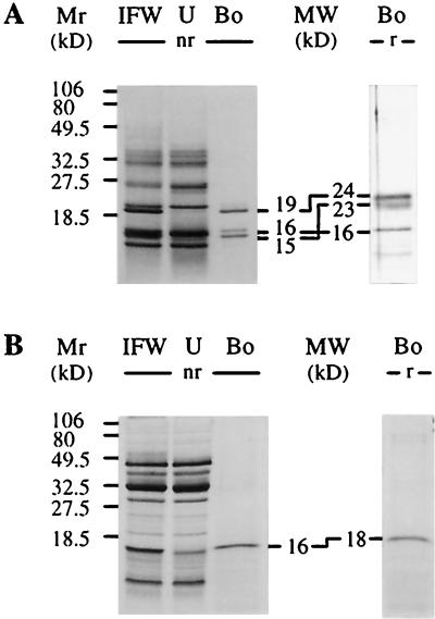
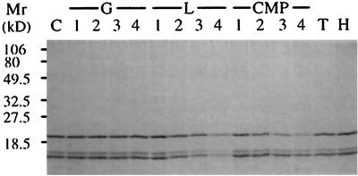
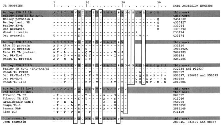
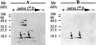
Pathogenesis-related proteins from intercellular fluid washings of stressed barley (Hordeum vulgare L.) leaves were analyzed to determine their binding to various water-insoluble polysaccharides. Three proteins (19, 16, and 15 kD) bound specifically to several water-insoluble beta-1,3-glucans. Binding of the barley proteins to pachyman occurred quickly at 22 degreesC at pH 5.0, even in the presence of 0.5 M NaCl, 0.2 M urea, and 1% (v/v) Triton X-100. Bound barley proteins were released by acidic treatments or by boiling in sodium dodecyl sulfate. Acid-released barley proteins could bind again specifically and singly to pachyman. Water-soluble laminarin and carboxymethyl-pachyman competed for the binding of the barley proteins to pachyman. The N-terminal sequence of the 19-kD barley beta-1,3-glucan-binding protein showed near identity to the barley seed protein BP-R and high homology to other thaumatin-like (TL) permatins. The 16-kD barley protein was also homologous to TL proteins, whereas the 15-kD barley protein N-terminal sequence was identical to the pathogenesis-related Hv-1 TL protein. Antifungal barley protein BP-R and corn (Zea mays) zeamatin were isolated by binding to pachyman. Two extracellular proteins from stressed pea (Pisum sativum L.) also bound to pachyman and were homologous to TL proteins.


Figures

Figure 1
Figure 1
Detection of barley and pea IFW proteins binding to pachyman. Chemically stressed barley (A) and pea (B) leaf IFW extracts (lanes IFW) containing 1 mg mL−1 proteins were subjected to SDS-PAGE before and after binding to pachyman. Binding assays in acetate buffer and SDS-PAGE were performed as in Methods. Proteins bound (lanes Bo) and unbound (lanes U) to pachyman were also analyzed after SDS-PAGE. Estimated molecular masses (MW) are given for the bound proteins separated under nonreducing (nr) or reducing (r) conditions. Numbers on the left refer to molecular mass markers (Mr).
Figure 2
Figure 2
Binding of the three barley IFW proteins to pachyman in the presence of soluble β-1,3-glucans. Sonicated and HCl-washed pachyman (2 mg) was incubated for 15 min at 22°C in 0.5 mL of 50 mm sodium acetate buffer, pH 5.0, with the three barley IFW proteins obtained after HCl elution of IFW proteins previously bound to pachyman. The binding was tested in the presence of Glc (G), laminarin (L), carboxymethyl-pachyman (CMP), laminaritetraose (lane T), and laminarihexaose (lane H) and in the absence of a competitor as a control (lane C). After incubation the insoluble pachyman was sedimented by centrifugation at 15,000g for 5 min at 22°C and washed twice, and proteins in the pellet were analyzed by SDS-PAGE under nonreducing conditions as in Figure 1. Lanes 1, 2, 3, and 4 marked “G,” 0.5, 2.5, 5 and 10 mg of Glc, respectively; lanes 1, 2, 3, and 4 marked “L,” 0.5, 2.5, 5, and 10 mg of laminarin, respectively; lanes 1, 2, 3, and 4 marked “CMP,” 0.5, 1, 2, and 4 mg of carboxymethyl-pachyman, respectively. Ten milligrams of laminaritetraose and laminarihexaose were used. Numbers on the left refer to molecular mass markers (Mr).
Figure 3
Figure 3
Comparison of N-terminal amino acid sequences of purified pachyman-binding proteins with several TL proteins. The seven proteins microsequenced in the present study are shaded and “This work” is indicated in the far-right column. The vertical rectangles indicate amino acids identical in all sequences. Dots at residue 20 correspond to one amino acid gap. Accession numbers on the right are from the National Center for Biotechnology Information (see Methods).
Figure 4
Figure 4
Detection of pea IFW proteins binding to pachyman after two-dimensional gel electrophoresis. Chemically stressed pea leaf IFW extract (A) and two proteins binding to pachyman (B) were subjected to two-dimensional PAGE. Binding to pachyman was as in Figure 1. The first-dimension separation (native 1st D) involved PAGE at pH 4.3 under native conditions (Reisfeld system) and was followed by a second-dimension PAGE (from top to bottom) in a denaturing (SDS) 15% polyacrylamide gel under nonreducing conditions. Proteins were stained as in Figure 1. Numbers on the left refer to molecular mass markers (Mr) and arrowheads 1 and 2 indicate the presence of the two TL proteins binding to pachyman (B) in the IFW extract (A).

References

    1. Abad LR, D'Urzo MP, Lin D, Narasimhan ML, Renveni M, Zhu JK, Niu X, Singh NK, Hasegawa PM, Bressan RA. Antifungal activity of tobacco osmotin has specificity and involves plasma membrane permeabilization. Plant Sci. 1996;118:11–23.
    1. Altschul SF, Gish W, Miller W, Myers EW, Lipman DJ. Basic local aligment search tool. J Mol Biol. 1990;215:403–410. - PubMed
    1. Audy P, Grenier J, Asselin A. Lysozyme activity in animal extracts after sodium dodecyl sulfate-polyacrylamide gel electrophoresis. Comp Biochem Physiol. 1989;92B:523–527. - PubMed
    1. Bol JF, Linthorst HJM, Cornelissen BJC. Plant pathogenesis-related proteins induced by virus infection. Annu Rev Phytopathol. 1990;28:113–138.
    1. Bussey H. K1 killer toxin, a pore-forming protein from yeast. Mol Microbiol. 1991;5:2339–2343. - PubMed

Publication types

LinkOut - more resources