. 1999 Jan;73(1):205-13.
doi: 10.1128/JVI.73.1.205-213.1999.

Microglia express CCR5, CXCR4, and CCR3, but of these, CCR5 is the principal coreceptor for human immunodeficiency virus type 1 dementia isolates

Affiliations

Microglia express CCR5, CXCR4, and CCR3, but of these, CCR5 is the principal coreceptor for human immunodeficiency virus type 1 dementia isolates

A V Albright et al. J Virol. 1999 Jan.

Abstract

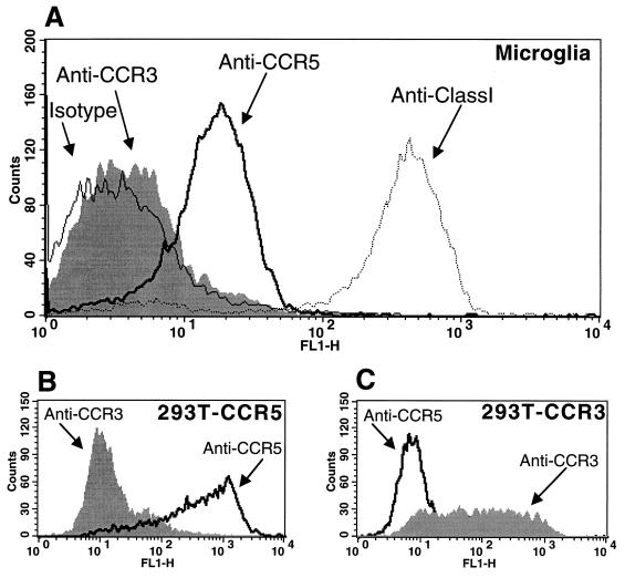
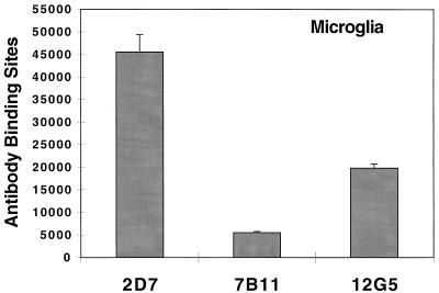
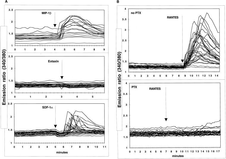
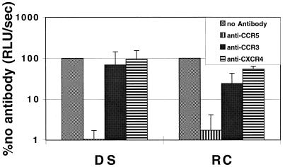
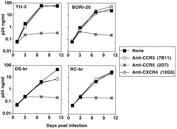
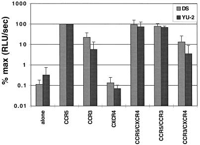
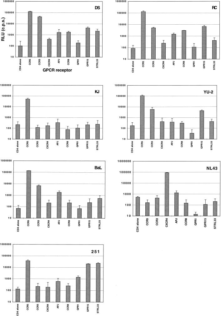
Microglia are the main human immunodeficiency virus (HIV) reservoir in the central nervous system and most likely play a major role in the development of HIV dementia (HIVD). To characterize human adult microglial chemokine receptors, we analyzed the expression and calcium signaling of CCR5, CCR3, and CXCR4 and their roles in HIV entry. Microglia expressed higher levels of CCR5 than of either CCR3 or CXCR4. Of these three chemokine receptors, only CCR5 and CXCR4 were able to transduce a signal in microglia in response to their respective ligands, MIP-1beta and SDF-1alpha, as recorded by single-cell calcium flux experiments. We also found that CCR5 is the predominant coreceptor used for infection of human adult microglia by the HIV type 1 dementia isolates HIV-1DS-br, HIV-1RC-br, and HIV-1YU-2, since the anti-CCR5 antibody 2D7 was able to dramatically inhibit microglial infection by both wild-type and single-round luciferase pseudotype reporter viruses. Anti-CCR3 (7B11) and anti-CXCR4 (12G5) antibodies had little or no effect on infection. Last, we found that virus pseudotyped with the DS-br and RC-br envelopes can infect cells transfected with CD4 in conjunction with the G-protein-coupled receptors APJ, CCR8, and GPR15, which have been previously implicated in HIV entry.


Figures

FIG. 1
FIG. 1
Surface expression of chemokine receptors CCR3 and CCR5 on microglia. (A) Adult human microglia were cultured for 6 days, detached, stained with a MAb for CCR5 (2D7) or CCR3 (7B11), an isotype-matched monoclonal control (807.09), or an anti-class I hybridoma supernatant, and analyzed by flow cytometry as described in Materials and Methods. Microglia were positive for CCR5, whereas levels of CCR3 staining were low or undetectable. Similar staining patterns were seen in microglia from three other donors. (B and C) As positive controls, 293T cells transiently transfected with CCR5 (B) and CCR3 (C) were stained with the same chemokine receptor MAbs.
FIG. 2
FIG. 2
Anti-chemokine receptor antibody binding sites on microglia cultured less than 24 h. To determine the number of antibody binding sites per cell, microglia were stained and analyzed with the MAbs 2D7 (anti-CCR5), 7B11 (anti-CCR3), and 12G5 (anti-CXCR4), as described in Materials and Methods. Error bars indicate the deviations from linearity obtained from the linear regression curve. The CCR3 quantification was repeated on microglia that had been cultured for 11 days with similar results.
FIG. 3
FIG. 3
Exposure to chemokines triggers changes in microglial intracellular free [Ca2+]. Microglia cultured for 5 to 10 days (3 × 10⁴/coverslip) were loaded with 2.5 μM fura-2/AM and prepared for single-cell calcium flux experiments as described in Materials and Methods. (A) Cells were exposed to 60 nM MIP-1β, 60 nM eotaxin, or 60 nM SDF-1α for 5 min, and changes in free Ca2+ were expressed as the emission ratio, at 510 nm, following excitation at 340 and 380 nm. These results are representative of experiments repeated multiple times with different microglial preparations. (B) Microglia that were or were not pretreated with 0.5 to 1.0 μg of pertussis toxin (PTX)/ml were exposed to 60 nM RANTES.
FIG. 4
FIG. 4
Inhibition of env-pseudotyped virus infection of microglia with antibodies to chemokine receptors. Microglia were preincubated with MAb to CCR5 (2D7), CCR3 (7B11), or CXCR4 (12G5) at a concentration of 20 μg/ml for 45 min at 4°C and were then infected with pseudotyped viruses. Cells were lysed 4 to 5 days postinfection, and infection was measured as luciferase activity (RLU per second). Data are expressed as percentages of luciferase activity obtained in the absence of antibody. DS, envelope from HIV-1DS-br; RC, envelope from HIV-1RC-br.
FIG. 5
FIG. 5
Inhibition of HIVD isolate infection of microglia in the presence of antibodies to chemokine receptors. Microglia were preincubated with MAb to CCR3 (7B11), CCR5 (2D7), or CXCR4 (12G5) at a concentration of 20 μg/ml for 45 min at 4°C and were then infected with 4 ng of p24gag of HIV-1YU-2, HIV-1BORI-20, HIV-1DS-br, or HIV-1RC-br. The following day, the inocula were removed, the cells were washed, and medium was replaced with the appropriate antibody (5 μg/ml). Infected cultures were maintained with the appropriate MAb at a constant concentration. Supernatants were collected over the course of infection and assayed for viral p24gag antigen, expressed in nanograms per milliliter.
FIG. 6
FIG. 6
HIVD env-pseudotyped virus infection of 293T cells transfected with two chemokine receptors. 293T cells were transiently transfected with CD4 alone or with CD4 and CCR5, CCR3, CXCR4, CCR5 plus CXCR4, CCR5 plus CCR3, or CCR3 plus CXCR4. pCDNA3.1 was used to equalize the quantity of DNA in each well. Data are expressed as percentages of infection of 293T cells with CD4 and CCR5. Error bars, standard deviations. DS, envelope from HIV-1DS-br; YU-2, envelope from HIV-1YU-2.
FIG. 7
FIG. 7
Alternative coreceptor use by HIVD viral envelopes. Pseudotyped viruses prepared with HIV envelopes DS-br, RC-br, and KJ-br and control envelopes YU-2, BaL, NL-43, and SIVmac251 were assayed for infectivity as measured by luciferase activity (in RLU per second) on U87 cells transiently transfected with CD4 and CCR5, CCR3, CXCR4, APJ, CCR8, GPR1, GPR15, or STRL33. Infections were performed in triplicate. Error bars, standard deviations.

References

    1. Albright A V, Strizki J, Harouse J M, Lavi E, O’Connor M, González-Scarano F. HIV-1 infection of cultured human adult oligodendrocytes. Virology. 1996;217:211–219. - PubMed
    1. Benveniste E N. Role of macrophages/microglia in multiple sclerosis and experimental allergic encephalomyelitis. J Mol Med. 1997;75:165–173. - PubMed
    1. Choe H, Farzan M, Konkel M, Martin K, Sun Y, Marcon L, Cayabyab M, Berman M, Dorf M E, Gerard N, Gerard C, Sodroski J. The orphan seven-transmembrane receptor Apj supports the entry of primary T-cell-line-tropic and dualtropic human immunodeficiency virus type 1. J Virol. 1998;72:6113–6118. - PMC - PubMed
    1. Cohen J. AIDS therapies: exploring how to get at—and eradicate—hidden HIV. Science. 1998;279:1854. - PubMed
    1. Deng H, Liu R, Ellmeier W, Choe S, Unutmaz D, Burkhart M, Di Marzio P, Marmon S, Sutton R E, Hill C M, Davis C B, Peiper S C, Schall T J, Littman D R, Landau N R. Identification of a major co-receptor for primary isolates of HIV-1. Nature. 1996;381:661–666. - PubMed

Publication types

LinkOut - more resources