. 1999 Jan;73(1):242-50.
doi: 10.1128/JVI.73.1.242-250.1999.

Matrix protein of rabies virus is responsible for the assembly and budding of bullet-shaped particles and interacts with the transmembrane spike glycoprotein G

Affiliations

Matrix protein of rabies virus is responsible for the assembly and budding of bullet-shaped particles and interacts with the transmembrane spike glycoprotein G

T Mebatsion et al. J Virol. 1999 Jan.

Abstract

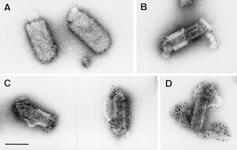
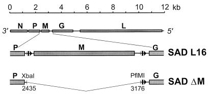
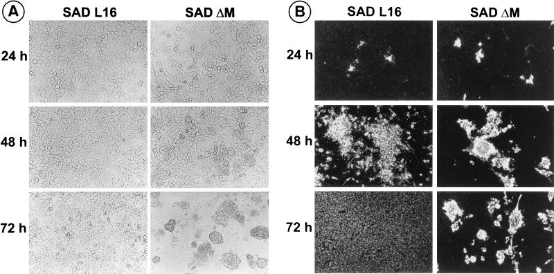
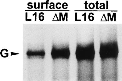
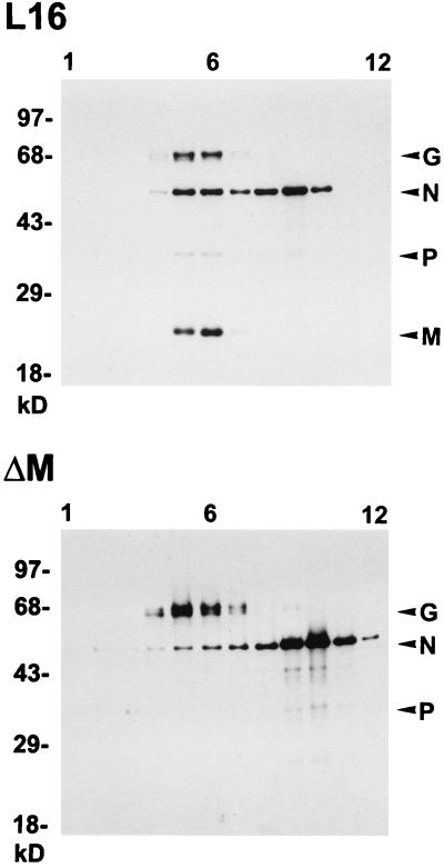
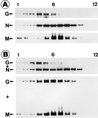
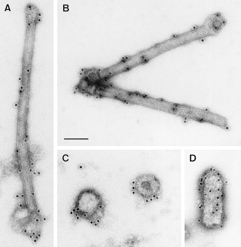
To elucidate the functions of rhabdovirus matrix (M) protein, we determined the localization of M in rabies virus (RV) and analyzed the properties of an M-deficient RV mutant. We provide evidence that M completely covers the ribonucleoprotein (RNP) coil and keeps it in a condensed form. As determined by cosedimentation experiments, not only the M-RNP complex but also M alone was found to interact specifically with the glycoprotein G. In contrast, an interaction of G with the nucleoprotein N or M-less RNP was not observed. In the absence of M, infectious particles were mainly cell associated and the yield of cell-free infectious virus was reduced by as much as 500,000-fold, demonstrating the crucial role of M in virus budding. Supernatants from cells infected with the M-deficient RV did not contain the typical bullet-shaped rhabdovirus particles but instead contained long, rod-shaped virions, demonstrating severe impairment of the virus formation process. Complementation with M protein expressed from plasmids rescued rhabdovirus formation. These results demonstrate the pivotal role of M protein in condensing and targeting the RNP to the plasma membrane as well as in incorporation of G protein into budding virions.


Figures

FIG. 1
FIG. 1
RV M protein is positioned between the lipid bilayer and the RNP coil. Virions treated with Triton X-100 (B to D) and untreated control samples (A) were purified as described in Materials and Methods and analyzed by electron microscopy. The absence of anti-M labeling on the outer membranes of untreated virions (90 to 100 nm in diameter) (A) and an intense anti-M labeling on the surfaces of the RNP coil or skeletons (50 to 60 nm in diameter) of detergent-treated virions (C) demonstrate that M surrounds the RNP coil. Only areas where the lipid bilayer was not completely removed (60 to 70 nm in diameter) are weakly labeled with anti-G MAb (B). In partially disassembled skeletons (D), only the uncoiled nucleocapsid, and not the surface of the skeleton, is labeled with anti-RNP serum. Bar, 100 nm.
FIG. 2
FIG. 2
Organization of the M-deficient RV genome. The entire RV genome with its five open reading frames (top), a detailed M cistron region (middle), and the region encompassing a deletion (bottom) are shown. Arrowheads and bars represent transcriptional start and stop signals, respectively. In SAD ΔM, the entire M cistron (SAD L16 nucleotides 2435 to 3176) was removed by a deletion starting within the nontranslated region of the P gene and ending close to the transcriptional stop signal of the M gene.
FIG. 3
FIG. 3
SAD ΔM induces increased cell-cell fusion and cell death. (A) Following infection of cultures with an MOI of 0.005, live cells were examined microscopically for morphological changes at the indicated times. Cell fusion is detected in SAD ΔM-infected cells after 48 h, and large syncytia are present at 72 h postinfection. (B) In cells infected in parallel, the spread of infection was monitored at the indicated times by direct immunofluorescence with a conjugate directed against RV N protein. Secondary infection of cells, as indicated by the appearance of fluorescing granules, is virtually absent in SAD ΔM. After 48 h, SAD ΔM N expression is found in large syncytia.
FIG. 4
FIG. 4
Analysis of cell surface expression of G proteins. Approximately 106 BSR cells were infected at an MOI of 1 and after 16 h were labeled with 100 μCi of [35S]methionine for 3 h. Anti-G MAb was incubated with live cells to bind G proteins expressed at the cell surface or with cell lysates containing total G protein. Phoshorimager quantitation revealed a 2.6-fold-higher level of G protein on the surfaces of cells infected by SAD ΔM.
FIG. 5
FIG. 5
Protein composition of cell-associated SAD L16 and SAD ΔM virions. Cell extracts from approximately 106 infected cells were prepared as described in Materials and Methods and purified by 10 to 70% sucrose gradient centrifugation. Twelve equal gradient fractions (numbered from top to bottom) were analyzed by Western blotting for the presence of viral proteins. The sucrose density was determined from the refractive index of each fraction. For both preparations, the peak of infectivity was present in fraction 6, which has a density of 1.14 g/cm3.
FIG. 6
FIG. 6
RV M interacts with G. Approximately 106 BSR cells were first infected with vTF7-3 and then transfected with 5 μg of plasmid DNA. After 16 h of infection, cell lysates were prepared, centrifuged to equilibrium, and analyzed as described for Fig. 5. (A) Density gradients from cells expressing G, N, or M protein individually; (B) cells coexpressing G and N proteins or G and M proteins. When G and M proteins were coexpressed, the G protein cosedimented with M protein (numbering is from the top to the bottom of the gradient).
FIG. 7
FIG. 7
Morphology of particles released from SAD ΔM-infected cells. SAD ΔM was grown in the absence of RV M protein (A to C) or in cells that express RV M protein from transfected plasmids (D). Purified supernatants from approximately 107 SAD ΔM-infected BSR cells were immunostained with a MAb specific for the RV G protein and analyzed by electron microscopy. In the absence of RV M protein, either long rod-shaped particles (A and B) or round vesicular structures (C), but no bullet-shaped particles, were detected. After expression of RV M protein, the majority of particles had the typical bullet-shaped rhabdovirus morphology (D). Bar, 100 nm.

Similar articles

Cited by

References

    1. Barge A, Gaudin Y, Coulon P, Ruigrok R W. Vesicular stomatitis virus M protein may be inside the ribonucleocapsid coil. J Virol. 1993;67:7246–7253. - PMC - PubMed
    1. Carroll A R, Wagner R R. Role of the membrane (M) protein in endogenous inhibition of in vitro transcription by vesicular stomatitis virus. J Virol. 1979;29:134–142. - PMC - PubMed
    1. Cartwright B, Smale C J, Brown F. Dissection of vesicular stomatitis virus into the infective ribonucleoprotein and immunizing components. J Gen Virol. 1970;7:19–32. - PubMed
    1. Chong L D, Rose J K. Membrane association of functional vesicular stomatitis virus matrix protein in vivo. J Virol. 1993;67:407–414. - PMC - PubMed
    1. Chong L D, Rose J K. Interactions of normal and mutant vesicular stomatitis virus matrix proteins with the plasma membrane and nucleocapsids. J Virol. 1994;68:441–447. - PMC - PubMed

Publication types