. 1999 Jan;73(1):377-87.
doi: 10.1128/JVI.73.1.377-387.1999.

Intracellular traffic of herpes simplex virus glycoprotein gE: characterization of the sorting signals required for its trans-Golgi network localization

Affiliations

Intracellular traffic of herpes simplex virus glycoprotein gE: characterization of the sorting signals required for its trans-Golgi network localization

A Alconada et al. J Virol. 1999 Jan.

Abstract

Herpes simplex virus (HSV) and varicella-zoster virus (VZV) are two pathogenic human alphaherpesviruses whose intracellular assembly is thought to follow different pathways. VZV presumably acquires its envelope in the trans-Golgi network (TGN), and it has recently been shown that its major envelope glycoprotein, VZV-gE, accumulates in this compartment when expressed alone. In contrast, the envelopment of HSV has been proposed to occur at the inner nuclear membrane, although to which compartment the gE homolog (HSV-gE) is transported is unknown. For this reason, we have studied the intracellular traffic of HSV-gE and have found that this glycoprotein accumulates at steady state in the TGN, both when expressed from cloned cDNA and in HSV-infected cells. In addition, HSV-gE cycles between the TGN and the cell surface and requires a conserved tyrosine-containing motif within its cytoplasmic tail for proper trafficking. These results show that VZV-gE and HSV-gE have similar intracellular trafficking pathways, probably reflecting the presence of similar sorting signals in the cytoplasmic domains of both molecules, and suggest that the respective viruses, VZV and HSV, could use the same subcellular organelle, the TGN, for their envelopment.


Figures

FIG. 1
FIG. 1
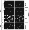
Colocalization of HSV-gE with TGN proteins. HeLa cells double transfected with mammalian expression vectors encoding HSV-gE and either VZV-gE (a and b), furin (c and d), or ST-VSVG (e and f) were visualized with the 7520 MAb against HSV-gE (a, c, and e) and the 1667 polyclonal antiserum against VZV-gE (b), a polyclonal serum against furin (d), and a polyclonal antiserum against the VSVG epitope (f), followed by fluorescein isothiocyanate- or rhodamine-coupled antirabbit and antimouse secondary antibodies. Bar, 5 μm.
FIG. 2
FIG. 2
Effect of BFA treatment on the subcellular distribution of HSV-gE. HeLa cells were double transfected with pSFFV-HSV-gE and either pSFFV-VZV-gE (a and b) or pSG5-furin (c and d) and treated for 5 min with 10 μg of BFA per ml. After fixation, the coverslips were processed for indirect immunofluorescence to detect HSV-gE (7520 MAb) (a and c) and VZV-gE (1667 polyclonal antiserum) (b) or furin (d). Bar, 5 μm.
FIG. 3
FIG. 3
Intracellular distribution of HSV-gE in HSV-2-infected cells. (a to f) HeLa cells grown on coverslips were transfected with plasmids encoding VZV-gE (a and b) or ST-VSVG (c to f) and, 40 h after transfection, exposed to a tissue culture supernatant of MRC-5 cells infected with HSV-2 for 2 h, and then the inoculum was removed and further incubated for another 2.5 h (a to d) or 10 h (e and f). Cells were fixed and processed for indirect immunofluorescence by using the 7520 anti-HSV-gE MAb (a, c, and e) and either the 1667 polyclonal antiserum against VZV-gE (b) or a polyclonal antiserum against the VSVG epitope (d and f). (g and h) A clone of MeWo cells stably expressing ST-VSVG was exposed to a tissue culture supernatant of MRC-5 cells infected with HSV-2 for 5 h, fixed, and stained with the 7520 anti-HSV-gE MAb (g) and a polyclonal antiserum against the VSVG epitope (h). Bar, 5 μm.
FIG. 4
FIG. 4
Recycling of HSV-gE through the plasma membrane. HeLa cells were double transfected with plasmids encoding VZV-gE and either HSV-gE (a and b) or HSV-gEΔ1 (c and d) and 48 h after transfection were incubated for 1 h at 37°C in culture medium containing 1:200 dilutions of the 1667 polyclonal rabbit antiserum against VZV-gE and the 7520 MAb against HSV-gE. Cells were fixed and processed for immunofluorescence by using a mixture of fluorescein isothiocyanate-coupled antirabbit IgG (to detect anti-VZV-gE [a and c]) and rhodamine (tetramethyl rhodamine isothiocyanate)-coupled antimouse IgG (to detect anti-HSV-gE [b and d]). Bar, 5 μm.
FIG. 5
FIG. 5
Multiple sequence alignment of the cytoplasmic tails of the gE homologs forms seven different members of the Alphaherpesvirinae subfamily. The sequences shown include those of HSV-1 (47), HEV-1 (72), BHV-1 (36), FHV-1 (48), VZV (15), SVV (21), and PRV (56). The sequences of the gE homologs from HSV-1, HEV-1, BHV-1, VZV, SVV, and PRV are under SwissProt accession no. vgle_hsv11, vgle_hsveb, vgle_hsvbs, vgle_vzvd, vgle_svvd, and vgl_prvri, respectively, and FHV-1 gE is under GenBank accession no. X98449. Identical amino acids found in at least three of the seven sequences are shown as white characters on a black background, and conserved substitutions are shown on a grey background. Brackets on top of the alignment indicate the conserved regions discussed in the text. The amino acid positions for every sequence (considering the full-length precursor proteins) at the end of each block in the alignment are shown in the right-hand margin. The last amino acids of the different HSV-gE truncation mutants (Δ1, Δ2, Δ3, and Δ4) are indicated by arrows.
FIG. 6
FIG. 6
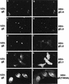
Intracellular distribution of HSV-gE mutants in the cytoplasmic domain. HeLa cells were double transfected with a plasmid encoding VZV-gE and either HSV-gEΔ4 (a and b), the HSV-gEΔ3 (c and d), the HSV-gEΔ2 (e and f), or the HSV-gEΔ1 (g and h) truncation mutants or the HSV-gE(Y463A) (i and j) point mutant and stained with a mixture of the 1667 polyclonal antiserum against VZV-gE (a, c, e, g, and i) and the 7520 MAb against HSV-gE (b, d, f, h, and j). Bar, 5 μm.

Similar articles

Cited by

References

    1. Alconada A, Bauer U, Baudoux L, Piette J, Hoflack B. Intracellular transport of the glycoproteins gE and gI of the varicella-zoster virus: gE accelerates the maturation of gI and determines its accumulation in the trans-Golgi network. J Biol Chem. 1998;273:13430–13436. - PubMed
    1. Alconada A, Bauer U, Hoflack B. A tyrosine-based motif and a casein kinase II phosphorylation site regulate the intracellular trafficking of the varicella-zoster virus glycoprotein I, a protein localized in the trans-Golgi network. EMBO J. 1996;15:6096–6110. - PMC - PubMed
    1. Avitabile E, Di Gaeta S, Torrisi M R, Ward P L, Roizman B, Campadelli-Fiume G. Redistribution of microtubules and Golgi apparatus in herpes simplex virus-infected cells and their role in viral exocytosis. J Virol. 1995;69:7472–7482. - PMC - PubMed
    1. Basu S, Dubin G, Basu M, Nguyen V, Friedman H M. Characterization of regions of herpes simplex virus type 1 glycoprotein E involved in binding the Fc domain of monomeric IgG and in forming a complex with glycoprotein I. J Immunol. 1995;154:260–267. - PubMed
    1. Bos K, Wraight C, Stanley K. TGN38 is maintained in the trans-Golgi network by a tyrosine-containing motif in the cytoplasmic domain. EMBO J. 1993;12:2219–2228. - PMC - PubMed

Publication types

Substances

LinkOut - more resources