. 1999 Jan;73(1):702-8.
doi: 10.1128/JVI.73.1.702-708.1999.

Sendai virus infection induces apoptosis through activation of caspase-8 (FLICE) and caspase-3 (CPP32)

Affiliations

Sendai virus infection induces apoptosis through activation of caspase-8 (FLICE) and caspase-3 (CPP32)

M Bitzer et al. J Virol. 1999 Jan.

Abstract

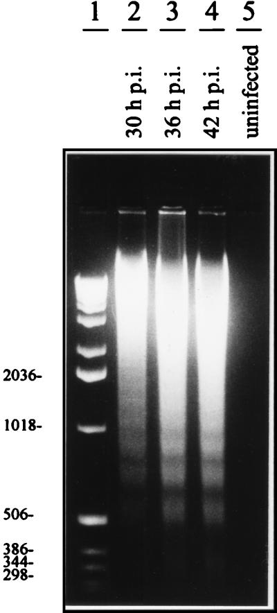
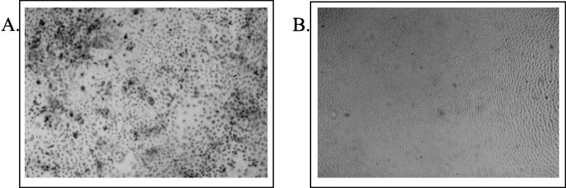
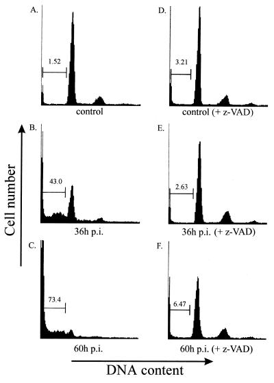
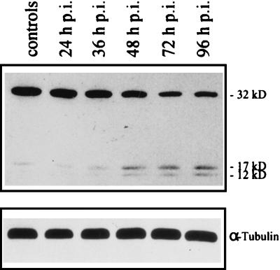
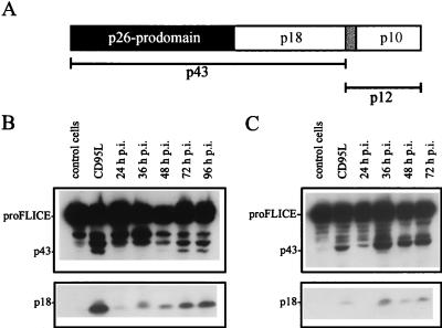
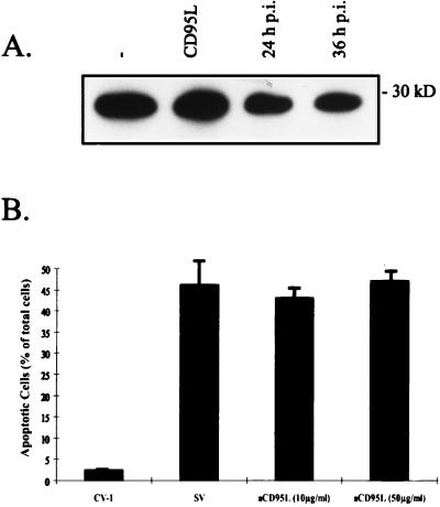
Sendai virus (SV) infection and replication lead to a strong cytopathic effect with subsequent death of host cells. We now show that SV infection triggers an apoptotic program in target cells. Incubation of infected cells with the peptide inhibitor z-VAD-fmk abrogated SV-induced apoptosis, indicating that proteases of the caspase family were involved. Moreover, proteolytic activation of two distinct caspases, CPP32/caspase-3 and, as shown for the first time in virus-infected cells, FLICE/caspase-8, could be detected. So far, activation of FLICE/caspase-8 has been described in apoptosis triggered by death receptors, including CD95 and tumor necrosis factor (TNF)-R1. In contrast, we could show that SV-induced apoptosis did not require TNF or CD95 ligand. We further found that apoptosis of infected cells did not influence the maturation and budding of SV progeny. In conclusion, SV-induced cell injury is mediated by CD95- and TNF-R1-independent activation of caspases, leading to the death of host cells without impairment of the viral life cycle.


Figures

FIG. 1
FIG. 1
DNA fragmentation induced by SV infection of CV-1 cells. Lanes 2 to 4, DNA preparations from productively infected cells at different time points p.i.; lane 1, DNA marker; lane 5, preparation from uninfected control cells.
FIG. 2
FIG. 2
In situ detection of apoptosis in infected CV-1 cells. Apoptotic DNA degradation was visualized by TUNEL staining and subsequent alkaline phosphatase staining as described in Materials and Methods. (A) Intense dark staining of CV-1 cells 24 h p.i.; (B) uninfected CV-1 cells.
FIG. 3
FIG. 3
Detection of hypodiploid DNA in SV-infected cells and inhibition by caspase inhibitor z-VAD. CV-1 cells were infected by SV (MOI, 10) and incubated with 0 (A, B, and C) or 100 (D, E, and F) μM of the caspase inhibitor z-VAD-fmk. Shown are the results of cytometric analysis at 36 and 60 h p.i. as well as the analysis of uninfected CV-1 cells (control) and CV-1 cells incubated with z-VAD-fmk for 60 h [control (+ z-VAD)] to exclude the toxic effects of z-VAD-fmk on these cells. The proportion of sub-2N DNA is indicated in the histograms.
FIG. 4
FIG. 4
SV infection triggers CPP32/caspase-3 activation. HepG2 cells were infected with SV (MOI, 10) and were prepared at the indicated time points p.i. Lane 1, preparation of uninfected control cells. The CPP32 proform and subunits p17 and p12 as well as tubulin were detected by Western blot analysis.
FIG. 5
FIG. 5
FLICE/caspase-8 cleavage in SV-infected host cells. (A) FLICE/caspase-8 cleavage products according to Medema et al. (28); initial cleavage generates p43 and p12 intermediates, followed by further processing to the active p18 and p10 subunits and the FLICE prodomain p26. (B and C) HepG2 (B) and CV-1 (C) cells were infected with SV (MOI, 10), and FLICE and cleavage products p43 and p18 were detected by Western blot analysis at the indicated times p.i. Lanes 1 and 2, preparations from uninfected cells (control cells) and, as a positive control, from uninfected cells incubated with CD95L, respectively.
FIG. 6
FIG. 6
CD95L expression in host cells is not responsible for SV-induced apoptosis. (A) CV-1 cells were infected with SV (MOI, 10), and FasL was detected by Western blot analysis at 24 and 36 h p.i. Lanes 1 and 2, preparations from uninfected controls (−) and from cells incubated with CD95L (at 12 h), respectively. (B) After infection with SV (MOI, 10), CV-1 cells were incubated with chimeric receptor decoy proteins consisting of the extracellular part of CD95 (aCD95L) fused to IgG-Fc. At 48 h p.i., the cells were analyzed by flow cytometry (propidium iodide staining). Cells with subgenomic DNA content were considered apoptotic. The bar labeled CV-1 indicates the results of the analysis of uninfected controls, and the bar labeled SV represents the results of the analysis of infected cells without chimeric receptor decoy proteins in the supernatant. In our setting concentrations of 5 to 10 μg of aCD95L/ml in the supernatant blocked CD95L-induced apoptosis completely. The error bars represent standard deviations.

References

    1. Antoni B A, Sabbatini P, Rabson A B, White E. Inhibition of apoptosis in human immunodeficiency virus-infected cells enhances virus production and facilitates persistent infection. J Virol. 1995;69:2384–2392. - PMC - PubMed
    1. Aragane Y, Kulms D, Metze D, Wilkes G, Pöppelmann B, Luger T A, Schwarz T. Ultraviolet light induces apoptosis via direct activation of CD95 (Fas/APO-1) independently from its ligand CD95L. J Cell Biol. 1998;140:171–182. - PMC - PubMed
    1. Badley A D, McElhinny J A, Leibson P J, Lynch D H, Alderson M R, Paya C V. Upregulation of Fas ligand expression by human immunodeficiency virus in human macrophages mediates apoptosis of uninfected T lymphocytes. J Virol. 1996;70:199–206. - PMC - PubMed
    1. Banki K, Hutter E, Gonchoroff N J, Perl A. Molecular ordering in HIV-induced apoptosis—oxidative stress, activation of caspases, and cell survival are regulated by transaldolase. J Biol Chem. 1998;273:11944–11953. - PubMed
    1. Bitzer M, Lauer U, Baumann C, Spiegel M, Gregor M, Neubert W J. Sendai virus efficiently infects cells via the asialoglycoprotein receptor and requires the presence of cleaved F0 precursor proteins for this alternative route of cell entry. J Virol. 1997;71:5481–5486. - PMC - PubMed

Publication types