

Fine structure and morphogenesis of Borna disease virus
- PMID: 9847384
- PMCID: PMC103885
- DOI: 10.1128/JVI.73.1.760-766.1999
Fine structure and morphogenesis of Borna disease virus
Abstract
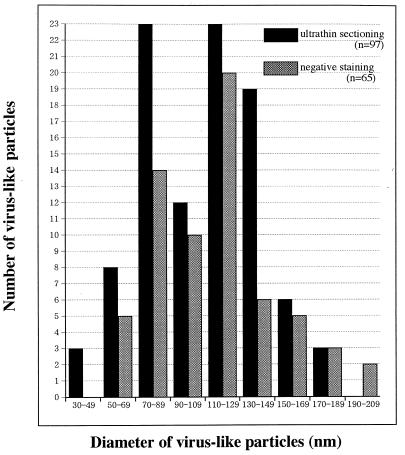
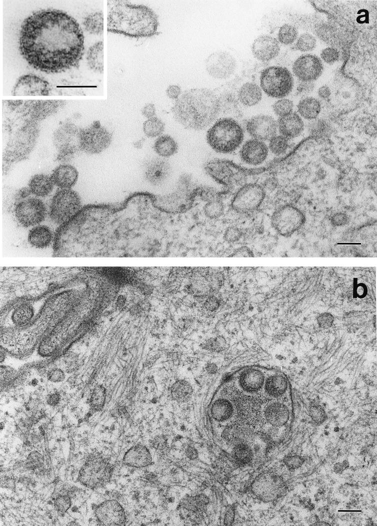
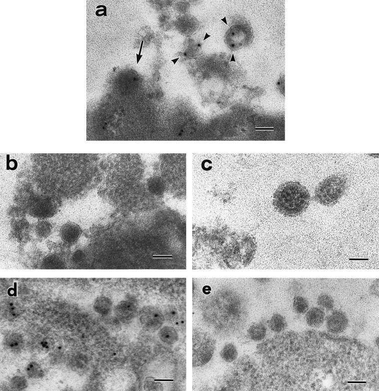
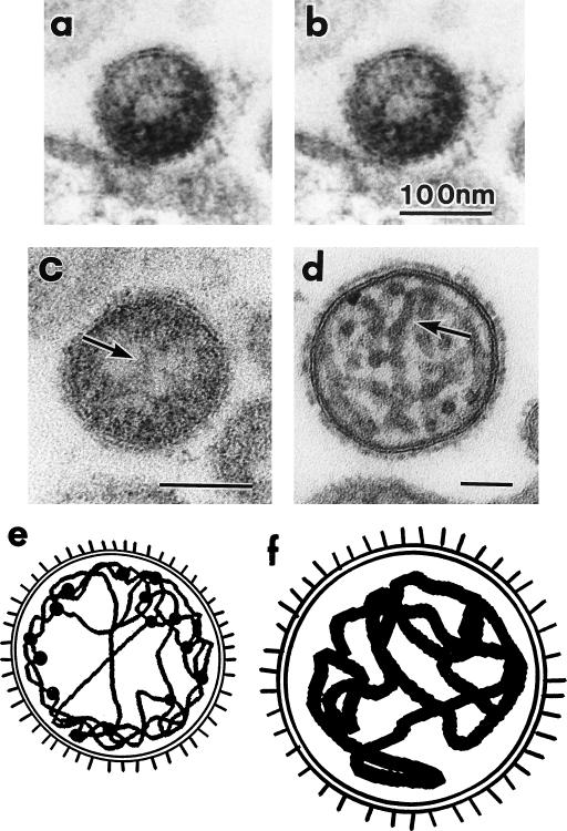
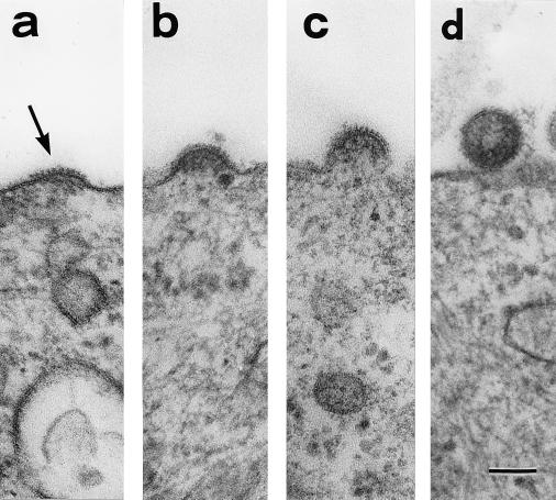
Borna disease virus (BDV), a negative nonsegmented single-stranded RNA virus, has not been fully characterized morphologically. Here we present what is to our knowledge the first data on the fine ultrastructure and morphogenesis of BDV. The supernatant of MDCK cells persistently infected with BDV treated with n-butyrate contained many virus-like particles and more BDV-specific RNA than that of untreated samples. The particles were spherical, enveloped, and approximately 130 nm in diameter; had spikes 7 nm in length; and reacted with BDV p40 antibody. A thin nucleocapsid, 4 nm in width, was present peripherally in contrast to the thick nucleocapsid of hemagglutinating virus of Japan. The BDV particles reproduced by budding on the cell surface.
Figures
References
-
- Bode L, Riegel S, Ludwig H, Amsterdam J D, Lange W, Koprowski H. Borna disease virus-specific antibodies in patients with HIV infection and with mental disorders. Lancet. 1988;ii:689. - PubMed
-
- Bode L, Riegel S, Lange W, Ludwig H. Human infections with Borna disease virus: seroprevalence in patients with chronic diseases and healthy individuals. J Med Virol. 1992;36:309–315. - PubMed
-
- Bode L, Ferszt R, Czech G. Borna disease virus infection and affective disorders in man. Arch Virol Suppl. 1993;7:159–167. - PubMed
-
- Bode L, Durrwald R, Ludwig H. Borna virus infections in cattle associated with fatal neurological disease. Vet Rec. 1994;135:283–284. - PubMed
-
- Bode L, Steinbach F, Ludwig H. A novel marker for Borna disease virus infection. Lancet. 1994;343:297–298. - PubMed
MeSH terms
Substances
LinkOut - more resources
Full Text Sources
