. 1998 Dec 1;12(23):3650-62.
doi: 10.1101/gad.12.23.3650.

The essential Gcd10p-Gcd14p nuclear complex is required for 1-methyladenosine modification and maturation of initiator methionyl-tRNA

Affiliations

The essential Gcd10p-Gcd14p nuclear complex is required for 1-methyladenosine modification and maturation of initiator methionyl-tRNA

J Anderson et al. Genes Dev. .

Abstract

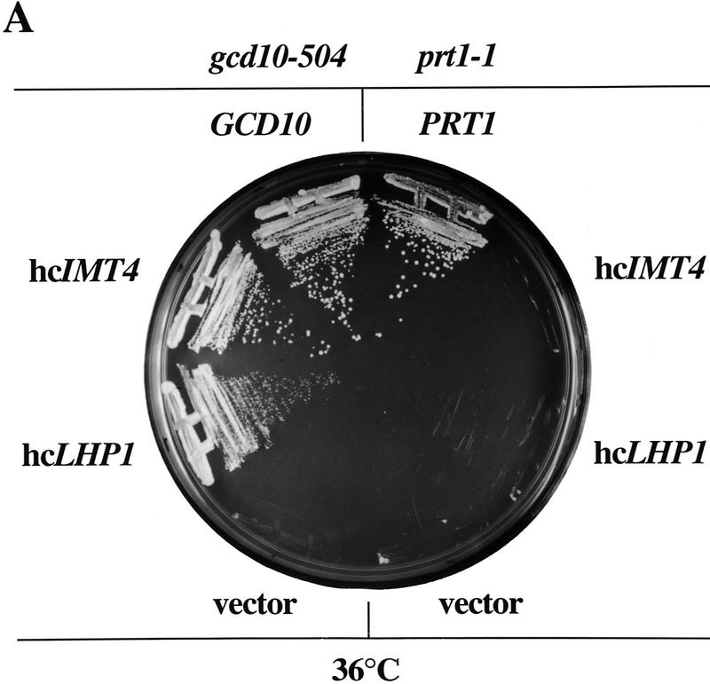
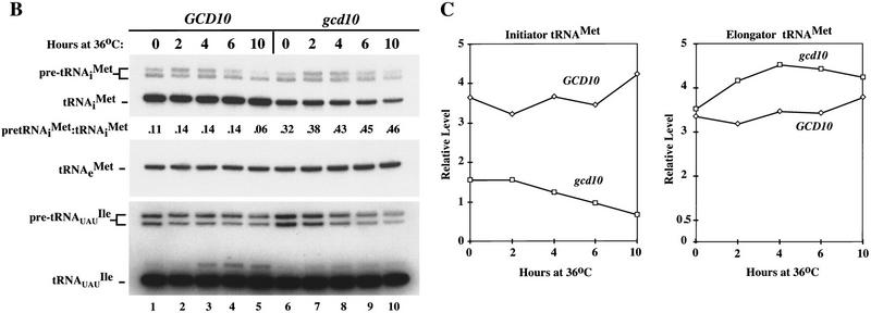
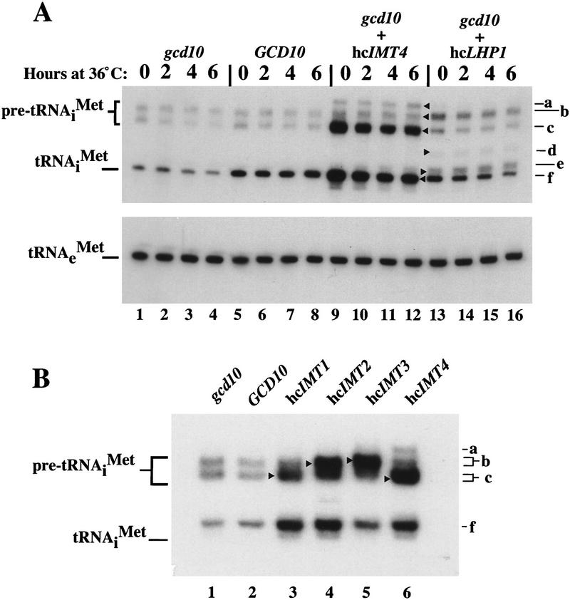
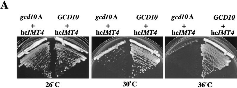
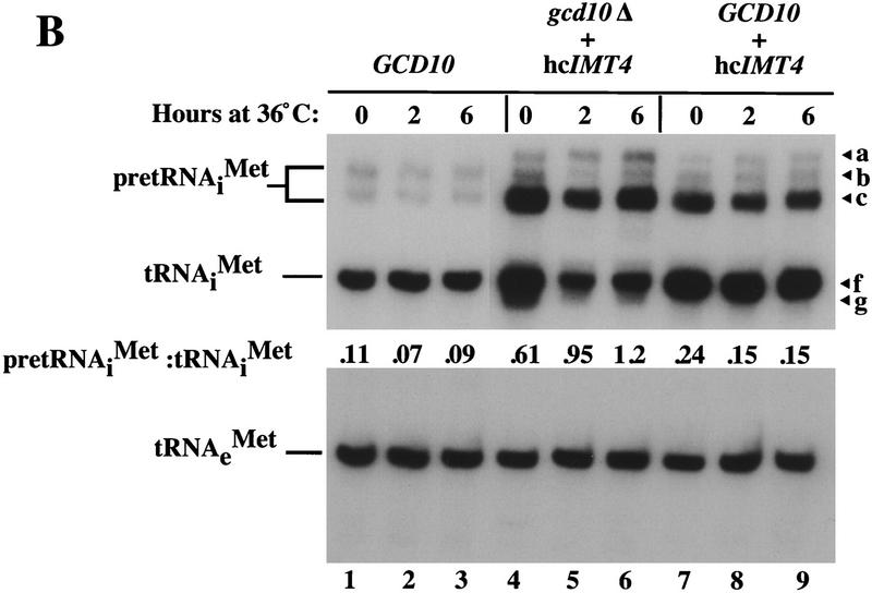
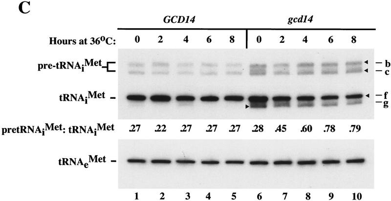
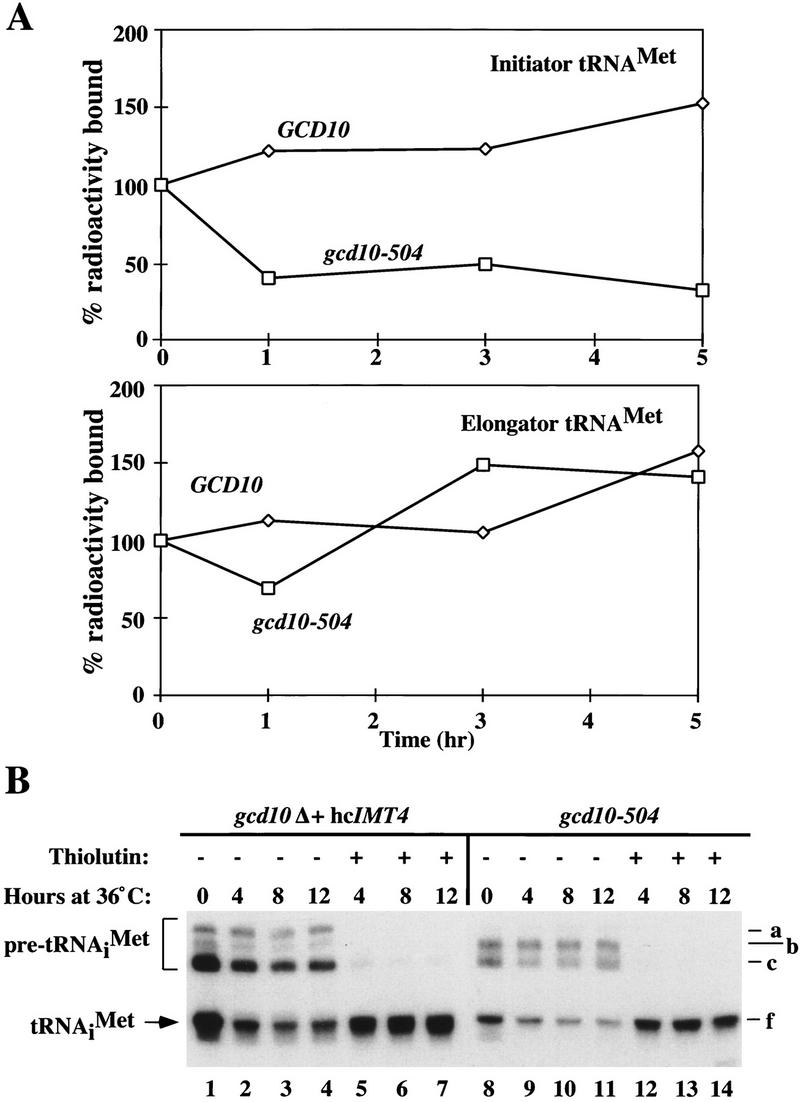
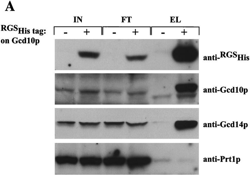
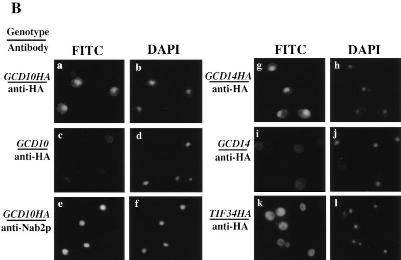
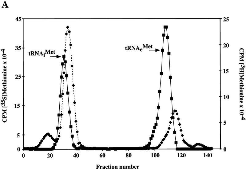
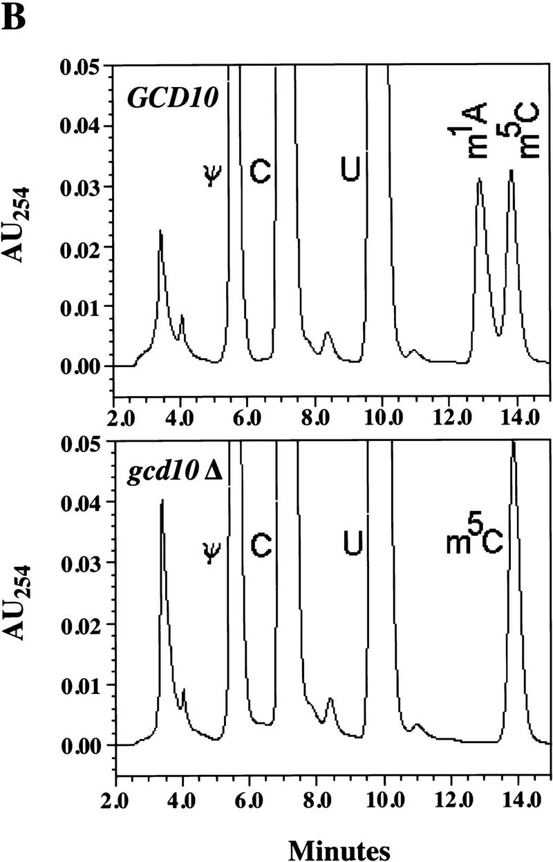
Gcd10p and Gcd14p are essential proteins required for the initiation of protein synthesis and translational repression of GCN4 mRNA. The phenotypes of gcd10 mutants were suppressed by high-copy-number IMT genes, encoding initiator methionyl tRNA (tRNAiMet), or LHP1, encoding the yeast homolog of the human La autoantigen. The gcd10-504 mutation led to a reduction in steady-state levels of mature tRNAiMet, attributable to increased turnover rather than decreased synthesis of pre-tRNAiMet. Remarkably, the lethality of a GCD10 deletion was suppressed by high-copy-number IMT4, indicating that its role in expression of mature tRNAiMet is the essential function of Gcd10p. A gcd14-2 mutant also showed reduced amounts of mature tRNAiMet, but in addition, displayed a defect in pre-tRNAiMet processing. Gcd10p and Gcd14p were found to be subunits of a protein complex with prominent nuclear localization, suggesting a direct role in tRNAiMet maturation. The chromatographic behavior of elongator and initiator tRNAMet on a RPC-5 column indicated that both species are altered structurally in gcd10Delta cells, and analysis of base modifications revealed that 1-methyladenosine (m1A) is undetectable in gcd10Delta tRNA. Interestingly, gcd10 and gcd14 mutations had no effect on processing or accumulation of elongator tRNAMet, which also contains m1A at position 58, suggesting a unique requirement for this base modification in initiator maturation.


Figures

Figure 1
Figure 1
High-copy suppressors of gcd10-504 overcome a defect in accumulation of tRNAiMet (A) Transformants of strains H2457 (gcd10-504) and H1676 (prt1-1) containing low-copy plasmids bearing GCD10 (pMG107) (Garcia-Barrio et al. 1995) or PRT1 (p2625), respectively, or high-copy plasmids bearing IMT4 (pC44) (Cigan et al. 1988), LHP1 (p2626), or empty vector YEp24 were streaked for single colonies on minimally supplemented SD plates and incubated at 36°C for 2 days. (B) Northern blot analysis of total RNA (5 μg) isolated as described (Kohrer and Domdey 1991) from strain H2457 (gcd10-504) bearing GCD10 on low-copy plasmid pMG107 (GCD10 lanes) or empty vector YEp24 (gcd10 lanes) grown in supplemented SD at 26°C to mid-exponential phase (0 hr at 36°C) and shifted to 36°C for 2, 4, 6, or 10 hr. The RNAs were separated on an 8% polyacrylamide-bis-acrylamide (19:1), 8.3 m urea gel by electrophoresis and transferred to Hybond-N+ membranes (Amersham). The blot was probed using a radiolabeled oligonucleotide that hybridized specifically to tRNAiMet, stripped and reprobed with radiolabeled oligonucleotides specific for tRNAeMet or tRNAUAUIle. Direct quantitation of all hybridized probes was conducted by PhosphorImager analysis using a Storm 860 apparatus (Molecular Dynamics) and ImageQuant software. The positions of pre-tRNAiMet species, mature tRNAiMet, tRNAeMet, pre-tRNAUAUIle, and mature tRNAUAUIle are indicated at left. The relative intensities of the hybridization signals were quantified for mature tRNAiMet and pre-tRNAiMet , and the ratios of pre-tRNAiMet to mature tRNAiMet are listed under the appropriate lanes. The species that migrated just above mature tRNAUAUIle and accumulated at high temperature most likely represent spliced precursors still bearing the 3′ extension (O’Connor and Peebles 1991). (C) The relative intensities of the hybridization signals in B were quantified by PhosphorImager analysis of the autoradiograph and plotted against the time of incubation at 36°C.
Figure 1
Figure 1
High-copy suppressors of gcd10-504 overcome a defect in accumulation of tRNAiMet (A) Transformants of strains H2457 (gcd10-504) and H1676 (prt1-1) containing low-copy plasmids bearing GCD10 (pMG107) (Garcia-Barrio et al. 1995) or PRT1 (p2625), respectively, or high-copy plasmids bearing IMT4 (pC44) (Cigan et al. 1988), LHP1 (p2626), or empty vector YEp24 were streaked for single colonies on minimally supplemented SD plates and incubated at 36°C for 2 days. (B) Northern blot analysis of total RNA (5 μg) isolated as described (Kohrer and Domdey 1991) from strain H2457 (gcd10-504) bearing GCD10 on low-copy plasmid pMG107 (GCD10 lanes) or empty vector YEp24 (gcd10 lanes) grown in supplemented SD at 26°C to mid-exponential phase (0 hr at 36°C) and shifted to 36°C for 2, 4, 6, or 10 hr. The RNAs were separated on an 8% polyacrylamide-bis-acrylamide (19:1), 8.3 m urea gel by electrophoresis and transferred to Hybond-N+ membranes (Amersham). The blot was probed using a radiolabeled oligonucleotide that hybridized specifically to tRNAiMet, stripped and reprobed with radiolabeled oligonucleotides specific for tRNAeMet or tRNAUAUIle. Direct quantitation of all hybridized probes was conducted by PhosphorImager analysis using a Storm 860 apparatus (Molecular Dynamics) and ImageQuant software. The positions of pre-tRNAiMet species, mature tRNAiMet, tRNAeMet, pre-tRNAUAUIle, and mature tRNAUAUIle are indicated at left. The relative intensities of the hybridization signals were quantified for mature tRNAiMet and pre-tRNAiMet , and the ratios of pre-tRNAiMet to mature tRNAiMet are listed under the appropriate lanes. The species that migrated just above mature tRNAUAUIle and accumulated at high temperature most likely represent spliced precursors still bearing the 3′ extension (O’Connor and Peebles 1991). (C) The relative intensities of the hybridization signals in B were quantified by PhosphorImager analysis of the autoradiograph and plotted against the time of incubation at 36°C.
Figure 2
Figure 2
High-copy suppressors of gcd10 mutations lead to increased amounts of initiator tRNAMet and identification of pre-tRNAiMet species. (A) Northern blot analysis of total RNA (5 μg) isolated from transformants of strains H2457 (gcd10-504) carrying empty vector YEp24 (gcd10 lanes), low-copy plasmid pMG107 bearing GCD10 (GCD10 lanes), high-copy plasmid pC44 bearing IMT4 (gcd10 + hcIMT4 lanes), or high-copy plasmid p2626 bearing LHP1 (gcd10 + hcLHP1 lanes), grown at 26°C or 36°C for the indicated times, as described in Fig. 1. The blot was probed for tRNAiMet and stripped and reprobed for tRNAeMet as described in Fig. 1. Indicated with arrowheads inside the blot and labeled to the right are the presumed positions of pre-tRNAiMet containing 5′ and 3′ extensions encoded by IMT4 and terminating downstream of the usual termination site (a), pre-tRNAiMet containing 5′ and 3′ extensions encoded by IMT2 and IMT3 (b), pre-tRNAiMet containing 5′ and 3′ extensions encoded by IMT1 and IMT4 (c), processing intermediate of IMT3 containing the 3′ extension (d), processing intermediate containing partial 3′ extension (e), and mature tRNAiMet (f). (B) Northern blot analysis of total RNA (10 μg) isolated from H2457 (gcd10-504) transformants containing empty vector YEp24 (gcd10), low-copy plasmid pMG107 bearing GCD10, or a high-copy plasmid bearing one of the four IMT genes, as indicated above the blot, grown at 26°C as described in Fig. 1. Initiator tRNAMet was detected by hybridization as described above and in Fig. 1. Indicated with arrowheads inside the blot and labeled to the right are the positions of pre-tRNAiMet containing 5′ and 3′ extensions (a–c) and mature tRNAiMet (f), as in A.
Figure 3
Figure 3
GCD10 is dispensable for cell viability in the presence of hcIMT4. (A) Growth of strain YJA146 (gcd10Δ + hcIMT4) and a transformant of its parental GCD10 strain BJ5464 bearing p1775 (GCD10 + hcIMT4) on YPD medium at 26°C for 3 days, and at 30°C or 36°C for 2 days. (B) Northern blot analysis of total RNA (7 μg) isolated from the same two strains described in A (gcd10Δ + hcIMT4 and GCD10 + hcIMT4 lanes) plus isogenic strain YJA143 containing the gcd10Δ chromosomal allele and single-copy GCD10 plasmid p2704 (GCD10). Strains were grown at 26°C or 36°C for 2 and 6 hr as described in Fig. 1. The membrane was probed for tRNAiMet and stripped and reprobed for tRNAeMet as described in Fig. 1. The different RNA species detected are indicated at left. The various forms of tRNAiMet species are labeled at right as in Fig. 2, with the addition of species g, which may be end-trimmed molecules lacking the CCA extension (see text). (C) Northern blot analysis of total RNA (5 μg) isolated from strain Hm296 (gcd14-2) containing wild-type GCD14 on single-copy plasmid pRC62 (GCD14 lanes) or empty vector YEp24 (gcd14 lanes), grown as described in Fig. 1. The membrane was probed for tRNAiMet and stripped and reprobed for tRNAeMet as described in Fig. 1. Indicated with arrowheads inside the blot and labeled to the right are the various tRNAiMet species described in Fig. 2 and above. The indicated ratios of pre-tRNAiMet to mature tRNAiMet were calculated from the relative intensities of hybridization signals quantitated by PhosphorImager analysis.
Figure 3
Figure 3
GCD10 is dispensable for cell viability in the presence of hcIMT4. (A) Growth of strain YJA146 (gcd10Δ + hcIMT4) and a transformant of its parental GCD10 strain BJ5464 bearing p1775 (GCD10 + hcIMT4) on YPD medium at 26°C for 3 days, and at 30°C or 36°C for 2 days. (B) Northern blot analysis of total RNA (7 μg) isolated from the same two strains described in A (gcd10Δ + hcIMT4 and GCD10 + hcIMT4 lanes) plus isogenic strain YJA143 containing the gcd10Δ chromosomal allele and single-copy GCD10 plasmid p2704 (GCD10). Strains were grown at 26°C or 36°C for 2 and 6 hr as described in Fig. 1. The membrane was probed for tRNAiMet and stripped and reprobed for tRNAeMet as described in Fig. 1. The different RNA species detected are indicated at left. The various forms of tRNAiMet species are labeled at right as in Fig. 2, with the addition of species g, which may be end-trimmed molecules lacking the CCA extension (see text). (C) Northern blot analysis of total RNA (5 μg) isolated from strain Hm296 (gcd14-2) containing wild-type GCD14 on single-copy plasmid pRC62 (GCD14 lanes) or empty vector YEp24 (gcd14 lanes), grown as described in Fig. 1. The membrane was probed for tRNAiMet and stripped and reprobed for tRNAeMet as described in Fig. 1. Indicated with arrowheads inside the blot and labeled to the right are the various tRNAiMet species described in Fig. 2 and above. The indicated ratios of pre-tRNAiMet to mature tRNAiMet were calculated from the relative intensities of hybridization signals quantitated by PhosphorImager analysis.
Figure 4
Figure 4
Evidence that newly synthesized initiator tRNAMet is unstable in gcd10 mutants. (A) Transformants of strain H2457 (gcd10-504) bearing the GCD10 plasmid pMG107 (GCD10) or vector YEp24 (gcd10-504) were grown in supplemented SD medium at 36°C for 2.25 hr before the addition of 5.0 mCi [5,6-3H]-uracil (37 Ci/mmole, 1 mCi/ml NEN). Cells were continuously labeled at 36°C for 60 min (pulse) after which 200-fold excess unlabeled uracil was added and incubation at 36°C was continued for 5 hr (chase). Total RNA was isolated from 2.0-ml aliquots at 0, 1, 3, and 5 hr after addition of unlabeled uracil and an amount of RNA representing equal cpms was hybridized to membrane-bound oligonucleotides complementary to full-length tRNAiMet (top) and tRNAeMet (bottom) in hybridization solution [500 mm NaCl, 24 mm NaH2PO4, 2.4 mm EDTA (pH 7.4), 30 % formamide, 5× Denhardt’s solution, 0.1% SDS] at 40°C for 2.5 days with constant mixing. After hybridization, filters were washed once in hybridization solution at 40°C for 30 min, once in 2× SSC, 0.1% SDS for 30 min at room temperature, once in 2× SSC for 30 min, and twice with 95% ethanol. Filters were dried and counted by liquid scintillation in Econo-fluor (Packard Chemical). The cpm bound to the membranes at each time point were corrected by subtracting the cpm bound to a third membrane containing a nonspecific oligonucleotide. The corrected counts per minute are expressed as the percentage of cpm bound to the membrane at the beginning of the chase (time = 0). (B) Strains YJA146 (gcd10Δ + hcIMT4) and a transformant of H2457 containing vector YEp24 (gcd10-504 + YEp24) were grown to mid-exponential phase at 26°C in SC or minimally supplemented SD medium and resuspended in the same medium prewarmed to 36°C containing 5 μg/ml thiolutin in DMSO or DMSO only. Northern blots of total RNA (10 μg) isolated from the strains at 26°C (0 hr at 36°C) or 36°C (4, 8, 12 hr at 36°C) with (+) or without (−) thiolutin treatment were probed with a labeled oligonucleotide that specifically hybridized to both pre-tRNAiMet and mature tRNAiMet, as described in Fig. 1. Labeled at right are various tRNAiMet species described in Figs. 2 and 3.
Figure 5
Figure 5
Gcd10p and Gcd14p form a stable nuclear complex in vivo. (A) Whole cell extracts were prepared from isogenic strains LPY251 (GCD10) and LPY252 (GCD10–His) containing wild-type and His-tagged Gcd10p, respectively, as described (Phan et al. 1998). Each clarified extract was batch-bound to 50 μl of Ni2+–agarose (Qiagen) in H2O (50% vol/vol) for 1 hr at 4°C. Proteins bound to Ni2+–agarose were collected by centrifugation at 3000 rpm for 2 min, washed four times with 300 μl of breaking buffer, and batch-eluted with 50 μl of breaking buffer containing 250 mm imidazole. Aliquots containing 10% of the input cell extracts (IN), 10% of the flowthrough wash (FT), and 100% of the eluate (EL) were resolved by SDS-PAGE and subjected to immunoblot analysis using monoclonal anti-RGSHis antibodies (1:500; Qiagen) directed against the tag on His–Gcd10p, and with polyclonal antibodies directed against Gcd10p (1:500), Gcd14p (1:500), or Prt1p(1:1000). (B) Indirect immunofluorescence was used to study the subcellular distribution of HA epitope-tagged forms of Gcd10p, Gcd14p, and Tif34p in strains YJA142 (GCD10–HA; a,b), YJA143 (GCD10; c,d), Hm296 bearing pRC64 (GCD14–HA; g,h), Hm296 bearing pRC62 (GCD14; i,j), and KAY8 (TIF34–HA; k,l), as described previously (Anderson et al. 1993). All antibodies were diluted in PBS, 5% non-fat dried milk. The affinity-purified 12CA5 monoclonal antibody against the HA epitope (at 20 μg/ml; Boehringer Mannheim) was used to probe strains expressing HA-tagged proteins and the isogenic control strains lacking tagged proteins (a,c,g,i,k). Monoclonal antibody 1E4 (at 1:750 dilution; Wilson et al. 1994) was used to detect Nab1p in strain YJA142 (e). Detection of the primary antibodies was accomplished using a fluorescein isothiocyanate (FITC)-conjugated secondary antibody (a,c,e,g,i,k) and the DNA distribution was visualized by DAPI (b,d,f,h,j,l).
Figure 5
Figure 5
Gcd10p and Gcd14p form a stable nuclear complex in vivo. (A) Whole cell extracts were prepared from isogenic strains LPY251 (GCD10) and LPY252 (GCD10–His) containing wild-type and His-tagged Gcd10p, respectively, as described (Phan et al. 1998). Each clarified extract was batch-bound to 50 μl of Ni2+–agarose (Qiagen) in H2O (50% vol/vol) for 1 hr at 4°C. Proteins bound to Ni2+–agarose were collected by centrifugation at 3000 rpm for 2 min, washed four times with 300 μl of breaking buffer, and batch-eluted with 50 μl of breaking buffer containing 250 mm imidazole. Aliquots containing 10% of the input cell extracts (IN), 10% of the flowthrough wash (FT), and 100% of the eluate (EL) were resolved by SDS-PAGE and subjected to immunoblot analysis using monoclonal anti-RGSHis antibodies (1:500; Qiagen) directed against the tag on His–Gcd10p, and with polyclonal antibodies directed against Gcd10p (1:500), Gcd14p (1:500), or Prt1p(1:1000). (B) Indirect immunofluorescence was used to study the subcellular distribution of HA epitope-tagged forms of Gcd10p, Gcd14p, and Tif34p in strains YJA142 (GCD10–HA; a,b), YJA143 (GCD10; c,d), Hm296 bearing pRC64 (GCD14–HA; g,h), Hm296 bearing pRC62 (GCD14; i,j), and KAY8 (TIF34–HA; k,l), as described previously (Anderson et al. 1993). All antibodies were diluted in PBS, 5% non-fat dried milk. The affinity-purified 12CA5 monoclonal antibody against the HA epitope (at 20 μg/ml; Boehringer Mannheim) was used to probe strains expressing HA-tagged proteins and the isogenic control strains lacking tagged proteins (a,c,g,i,k). Monoclonal antibody 1E4 (at 1:750 dilution; Wilson et al. 1994) was used to detect Nab1p in strain YJA142 (e). Detection of the primary antibodies was accomplished using a fluorescein isothiocyanate (FITC)-conjugated secondary antibody (a,c,e,g,i,k) and the DNA distribution was visualized by DAPI (b,d,f,h,j,l).
Figure 6
Figure 6
Evidence that methionine-accepting and other tRNAs from a gcd10Δ strain are hypomethylated. (A) Total tRNA isolated from YJA146 (gcd10Δ) and the p1775 transformant of Bj5464 (GCD10) was aminoacylated with [35S]methionine and [3H]methionine, respectively, and 500,000 cpm of each were chromatographed on a RPC-5 column. The radioactivity in each fraction (0.6 ml) was measured by liquid scintillation in 7 ml of Ecoscint A (National Diagnostics) and is plotted on different y-axes, as shown, against the fraction number. (█) Results obtained with wild-type tRNA (GCD10); (♦) results with gcd10Δ tRNA. The elution positions of the methionine-accepting tRNAs are indicated at the appropriate positions. (B) Transfer RNAs were digested to nucleosides and chromatographed by HPLC on a Supelcosil LC-18S column. Only the portion of the chromatogram (corresponding to retention times of 2–14.5 min) showing a difference between the GCD10 and gcd10Δ samples is shown here. The peak that is absent in the gcd10Δ sample was identified as m1A by several means (see text). The identities of other peaks in this portion of the chromatogram are indicated: (Θ) Pseudouridine; (C) cytidine; (U) uridine; (m1A) 1-methyladenosine; (m5C) 5-methylcytidine. (AU254) Absorbance units at 254 nm.
Figure 6
Figure 6
Evidence that methionine-accepting and other tRNAs from a gcd10Δ strain are hypomethylated. (A) Total tRNA isolated from YJA146 (gcd10Δ) and the p1775 transformant of Bj5464 (GCD10) was aminoacylated with [35S]methionine and [3H]methionine, respectively, and 500,000 cpm of each were chromatographed on a RPC-5 column. The radioactivity in each fraction (0.6 ml) was measured by liquid scintillation in 7 ml of Ecoscint A (National Diagnostics) and is plotted on different y-axes, as shown, against the fraction number. (█) Results obtained with wild-type tRNA (GCD10); (♦) results with gcd10Δ tRNA. The elution positions of the methionine-accepting tRNAs are indicated at the appropriate positions. (B) Transfer RNAs were digested to nucleosides and chromatographed by HPLC on a Supelcosil LC-18S column. Only the portion of the chromatogram (corresponding to retention times of 2–14.5 min) showing a difference between the GCD10 and gcd10Δ samples is shown here. The peak that is absent in the gcd10Δ sample was identified as m1A by several means (see text). The identities of other peaks in this portion of the chromatogram are indicated: (Θ) Pseudouridine; (C) cytidine; (U) uridine; (m1A) 1-methyladenosine; (m5C) 5-methylcytidine. (AU254) Absorbance units at 254 nm.

Similar articles

Cited by

References

    1. Alani E, Cao L, Kleckner N. A method for gene disruption that allows repeated use of URA3 selection in the construction of multiply disrupted yeast strains. Genetics. 1987;116:541–545. - PMC - PubMed
    1. Anderson JT, Paddy MR, Swanson MS. PUB1 is a major nuclear and cytoplasmic polyadenylated RNA-binding protein in Saccharomyces cerevisiae. Mol Cell Biol. 1993;13:6102–6113. - PMC - PubMed
    1. Asano K, Phan L, Anderson J, Hinnebusch AG. Complex formation by all five homologues of mammalian translation initiation factor 3 subunits from yeast Saccharomyces cerevisiae. J Biol Chem. 1998;273:18573–18585. - PubMed
    1. Basavappa R, Sigler PB. The 3 Å crystal structure of yeast initiator tRNA: Functional implications in initiator/elongator discrimination. EMBO J. 1991;10:3105–3111. - PMC - PubMed
    1. Boeke JD, Trueheart J, Natsoulis G, Fink GR. 5-Fluoroorotic acid as a selective agent in yeast molecular genes. Methods Enzymol. 1987;154:164–175. - PubMed

Publication types

MeSH terms