. 1998 Dec 1;12(23):3663-74.
doi: 10.1101/gad.12.23.3663.

c-Cbl/Sli-1 regulates endocytic sorting and ubiquitination of the epidermal growth factor receptor

Affiliations

c-Cbl/Sli-1 regulates endocytic sorting and ubiquitination of the epidermal growth factor receptor

G Levkowitz et al. Genes Dev. .

Abstract

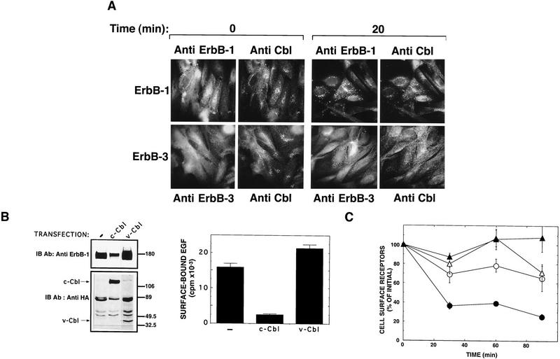
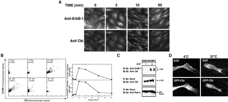
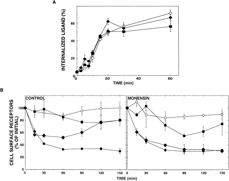
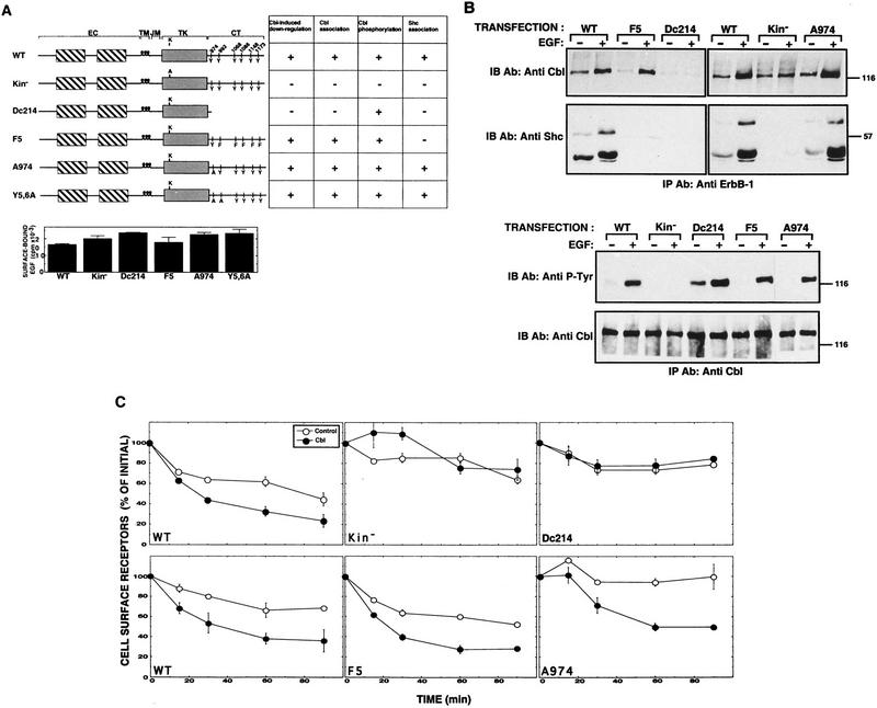
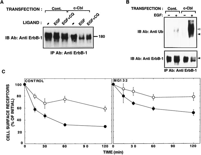
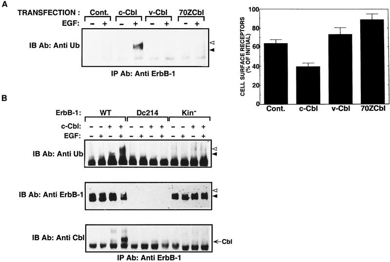
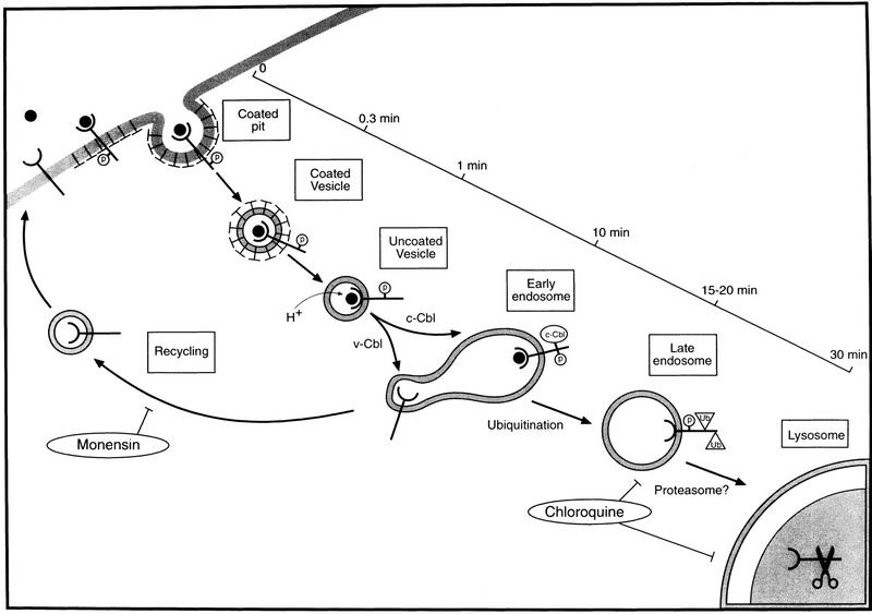
Ligand-induced down-regulation of two growth factor receptors, EGF receptor (ErbB-1) and ErbB-3, correlates with differential ability to recruit c-Cbl, whose invertebrate orthologs are negative regulators of ErbB. We report that ligand-induced degradation of internalized ErbB-1, but not ErbB-3, is mediated by transient mobilization of a minor fraction of c-Cbl into ErbB-1-containing endosomes. This recruitment depends on the receptor's tyrosine kinase activity and an intact carboxy-terminal region. The alternative fate is recycling of internalized ErbBs to the cell surface. Cbl-mediated receptor sorting involves covalent attachment of ubiquitin molecules, and subsequent lysosomal and proteasomal degradation. The oncogenic viral form of Cbl inhibits down-regulation by shunting endocytosed receptors to the recycling pathway. These results reveal an endosomal sorting machinery capable of controlling the fate, and, hence, signaling potency, of growth factor receptors.


Figures

Figure 1
Figure 1
c-Cbl, but not v-Cbl, increases degradation of ErbB-1 by its removal from the cell surface. (A) Shown are the domain structures of c-Cbl and three derivative proteins. The following structural motifs are represented: A 7 residue-long histidine stretch (7 His), a positively charged basic domain (Basic), a ring-finger domain (RF), a proline-rich domain (Pro-Rich), and a leucine-zipper (LZ). An influenza virus hemagglutinin (HA) epitope tag was added to the amino-terminal end of each protein. cDNAs corresponding to the three natural forms of Cbl were transiently transfected into CHO cells. Forty-eight hours after transfection, cells were lysed and whole cell lysates subjected to immunoblotting (IB) with an anti-HA antibody. (B) ErbB-1 (left) or ErbB-3 (right) were transiently expressed in CHO cells by cotransfection with plasmids encoding the indicated Cbl proteins, or with a control empty plasmid (Cont.). Cells were incubated for 45 min at 37°C with EGF or NDF (each at 100 ng/ml). Thereafter, whole cell lysates were subjected to immunoprecipitation (IP) and immunoblotting (IB) with the indicated antibodies. (C) CHO cells were co-transfected with an ErbB-1 expression vector together with a vector directing expression of c-Cbl or a control empty vector (Cont.) Cell surface-exposed proteins were covalently labeled with biotin at 4°C. Soluble biotin was then removed and cells incubated at 37°C for 45 min in the presence or absence of EGF. Cell lysates were subjected to immunoprecipitation (IP) with an anti ErbB-1 antibody and the electrophoretically resolved proteins were probed either with horseradish peroxidase-conjugated Streptavidin, or with an anti-Shc antibody. The locations of molecular mass markers are indicated in kilodaltons. Note that the three Shc isoforms (p66, p52, and p46) associate with ErbB-1.
Figure 2
Figure 2
c-Cbl colocalizes with ErbB-1, but not with ErbB-3, and accelerates its down-regulation. (A) CHO cells that stably co-overexpress ErbB-1 and ErbB-3 (Tzahar et al. 1996) were plated on cover-slips and treated at 37°C with EGF (top) or NDF (bottom) for the indicated periods of time. After fixation and permeabilization, cover slips were simultaneously stained with a polyclonal anti-Cbl antibody and a mAb to ErbB-1 or to ErbB-3, as indicated. Antibody detection by immunofluorescence was performed as described (Materials and Methods). (B) Stable CHO transfections expressing c-Cbl or v-Cbl were established by cotransfection of Cbl constructs together with the pBABE/puro plasmid into CHO cells overexpressing ErbB-1 (Tzahar et al. 1996). Individual clones were screened by immunoblotting with antibodies to ErbB-1 and to the HA tag of Cbl proteins (left, the locations of c-Cbl and v-Cbl protein bands are indicated by arrows). To assess cell surface expression of ErbB-1, clones expressing c-Cbl or v-Cbl, as well as the parental ErbB-1 overexpressing cell line (−), were incubated for 90 min at 4°C with radiolabeled EGF (10 ng/ml). Cell-bound radioactivity is shown as the average and range (bars) of duplicate determinations. (C) CHO cells were cotransfected with pairs of two plasmids: an ErbB-expression vector and either a control empty pcDNA3 plasmid (○; ErbB-1; ▵, ErbB-3), or a c-Cbl expression vector (•, ErbB-1; ▴, ErbB-3). Cell monolayers were subjected to a down-regulation assay 48 hr post-transfection. The results are expressed as the average fraction (and range, bars) of original binding sites that remained on the cell surface after exposure to the nonlabeled ligand at 37°C.
Figure 3
Figure 3
Time dependence of EGF-induced ErbB-1 and c-Cbl co-localization in endosomes. (A) CHO cells that coexpress ErbB-1 and ErbB-3 were either fixed (0 min) or first incubated with EGF for 5 min at 37°C. Thereafter, EGF was removed and incubation continued for the indicated time intervals. Double staining of ErbB-1 and c-Cbl was performed and visualized as described in Materials and Methods. (B, left) For each time point, digital images of three representative cells were segmented according to the labeling of Cbl. The fluorescence of c-Cbl and ErbB-1 labeling was calculated for each segmented vesicle. The scatter plots (arbitrary units) present ErbB-1 fluorescence vs. the c-Cbl fluorescence in each segmented vesicle. The correlation coefficient (r) indicates the strength of a linear correlation between ErbB-1 and c-Cbl fluorescence. (B, right) Cbl-segmented vesicles, which showed ErbB-1 positivity above a threshold, were considered Cbl-vesicles with ErbB1. (Top) Percentage of those vesicles from the total number of Cbl-segmented vesicles at each time point. (Bottom) Percentage of ErbB-1 vesicles with Cbl from the total number of ErbB1-segmented vesicles. (C) CHO cells were cotransfected with ErbB-1 and c-Cbl expression vectors and cells were incubated for 15 min at 37°C with EGF. Control monolayers were mock stimulated (−). Endosomes were prepared as described in Materials and Methods and solubilized (1% Triton X-100) for 30 min at 4°C. Cell lysates were cleared and subjected to immunoprecipitation (IP) and immunoblotting (IB) with the indicated antibodies. Both the endosomal marker protein Rab-5 and c-Cbl were significantly enriched in the isolated fraction relative to other fractions that were collected (bottom; data not shown). (D) CHO cells were cotransfected with ErbB-1 and an expression vector encoding c-Cbl fused in frame to a green fluorescence protein (GFP-Cbl). Forty-eight hours after transfection, cells were incubated with Texas-red-labeled EGF (0.5 μg/ml) for 30 min at 4°C and then either transferred to 37°C (right), or left at 4°C (left), for an additional incubation of 15 min.
Figure 4
Figure 4
v-Cbl promotes receptor recycling, whereas c-Cbl induces receptor down-regulation. (A) Ligand internalization analyses. CHO cells were cotransfected with an ErbB-1 vector along with one of the following plasmids: pcDNA3 (control, ○), c-Cbl expression vector (•), or a plasmid directing v-Cbl expression (█). Cell monolayers were treated for 2 hr at 4°C with 125I-labeled EGF (at 10 ng/ml) and then transferred to 37°C for the indicated periods of time. The fraction of internalized ligand was determined by use of a low-pH wash. Each data point represents the average ± s.e. (bars) of triplicate measurements. (B) CHO cells were cotransfected with an ErbB-1-encoding plasmid along with an expression vector encoding v-Cbl (█), c-Cbl (•), both v- and c-Cbl (♦), or with an empty vector (control, ○). Cells were rinsed and incubated at 37°C for the indicated periods of time with EGF (at 250 ng/ml). Sister cultures were similarly treated, except that monensin (100 μm) was added to the medium. Down-regulation assays were performed as described in Materials and Methods.
Figure 5
Figure 5
Structural determinants of ErbB-1 that are essential for functional interactions with c-Cbl. (A) Schematic representation of ErbB-1 mutants and their interactions with c-Cbl. The domain structure of ErbB-1 is shown by boxes that correspond to the double cysteine-rich domain of the extracellular (EC) region, the transmembrane domain (TM), the juxtamembrane domain (JM), the tyrosine kinase (TK) domain, and the carboxy-terminal tail (CT). The five major tyrosine autophosphorylation sites, along with the α-adaptin tyrosine-based internalization signal [Y974; (Sorkin et al. 1996)] are indicated. The ATP-binding lysine residue (K721) was mutated to an alanine residue in the kinase-defective mutant (Kin). A carboxy-terminal deletion mutant (Dc214) lacking 214 carboxy-terminal amino acids, and an ErbB-1 mutant in which the five major tyrosine phosphorylation sites were mutated to phenylalanine (F5) have been described previously (Sorkin et al. 1996). A double tyrosine to alanine mutant (Y5,6) is also shown. A summary of the results shown in B and C is presented in the table. The histogram presents the results of an assay that determined the binding of radioactive EGF to the surface of cells transiently expressing the indicated mutants. (B) Monolayers of CHO cells were separately cotransfected with plasmids encoding the indicated ErbB-1 mutants together with a c-Cbl-encoding vector. Sister plates were incubated for 15 min at 37°C with or without EGF (at 100 ng/ml). Thereafter, whole cell lysates were subjected to immunoprecipitation (IP) and immunoblotting (IB) with the indicated antibodies. (C) The indicated ErbB-1 mutants were introduced into CHO cells by cotransfection with a control vector (○) or a c-Cbl plasmid (•). EGF-induced down-regulation assay was then performed.
Figure 6
Figure 6
c-Cbl-induced down-regulation involves an increase in ErbB-1 ubiquitination. (A) Chloroquine sensitivity. The wild-type form of ErbB-1 was expressed in CHO cells by cotransfection of an erbB-1-encoding plasmid together with either a c-Cbl-expression vector or an empty vector (Cont.). Cells were incubated for 45 min at 37°C in the absence or presence of EGF (at 100 ng/ml) and chloroquine (CQ, 0.1 mm). Cell lysates were prepared and subjected to immunoprecipitation (IP) and immunoblotting (IB) with anti-ErbB-1 antibodies. (B) CHO cells were transfected and treated as in A. Cell lysates were subjected to immunoprecipitation (IP) with antibodies to ErbB-1 and immunoblotting (IB) with antibodies to either ubiquitin (Ub) or ErbB-1. (Closed arrowheads) The major band of ErbB-1; (open arrowheads) the minor fraction that underwent ubiquitination. (C) ErbB-1 was transiently expressed in CHO cells by cotransfection with either an empty expression vector (control, ○) or a c-Cbl expression vector (•). EGF-induced down-regulation of ErbB-1 was determined in the presence or absence of the proteasomal inhibitor MG132 (10 μm).
Figure 7
Figure 7
Effect of Cbl proteins on ubiquitination of ErbB-1 and its mutants. (A) CHO cells were cotransfected with a plasmid encoding ErbB-1 together with vectors directing the expression of the indicated Cbl proteins. An empty vector was used for control (Cont.). Cell monolayers were treated for 15 min at 37°C with EGF (100 ng/ml). Thereafter, we either analyzed cell lysates by immunoprecipitation (IP) and immunoblotting (IB) with the indicated antibodies (left, arrowheads are as in Fig. 6) or performed a ligand-binding assay as described in the legend to Fig. 2B. (B) Monolayers of CHO cells were transfected with a plasmid expressing c-cbl or a control empty vector (−) together with vectors encoding the wild-type form (WT) of ErbB-1, or the indicated mutants. Cell monolayers were treated with EGF as in A and their whole lysates subjected to immunoprecipitation (IP) with an antibody directed to the extracellular portion of ErbB-1. Immunoblotting (IB) was performed with an antiserum to ubiquitin, or with an antibody directed to the most carboxy-terminal 14 amino acids of ErbB-1. To confirm expression of the Dc214 mutant of ErbB-1, which was not recognized by the immunoblotting antibody, we performed a ligand-binding assay on living cells (data not shown).
Figure 8
Figure 8
Proposed model of ligand-induced endocytosis of ErbB-1. The model summarizes the major steps of receptor endocytosis and indicates their presumed time scale. Ligand binding to ErbB-1 molecules, probably by elevating autophosphorylation (encircled P), induces their interactions with clathrin-coated areas of the plasma membrane, which rapidly invaginate to form coated pits. c-Cbl may not affect excision of the pit to form a coated vesicle and the subsequent rapid uncoating process. c-Cbl recruitment to endosome-located ErbB-1 molecules tags them for ubiquitination (Ub) and subsequent degradation through the combined action of prelysosomal/lysosomal acid hydrolases, as well as by proteasomal proteinases. v-Cbl shunts receptors to the default pathway, which involves recycling of vesicles back to the cell surface. This step is inhibitable by monensin.

Similar articles

Cited by

References

    1. Alroy I, Yarden Y. The ErbB signaling network in embryogenesis and oncogenesis: Signal diversification through combinatorial ligand-receptor interactions. FEBS Lett. 1997;410:83–86. - PubMed
    1. Basu SK, Goldstein JL, Anderson RGW, Brown MS. Monensin interrupts the recycling of low density lipoprotein receptors in human fibroblasts. Cell. 1981;24:493–502. - PubMed
    1. Boussiotis VA, Freeman GJ, Berezovskaya A, Barber DL, Nadler LM. Maintenance of human T cell anergy: Blocking of IL-2 gene transcription by activated Rap1. Science. 1997;278:124–128. - PubMed
    1. Bowtell DD, Langdon WY. The protein product of the c-cbl oncogene rapidly complexes with the EGF receptor and is tyrosine phosphorylated following EGF stimulation. Oncogene. 1995;11:1561–1567. - PubMed
    1. Chang C-P, Lazar CS, Walsh BJ, Komuro M, Collawn JF, Kuhn LA, Tainer JA, Trowbridge IS, Farquhar MG, Rosenfeld MG, Wiley HS, Gill GN. Ligand-induced internalization of the epidermal growth factor receptor is mediated by multiple endocytic codes analogous to the tyrosine motif found in constitutively internalizing receptors. J Biol Chem. 1993;268:19312–19320. - PubMed

Publication types

MeSH terms