. 1998 Dec 1;12(23):3693-702.
doi: 10.1101/gad.12.23.3693.

Deletion of the H19 differentially methylated domain results in loss of imprinted expression of H19 and Igf2

Affiliations

Deletion of the H19 differentially methylated domain results in loss of imprinted expression of H19 and Igf2

J L Thorvaldsen et al. Genes Dev. .

Abstract

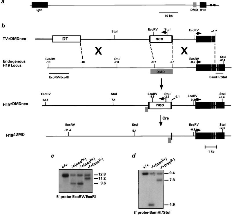
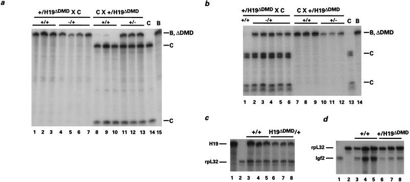
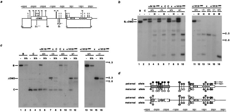
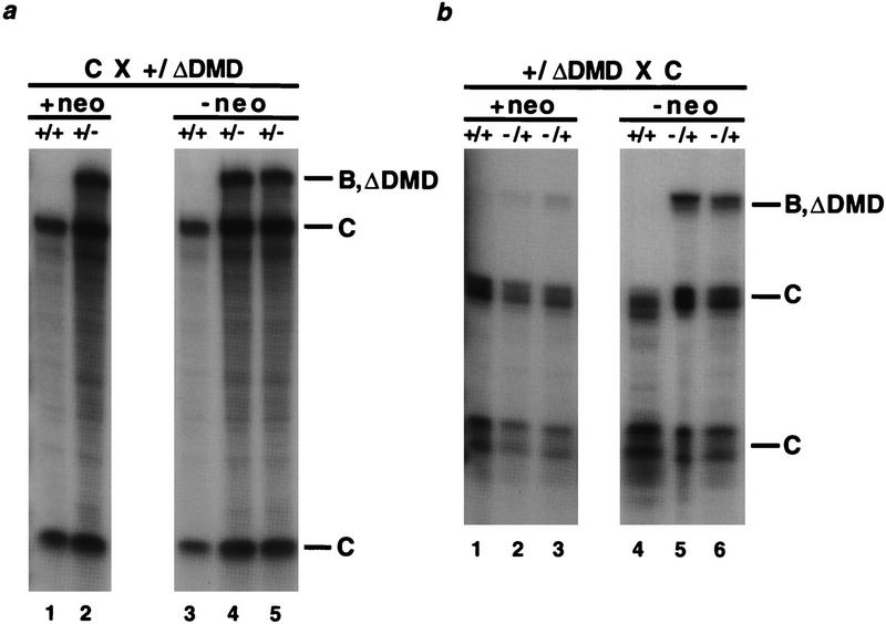
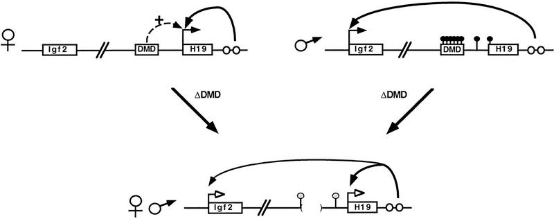
Differentially methylated sequences associated with imprinted genes are proposed to control genomic imprinting. A 2-kb region located 5' to the imprinted mouse H19 gene is hypermethylated on the inactive paternal allele throughout development. To determine whether this differentially methylated domain (DMD) is required for imprinted expression at the endogenous locus, we have generated mice harboring a 1.6-kb targeted deletion of the DMD and assayed for allelic expression of H19 and the linked, oppositely imprinted Igf2 gene. H19 is activated and Igf2 expression is reduced when the DMD deletion is paternally inherited; conversely, upon maternal transmission of the mutation, H19 expression is reduced and Igf2 is activated. Consistent with the DMD's hypothesized role of setting up the methylation imprint, the mutation also perturbs allele-specific methylation of the remaining H19 sequences. In conclusion, these experiments show that the H19 hypermethylated 5' flanking sequences are required to silence paternally derived H19. Additionally, these experiments demonstrate a novel role for the DMD on the maternal chromosome where it is required for the maximal expression of H19 and the silencing of Igf2. Thus, the H19 differentially methylated sequences are required for both H19 and Igf2 imprinting.


Figures

Figure 1
Figure 1
Deletion of the H19 differentially methylated domain in ES cells. (a) The positions of Igf2 and H19 relative to the DMD on mouse chromosome 7 are indicated. The gray box corresponds to the 2-kb DMD, which is located −2 to −4 kb relative to the start of H19 transcription. (•) The endodermal enhancers located at +9 and +11 kb. (b) From top to bottom: the linearized targeting vector, the endogenous H19 locus, the targeted H19 locus (H19ΔDMDneo), and the targeted H19 locus after removal of PGK–neo using CRE–loxP recombination (H19ΔDMD). The linearized vector includes Bluescript II KS (thick line), the diphtheria toxin A gene (open box, DT), PGK–neo (open box, neo) flanked by loxP sites (black vertical bars), 5′ H19 sequence (thin black line) and H19 gene sequence extending into the third exon (solid boxes). Arrows indicate direction of transcriptional orientation of PGK–neo and H19. The positions of the restriction sites and the external probes (EcoRV/EcoRI and BamHI/StuI) used to analyze the targeted clones are indicated. (c,d) ES cell clones were screened by Southern blot analysis for the targeting event. Genomic DNA was digested with EcoRV and hybridized to the 5′ probe EcoRV/EcoRI (c) or with StuI and hybridized to the 3′ probe BamHI/StuI (d). The DNA samples shown include the parental ES cell DNA (+/+), targeted clones with the neor gene (neoR+), and a targeted clone in which the neor gene was excised (neoR−). Molecular sizes (in kb) are indicated to the right. Of the 85 G418-resistant clones analyzed, four were correctly targeted to the H19 locus.
Figure 2
Figure 2
Expression of H19 and Igf2 in H19ΔDMD heterozygous mice. Livers were isolated from neonates generated from reciprocal crosses of B6(CAST–H19) (C) and F1 H19ΔDMD heterozygotes maintained in a C57BL/6 background (B). Three micrograms of total RNA was analyzed using an allele-specific RNase protection assay. The C-, B-, and ΔDMD-specific protected fragments are designated. (a) H19 expression when H19ΔDMD was inherited from the mother (lanes 1–7) or the father (lanes 8–13). When the mother was heterozygous for the mutation, the maternal allele (B, ΔDMD) was expressed in wild-type newborn mice (+/+, lanes 1–3) and expressed at a reduced level in mutant (−/+, lanes 4–7) newborn mice. In 5-day-old littermates generated from paternal heterozygotes, the maternal allele (C) is expressed in wild-type mice (+/+, lanes 8–10), whereas mutant mice express both alleles (+/−, lanes 11–13). The relative ratio of paternally to maternally derived H19 RNA is 0.615, 0.586, and 0.575 (lanes 11–13, respectively). Note that a low level of expression of the paternal allele (<0.5%) was detected in wild-type mice (lanes 8,9) as observed previously (Leighton et al. 1995a). Control liver RNA isolated from 4-day-old B6(CAST–H19) and 5-day-old C57BL/6 mice was analyzed in lanes 14 and 15, respectively. (b) Igf2 expression when the H19ΔDMD allele is maternally (lanes 1–6) or paternally derived (lanes 7–12). Three-day-old mice generated from maternal heterozygotes expressed the paternal allele (C) when wild-type for the mutation (+/+, lane 1) and expressed both alleles when heterozygous for the mutation (−/+, lanes 2–6). The relative ratio of maternally to paternally derived Igf2 RNA is 0.373, 0.315, 0.317, 0.338, and 0.370 (lanes 2–6, respectively). In 5-day-old littermates generated from paternal heterozygotes, the paternal allele is expressed exclusively in wild-type mice (+/+, lanes 7–9) and expressed at a reduced level in mutant mice (+/−, lanes 10–12). Control liver RNAs as described in a are assayed in lanes 13 and 14. (c) The expression of H19 is analyzed relative to the expression of rpL32 when the H19ΔDMD allele is transmitted by the mother. RNA from wild-type (+/+, lanes 3–5) and mutant (H19ΔDMD/+, lanes 6–8) 5-day-old littermates was assayed with H19 and rpL32 probes. The ratio of H19 to rpL32 RNA is as follows: 2.72, 2.85, 1.60, 1.14, 1.34, and 1.04 (lanes 3–8, respectively). Neonatal liver RNA is assayed with the H19 or rpL32 probes alone in lanes 1 and 2, respectively. (d) The expression of Igf2 was analyzed relative to rpL32 when the H19ΔDMD allele was transmitted by the father. RNA from wild-type (+/+, lanes 3–5) and mutant (+/H19ΔDMD, lanes 6–8) newborn littermates is assayed with the Igf2 and rpL32 probes. The ratio of Igf2 to rpL32 RNA is as follows: 0.465, 0.502, 0.536, 0.150, 0.189, and 0.165 (lanes 3–8, respectively). Neonatal liver RNA was assayed with the Igf2 or rpL32 probes alone in lanes 1 and 2, respectively. Total RNA levels for the wild-type and heterozygous mutants were confirmed by Northern analysis (data not shown).
Figure 3
Figure 3
Methylation analysis of H19 in heterozygous and homozygous DMD mutant mice. (a) The location of the HpaII (H) and HhaI (Hh) sites with respect to the deleted DMD sequence. The 5′ H19 DMD sequence, deleted between the KpnI (K) and HindIII (Hd) sites, and H19 are represented by boxed regions. (P) PvuII; (R) EcoRI; (St) StuI; (Sc) SacI. (*) The polymorphic PvuII site that is found in C57BL/6J and H19ΔDMD; (**) the polymorphic M. castaneus SacI site. The RSt probe used in b and the ScK probe used in c are indicated beneath the line. The position of the sites, relative to the start of transcription, are indicated (in bp) above the gene line. (b) The methylation status of HpaII sites in the H19 PvuII/StuI fragment is assessed with the RSt probe. DNA from C57BL/6J (B) mice, B6(CAST–H19) mice (C), progeny of a H19ΔDMD heterozygous female mated to a B6(CAST–H19) male (lanes 3–6), progeny from a B6(CAST–H19) female mated to a H19ΔDMD heterozygous male (lanes 7–10) and progeny of a H19ΔDMD heterozygous female mated to a H19ΔDMD heterozygous male (lanes 11–16) were analyzed. DNA was digested with PvuII and StuI and, in lanes indicated, HpaII (H) or MspI (M). The genotypes of the assayed DNA from the specific mating are indicated above the lanes. DNA from neonatal livers (lanes 1–13,16) and adult sperm (lanes 14,15) were assayed. The M. castaneus-specific PvuII/StuI fragment (C, 3.4 kb) and the C57BL/6 and H19ΔDMD-specific PvuII/StuI fragment (B, ΔDMD, 3.2 kb) are noted at left, with size markers in kb shown at right. (c) DNA from mice described in b was analyzed for HhaI methylation in the 5′ H19 SacI fragment using the ScK probe. DNA was digested with SacI and, in lanes indicated, HhaI (Hh). The genotypes of the DNA samples are indicated above the lanes. The C57BL/6J (3.8 kb), B6(CAST–H19) (1.5 kb) and the H19ΔDMD (2.2 kb)-specific SacI fragments are indicated at left. (d) Summary of the parental-specific methylation status of HpaII and HhaI sites on wild-type (top line) vs. H19ΔDMD (middle line) alleles. The taller and shorter lollipops represent the HhaI and the HpaII sites, respectively. (Solid circles) Fully methylated sites; (open circles) unmethylated sites; (striped circles) sites that are methylated on a subset of the alleles, as determined from previous studies (bisulfite sequence and Southern analyses) and data presented herewith; (shaded circles) sites that are partially methylated. The paternal-specific methylation status is presented above each allele; the maternal-specific methylation status is presented below each allele.
Figure 4
Figure 4
Expression of H19 and Igf2 in H19ΔDMD and H19ΔDMDneo mice. The genotypes are noted above the lanes and the parental identity of protected fragments are indicated to the right. Neonatal liver RNA (3 μg) is analyzed using allele-specific RNase protection assays. (a) H19 expression in paternal heterozygotes. The RNA isolated from a wild-type and a mutant littermate, generated from mating a B6(CAST–H19) female with a heterozygous H19ΔDMDneo male, is assayed in lanes 1 and 2, respectively. The RNA isolated from the progeny of a mating between a B6(CAST–H19) female and a heterozygous H19ΔDMD male is assayed in lanes 3–5. (b) Igf2 expression in maternal heterozygotes. RNA from wild-type and mutant +neo littermates (lanes 1 and 2 and 3, respectively) and wild-type and mutant −neo littermates (lanes 4 and 5 and 6, respectively) produced from the reciprocal mating performed in a is assayed.
Figure 5
Figure 5
A model for DMD-regulated H19 and Igf2 imprinting based on the analysis of neonatal liver. The maternal and paternal wild-type alleles are represented with parental-specific gene expression (horizontal arrows) and H19 paternal-specific methylation (filled-in circles above the locus). The H19 and Igf2 genes and the DMD are depicted as boxes and endodermal enhancers as open circles. The deletion of the DMD (bordered by parentheses) on the maternal or the paternal allele results in expression of both genes as depicted at bottom. In addition, 5′ H19 sequence is similarly methylated on both alleles (shaded circles above 5′ H19 sequence). The model illustrates the following properties of the DMD. (1) The DMD positively affects maternal H19 expression (broken arrow); when it is deleted, maternal H19 expression is reduced (open arrowheads). (2) The DMD contributes to silencing of the paternal H19 allele. (3) Presence of the DMD is required for reciprocal imprinting of Igf2. In ΔDMD mice, reduced H19 expression on the maternal allele is accompanied by activated Igf2 expression, and activated H19 expression from the paternal allele is accompanied by reduced Igf2 expression, consistent with previous proposals that the endodermal enhancers are shared. (4) Paternal-specific methylation is maintained in the presence of DMD. (5) The DMD serves as the mark that distinguishes the parental alleles of H19.

Similar articles

Cited by

References

    1. Auffray C, Rougeon F. Purification of mouse immunoglobulin heavy-chain messenger RNAs from total myeloma tumor RNA. Eur J Biochem. 1980;107:303–314. - PubMed
    1. Barlow DP. Competition—a common motif for the imprinting mechanism. EMBO J. 1997;16:6899–6905. - PMC - PubMed
    1. Bartolomei MS, Tilghman SM. Parental imprinting of mouse chromosome 7. Semin Dev Biol. 1992;3:107–117.
    1. ————— Genomic imprinting in mammals. Annu Rev Genet. 1997;31:493–525. - PubMed
    1. Bartolomei MS, Zemel S, Tilghman SM. Parental imprinting of the mouse H19 gene. Nature. 1991;351:153–155. - PubMed

Publication types

LinkOut - more resources