. 1998 Dec 1;12(23):3728-40.
doi: 10.1101/gad.12.23.3728.

Ligand-induced cleavage and regulation of nuclear entry of Notch in Drosophila melanogaster embryos

Affiliations

Ligand-induced cleavage and regulation of nuclear entry of Notch in Drosophila melanogaster embryos

S Kidd et al. Genes Dev. .

Abstract

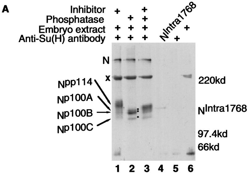
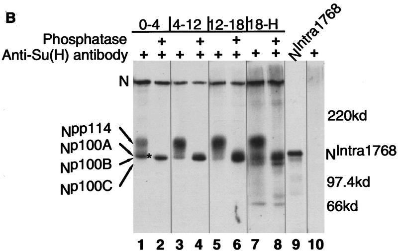
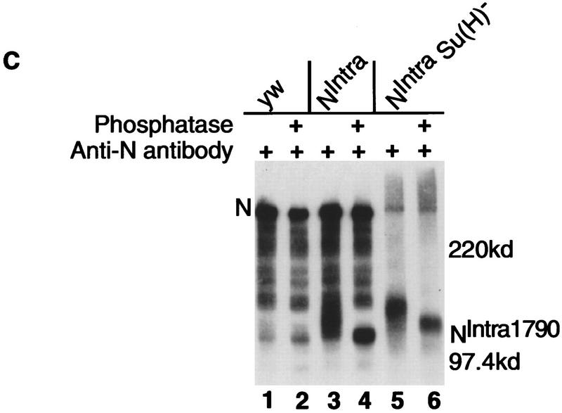
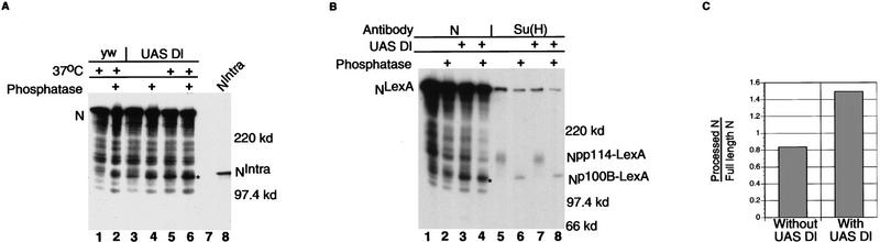
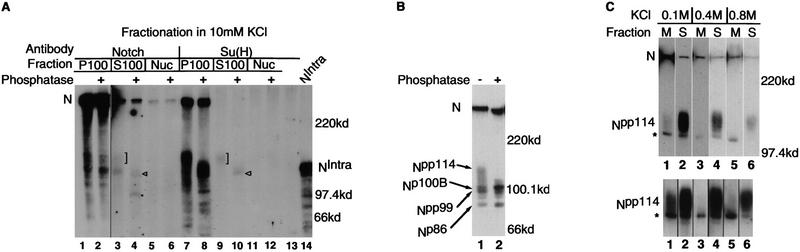
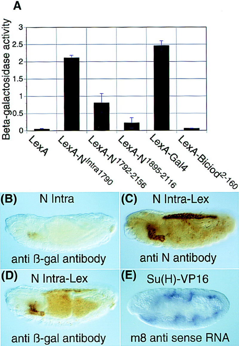
Notch, a transmembrane protein found in a wide range of organisms, is a component of a pathway that mediates cell-fate decisions that involve intercellular communication. In this paper, we show that in Drosophila melanogaster, Notch (N) is processed in a ligand-dependent fashion to generate phosphorylated, soluble intracellular derivatives. Suppressor of Hairless [Su(H)] is predominantly associated with soluble intracellular N. It has been demonstrated by others that N has access to the nucleus, and we show that when tethered directly to DNA, the cytoplasmic domain of N can activate transcription. Conversely, a viral activator fused to Su(H) can substitute for at least some N functions during embryogenesis. We suggest that one function of soluble forms of N is to bind to Su(H), and in the nucleus, to act directly as a transcriptional transactivator of the latter protein. Although N has functional nuclear localization signals, the N/Su(H) complex accumulates in the cytoplasm and on membranes suggesting that its nuclear entry is regulated. Localization studies in cultured cells and embryos suggest that Su(H) plays a role in this regulation, with the relative levels of Delta, N and Su(H) determining whether a N/Su(H) complex enters the nucleus.


Figures

Figure 1
Figure 1
Constructs and antibodies. (A) A diagram of the N protein (N). (EGF) Epidermal growth factor-like repeats; (LNG) lin-12, Notch, Glp-1 repeats; (cc) evolutionary conserved cysteine residues; (CDC10) CDC10 or ankyrin repeats; (nls1, nls2) putative nuclear localization signals; (polyQ) polymeric glutamines; (PEST) PEST sequence thought to be involved in protein stability (Rogers et al. 1986). The bars above the diagram of N indicate the various regions used as antigens to create anti-N antibodies. Beneath the diagram of N are the N constructs used in this paper. (B) Su(H) constructs used in this paper.
Figure 2
Figure 2
Su(H) is associated with phosphorylated N proteins. (A,B) Western blot analysis of anti-Su(H) immunoprecipitates from Drosophila embryos probed with anti-N (NPCR) antibody. For A, an overnight collection of embryos was used and in B, staged embryos (age indicated in hours at top of lanes; H denotes hatching) were used for detergent extracts. Prior to electrophoresis, some of the samples were treated with alkaline phosphatase with or without inhibitor. (+) Presence of phosphatase and/or inhibitor. (A, Lane 4 and B, lane 9) In vitro-translated NIntra1768, the size of which is not affected by phosphatase treatment; (A, lane 5) Antibody but no extract; (A, lane 6) Embryo extract recovered on protein A–Sepharose beads that were not conjugated to antibody. The locations of N (N), NIntra1768, Npp114, Np100A, Np100B, and Np100C proteins are shown along side each blot, and also by dots on the blot. The × in A indicates a cross-reacting protein nonspecifically bound to the protein A–Sepharose beads; the asterisk (*) in B indicates a hypophosphorylated N protein that comigrates with Np100B. (A, all lanes with extract; B, lanes 3–6) 500 μg of protein was used for each immunoprecipitation; the remaining lanes in B contain 2.5 mg(lanes 1,2) and 1.75 mg (lanes 7,8). (C) Western blot analysis of anti-N (NI) immunoprecipitates from Drosophila embryos probed with anti-N (NPCR) antibody. (Lanes 1, 2) Immunoprecipitates from yw embryos; (lanes 3,4) immunoprecipitates of embryos resulting from a cross of Su(H) hsNintra1790/CyO males to Su(H)SF8 FRT40A/CyO virgins; (lanes 5,6) immunoprecipitates of embryos resulting from a cross of Su(H) hsNintra1790/CyO males to hsflp12/yw; Su(H)SF8 FRT40A/ovoD FRT40A virgins. All NIntra expressing embryos are both zygotically and maternally Su(H) null. For all genotypes, 3- to 5 hr-old embryos were subjected to a 30 min heat shock at 37°C and allowed to recover for 15–30 min prior to collection. (+) Phosphatase treatment. The locations of N (N) and NIntra are indicated next to the blot.
Figure 2
Figure 2
Su(H) is associated with phosphorylated N proteins. (A,B) Western blot analysis of anti-Su(H) immunoprecipitates from Drosophila embryos probed with anti-N (NPCR) antibody. For A, an overnight collection of embryos was used and in B, staged embryos (age indicated in hours at top of lanes; H denotes hatching) were used for detergent extracts. Prior to electrophoresis, some of the samples were treated with alkaline phosphatase with or without inhibitor. (+) Presence of phosphatase and/or inhibitor. (A, Lane 4 and B, lane 9) In vitro-translated NIntra1768, the size of which is not affected by phosphatase treatment; (A, lane 5) Antibody but no extract; (A, lane 6) Embryo extract recovered on protein A–Sepharose beads that were not conjugated to antibody. The locations of N (N), NIntra1768, Npp114, Np100A, Np100B, and Np100C proteins are shown along side each blot, and also by dots on the blot. The × in A indicates a cross-reacting protein nonspecifically bound to the protein A–Sepharose beads; the asterisk (*) in B indicates a hypophosphorylated N protein that comigrates with Np100B. (A, all lanes with extract; B, lanes 3–6) 500 μg of protein was used for each immunoprecipitation; the remaining lanes in B contain 2.5 mg(lanes 1,2) and 1.75 mg (lanes 7,8). (C) Western blot analysis of anti-N (NI) immunoprecipitates from Drosophila embryos probed with anti-N (NPCR) antibody. (Lanes 1, 2) Immunoprecipitates from yw embryos; (lanes 3,4) immunoprecipitates of embryos resulting from a cross of Su(H) hsNintra1790/CyO males to Su(H)SF8 FRT40A/CyO virgins; (lanes 5,6) immunoprecipitates of embryos resulting from a cross of Su(H) hsNintra1790/CyO males to hsflp12/yw; Su(H)SF8 FRT40A/ovoD FRT40A virgins. All NIntra expressing embryos are both zygotically and maternally Su(H) null. For all genotypes, 3- to 5 hr-old embryos were subjected to a 30 min heat shock at 37°C and allowed to recover for 15–30 min prior to collection. (+) Phosphatase treatment. The locations of N (N) and NIntra are indicated next to the blot.
Figure 3
Figure 3
The presence of Notch ligand and the extracellular domain of N are required for the isolation of Np100 bound to Su(H). (A) Detergent extracts from embryos expressing either NLexA (lanes 1,3) or NΔEGF1-36–LexA [lanes 2,4 (see Fig. 1 for structures of NLexA and NΔEGF1-36–LexA] were immunoprecipitated with antibodies against either N (NI) (lanes 1,2) or Su(H) (lanes 3,4) treated with phosphatase, and then probed with antibodies against LexA. Antibodies against N immunoprecipitate NLexA (lane 1) and NΔEGF1-36–LexA (lane 2); the smaller proteins seen in lane 2 are missing from lane 1 and, are most likely nonspecific breakdown products visible because of massive overexpression of NΔEGF1-36–LexA compared with NLexA. NLexA and NΔEGF1-36–LexA are coimmunoprecipitated by anti-Su(H) antibodies as is a smaller, processed form of NLexA (Np100–LEXA, lane 3, *). No such processed protein is seen when the extracellular ligand binding domain of N is deleted (lane 4). (B) Dl temperature-sensitive mutants reduce the level of processed N protein. yw (wt), Dl6B/TM6 (a strong) or DlRF/TM6 (a weak temperature-sensitive Dl allele) males were mated to Dlx/TM6 (an amorphic Dl allele) females. All the embryos resulting from the above crosses were incubated at either room temperature (lanes 1,3,5) or at the nonpermissive temperature, 29°C (lanes 2,4,6). N proteins coimmunoprecipitated from detergent extracts by anti-Su(H) antibody were detected with the NPCR anti-N antibody. Beneath each lane the ratio of processed N to N (termed Npp114/N) is shown. The strong temperature-sensitive Dl allele (lanes 3,4) has a considerably greater effect on the level of processed N than the weaker allele (lanes 5,6)
Figure 4
Figure 4
Ligand-induced cleavage of N. (A) Anti-N antibodies immunoprecipitate a N protein, the size of Np100B (*) from embryos exposed to ectopic Dl (lane 5). Embryos aged from 2 to 4 hr were heat-shocked at 37°C for 30 min and allowed to recover at 30°C for 2 hr prior to detergent extraction. Extracts from either heat-shocked yw (lanes 1,2) or heat-shocked GAL4; UAS Dl with (lanes 5,6), or without heat shock (lanes 3,4) were immunoprecipitated with anti-N NI antibody. Lane 7 lacks embryo extract; lane 8 contains NIntra1768 in vitro translation products. After immunoprecipitation, lanes marked with a + were treated with alkaline phosphatase prior to electrophoresis. N proteins were detected with the anti-N antibody NPCR. (Labels) Locations of N (N) and NIntra1768(NIntra). (B) Coexpression of NLexA and Dl results in an increase in the level of Npp114–LexA. Detergent extracts from 4–8 hr UASNlexA × hGAL4 embryos (lanes 1,2,5,6) or UAS Dl30B; UAS NlexA × hGAL4 embryos (lanes 3,4,7,8) were immunoprecipitated with either anti-N antibody (NI) (lanes 1–4) or anti-Su(H) antibody (lanes 5–8). Samples in lanes 2, 4, 6, and 8 were treated with alkaline phosphatase prior to electrophoresis. After blotting, proteins were detected with an anti-LexA monoclonal antibody. Coexpression of Dl and NLexA causes the appearance of a novel hypophosphorylated NLexA protein in the anti-N immunoprecipitations (*, cf. lanes 1 and 3). In this experiment, there was an ∼40%–50% increase in the amount of Npp114–LexA bound to Su(H) (cf. the ratios of Npp114LexA to NLexA in lane 5 with lane 7, and in lane 6 with lane 8). (C) A histogram comparing the relative amounts of processed vs. full-length N associated with Su(H) in the absence or presence of ectopic Dl. The average of the results of four immunoprecipitations are plotted. The fold increase in the amount of processed N compared with full-length N in the presence of UAS Dl for each of the four experiments was as follows: 1.7, 1.5, 2.0, and 1.8.
Figure 5
Figure 5
Npp114 is a soluble protein. (A) Subcellular distribution of Npp114. Subcellular fractions were prepared from Drosophila embryos that had been lysed under hypotonic conditions (10 mm KCl). (Nuc) The nuclear fraction; (P100) the membrane fraction; (S100) the soluble fraction. The position of Npp114 is indicated by a square bracket, Np100B by an arrowhead, and Np86 by a dot. Equal proportions of each fraction were immunoprecipitated with antibodies against either N (NI) (lanes 1–6) or Su(H) (lanes 7–12) and then detected with the anti-NPCR antibody. The autoradiograph has been overexposed to show the presence of processed N in the soluble fraction. Because the amount of N in the membrane fraction is so high, lanes 1 and 2 are from a shorter exposure. (B) Evidence for additional processing of the cytoplasmic domain of N. Soluble proteins extracted in 0.4 m KCl were immunoprecipitated with anti-NI antibody and detected with anti-NPCR antibody. As well as immunoprecipitating the same N proteins as those coimmunoprecipitated by anti-Su(H) antibodies, anti-N antibodies immunoprecipitate two novel proteins of ∼99 kD (Npp99) and 86 kD (Np86) (lane 1), treatment with alkaline phosphatase (lane 2) reduces the amount of Npp99 and increases the amount of Np86, suggesting that Npp99 is a phosphorylated form of Np86. (C) Some Npp114 is associated, but not stably, with membranes. Postnuclear supernatants were incubated with increasing amounts of KCl prior to fractionation by centrifugation into membrane bound (lanes marked M) or soluble proteins (lanes marked S). Fractionated proteins were then immunoprecipitated with anti-Su(H) antibody and detected with the anti-N NPCR antibody, a longer exposure of the relevant region of the resulting autoradiograph is shown at bottom. Some Npp114 can be seen to be associated with membranes when extracted at 0.1 m KCl but is removed at higher salt concentrations. The protein that comigrates with Np100B and appears to be stably associated with membranes is marked by an asterisk.
Figure 6
Figure 6
NIntra is retained in the cytoplasm in embryos. Confocal images showing the localization of NIntra in embryos and Drosophila S2 cells as detected by immunofluorescence. With the exception of the left panel in C, NIntra protein is represented in green and the nuclei in red. This panel is a pseudocolored representation to illustrate the relative amounts of NIntra1768. The correspondence of the colors with the intensity of the signal is indicated by the pseudocolor bar, with the more intense signals being depicted by colors higher up the bar. (A) Heat shock-induced NIntra1768 appears to be totally nuclear in S2 cells. UAS NIntra1768 was cotransfected along with HS GAL4 and detected with a rabbit anti-N (NI) antibody. The nucleus was detected with SYTOX Green. (B,C) In embryos (hGAL4; UAS Nintra1768) there is retention of NIntra1768 in the cytoplasm. (B,C, right) The merged images of the N signal in green and the nuclei in red. (C, left) NIntra1768 is primarily nuclear in those cells in which it is most highly expressed. Mouse anti-NPCR antibody was used to detect NIntra1768. (B) Mouse anti-Flag antibody was used to detect Flag-tagged NIntra1768. The nuclei were detected by propidium iodide. (D) NIntra1768 is still retained in the cytoplasm in embryos that are maternally and zygotically N null (N264–47 FRT/ovoD FRT; hGAL4/hsflp X FM7/Y; Nintra1768. (D, left) Mouse NPCR antibody was used to detect NIntra1768; (D, right) a merged image of N (green) and nuclei (red). (E) NIntra1790 is predominantly nuclear in embryos with reduced levels of Su(H). Anti- myc antibody was used to detect myc-tagged Nintra1790 in embryos that are maternally Su(H) (hsflp/yw;Su(H)SF8 FRT/ovoD FRT;hGAL4 X Su(H)/CyO; UASNIntra1790). In embryos that are maternally Su(H)+ (hGAL4 X Su(H)/CyO;UASNIntra1790) there is retention of NIntra1790 in the cytoplasm (data not shown).
Figure 7
Figure 7
Su(H) can retain NIntra in the cytoplasm. Confocal images showing the localization of Su(H) and NIntra1768. With the exception of F, which is a pseudocolored image, Su(H) is represented in blue, NIntra in green, and the nuclei in red. (A) Su(H) is expressed in both the cytoplasm and nuclei of transfected S2 cells. S2 cells were transfected with a construct expressing Su(H) under the control of an actin promoter. Su(H) is detected with rat anti-Su(H) antibody and the DNA with SYTOX Green. (B,C) Coexpression of high levels of NIntra1768 along with Su(H) results in both being found in nuclei of S2 cells. (B) Two cells are depicted showing Su(H) expression in blue and N expression in green. In the lower cell, no NIntra1768 is present and Su(H) is localized ubiquitously. In contrast, the upper cell expresses high levels of NIntra1768 along with Su(H) and both are localized in the nucleus. A merged image of the first two panels is shown at right. The cell shown in C was probed with a DNA marker as well as with anti-N and Su(H) antibodies. Actin driven Su(H) was cotransfected with UAS NIntra1768 and HS GAL4. Su(H) protein was detected by rat anti-Su(H) antibody, N protein was detected by a mouse anti-Flag antibody, and the DNA with SYTOX Green. (D,E,G) The difference in localization of NIntra1768 promoted by expression of an excess of Su(H). NIntra is represented in green and the nuclei in red. (D) UAS NIntra1768 and HS GAL4 were transfected into S2 cells. (E,G) A 20× mass excess of actin Su(H) was cotransfected along with UAS NIntra1768 and HS GAL4. (D,E) Localization of NIntra1768 in nuclei is indicated by dots and retention in the cytoplasm by asterisks. Two cells from those shown in E are depicted at higher magnification in G. As cytoplasmic localization of NIntra1768 is never seen in cells lacking ectopic Su(H) (D), the upper cell in G must have received more Su(H) relative to NIntra1768 than the lower one, resulting in NIntra1768 being retained in the cytoplasm. NIntra1768 was detected with a rabbit anti-N (NI) antibody and the nuclei with SYTOX Green. (F) When low levels of NIntra1768 are expressed along with Su(H), NIntra1768 is retained in the cytoplasm. A pseudocolored confocal image showing the relative levels on N at right and Su(H) at left is portrayed. The intensity of staining is depicted by the pseudocolor bar with the colors representing the more intensely stained regions being higher up the bar. In the lower cell, NIntra1768 is expressed at relatively high levels and both NIntra1768 and Su(H) are found in the nucleus. In the upper cell, NIntra1768 is expressed at relatively low levels and both NIntra1768 and Su(H) are found in the cytoplasm. When cells are doubly stained for DNA and NIntra1768, the two stains converge only when levels of NIntra1768 are high compared with Su(H) (data not shown). Su(H) was detected with a rat anti-Su(H) antibody and NIntra1768 with a mouse anti-Flag antibody.
Figure 8
Figure 8
The cytoplasmic domain of N can behave as a transcriptional activator. (A) The cytoplasmic domain of N is a transcriptional activator in yeast. The transcriptional activation activity of various N constructs fused to the DNA-binding domain of LexA (Fig. 1) was determined by their ability to drive expression from a LexA–β-galactosidase reporter. The bar marked LexA is the β-galactosidase activity of yeast expressing the vector alone, LexA–Bicoid2-160 is a negative control. The height of the bar is the average of three samples, standard deviations are shown by the error bars. (B) NIntra1790 cannot activate transcription from LexA–β-galactosidase reporter. A lexA–β-gal; UAS Nintra1790; HS GAL4 embryo is stained with anti β-galactosidase antibody. There is anti β-galactosidase reactivity in the secretory cells and the anal pads that results from leakiness of the reporter. (C) NIntra1790–LexA accumulates to its highest levels in the salivary glands, amnioserosa, and midgut. A lexA–β-gal; UAS Nintra1790–lexA; HS GAL4 embryo is stained with anti-NPCR antibody. (D) NIntra1790–LexA induces expression of β-gal from the LexA–β-gal reporter. A lexA–β-gal; UAS Nintra1790–LexA; HS GAL4 embryo is stained with anti β-galactosidase antibody. β-Galactosidase accumulates in the salivary glands, amnioserosa and midgut, which correspond with regions that accumulate the highest levels of NIntra1790–LexA in C. (B,C,D) Embryos were fixed 2 hr after a 30-min heat shock. (E) A heterologous activator fused to Su(H) can substitute for N function and activate transcription of m8. N264-47 FRT/ovoD FRT; h GAL4/hsflp × FM7 lac-Z/Y; myc Su(H)–VP16 embryos are stained with an m8 probe. m8 expression is induced in seven stripes, in which h is expressed. Overexpression of Su(H) alone does not result in induction of m8 expression (data not shown). Anti β-galactosidase antibody was used to distinguish the N null embryos.

Similar articles

Cited by

References

    1. Andrews NC, Faller DV. A rapid micropreparation technique for extraction of DNA-binding proteins from limiting numbers of mammalian cells. Nucleic Acids Res. 1991;19:2499. - PMC - PubMed
    1. Artavanis-Tsakonas S, Matsuno K, Fortini ME. Notch signaling. Science. 1995;268:225–232. - PubMed
    1. Azpiazu N, Frasch M. Tinman and bagpipe: Two homeo box genes that determine cell fates in the dorsal mesoderm of Drosophila. Genes & Dev. 1993;7:1325–1340. - PubMed
    1. Bailey AM, Posakony JW. Suppressor of Hairless directly activates transcription of enhancer of split complex genes in response to Notch receptor activity. Genes & Dev. 1995;9:2609–2622. - PubMed
    1. Blaumueller CM, Qi H, Zagouras P, Artavanis-Tsakonas S. Intracellular cleavage of Notch leads to a heterodimeric receptor on the plasma membrane. Cell. 1997;90:281–291. - PubMed

Publication types

LinkOut - more resources