. 1998 Dec 1;12(23):3752-63.
doi: 10.1101/gad.12.23.3752.

The bHLH protein PTF1-p48 is essential for the formation of the exocrine and the correct spatial organization of the endocrine pancreas

Affiliations

The bHLH protein PTF1-p48 is essential for the formation of the exocrine and the correct spatial organization of the endocrine pancreas

A Krapp et al. Genes Dev. .

Abstract

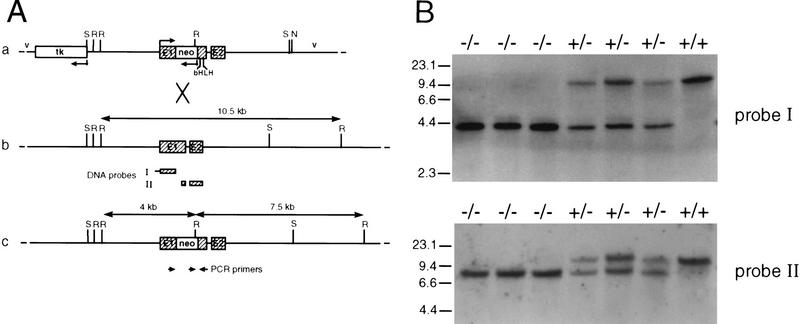
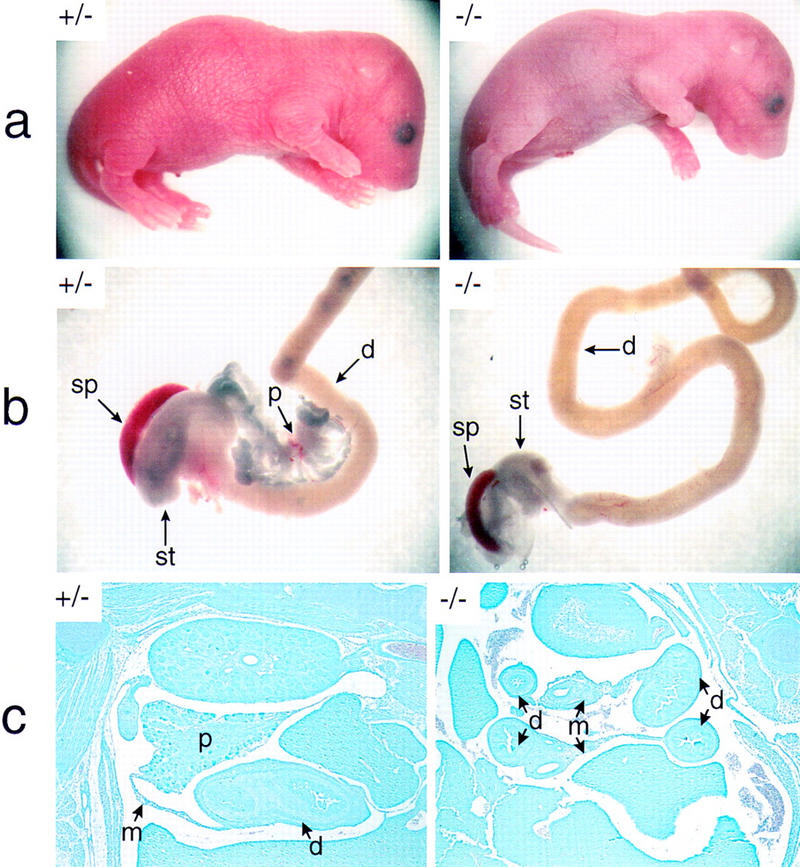
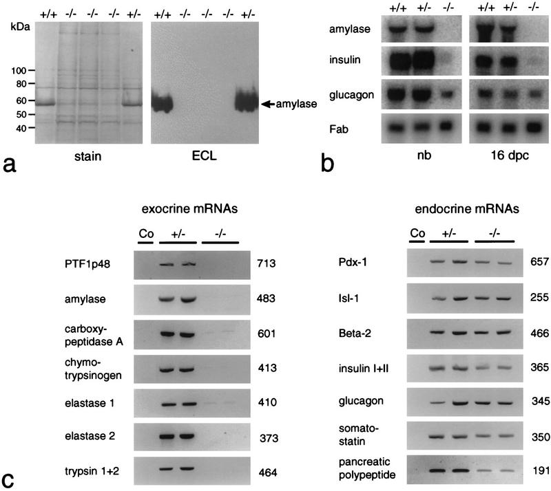
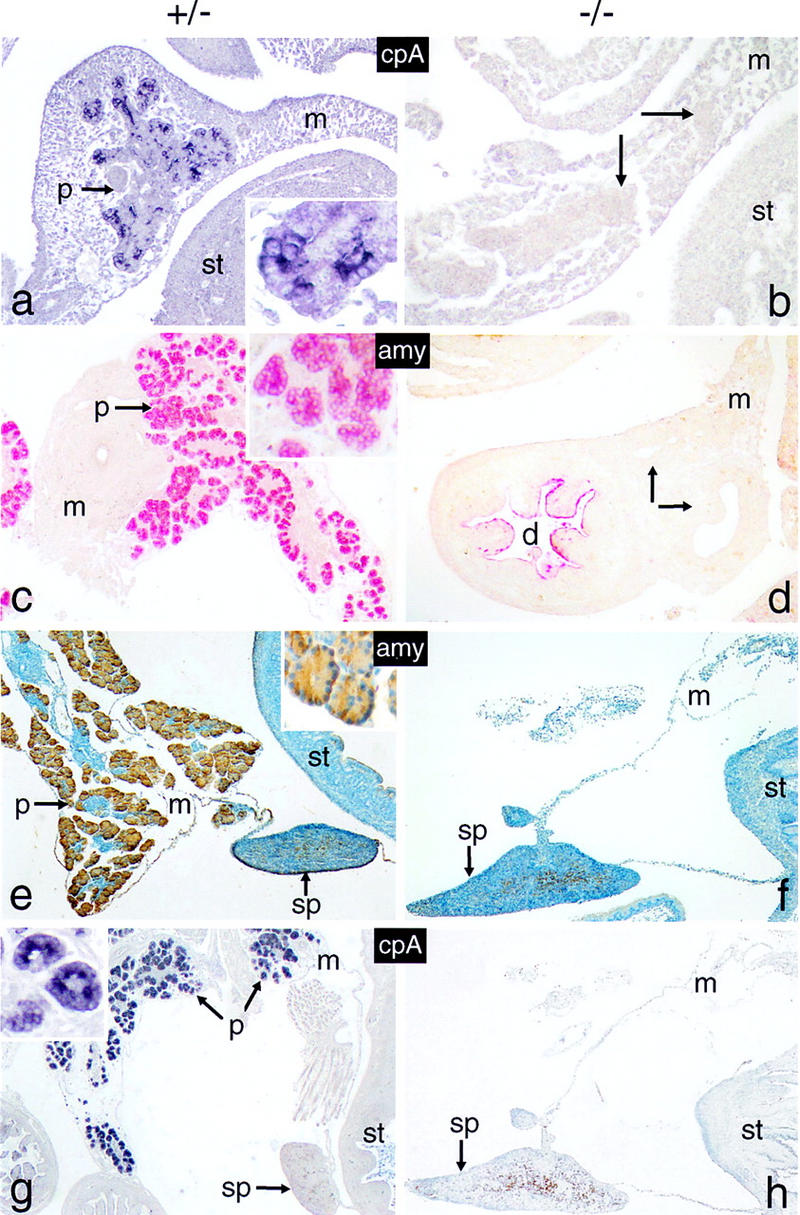
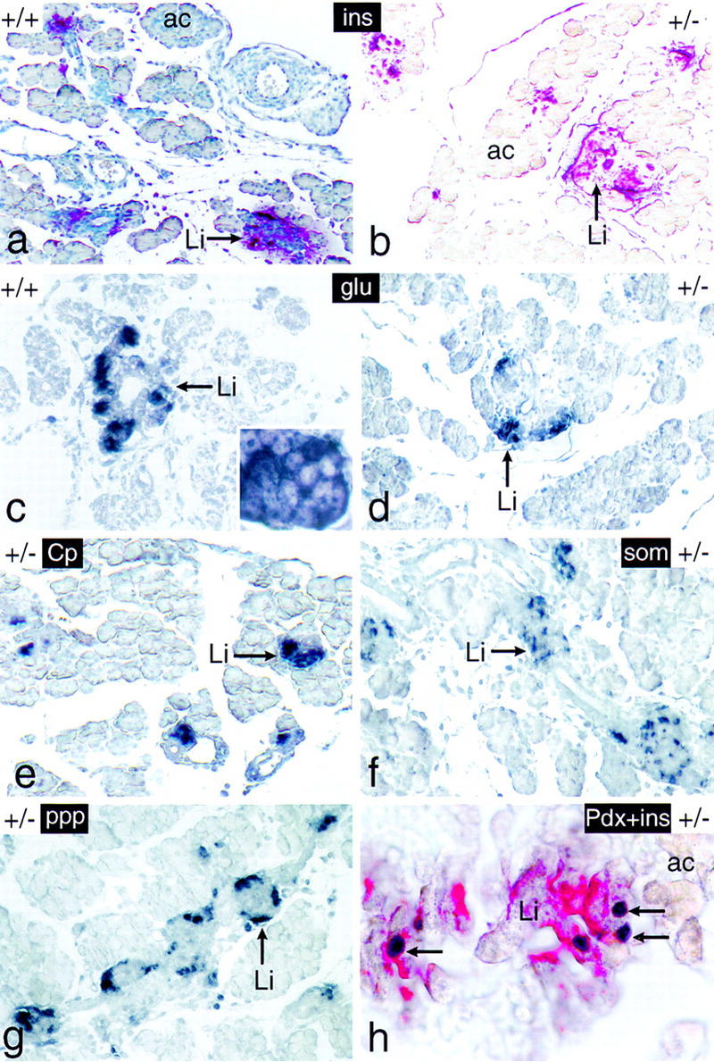
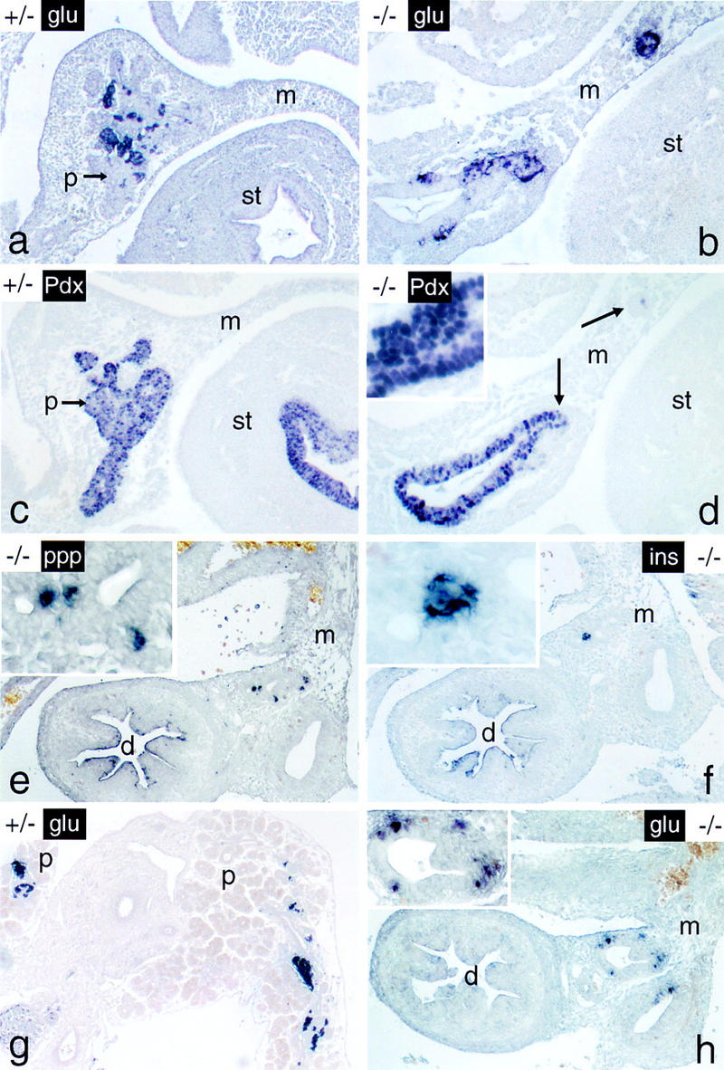
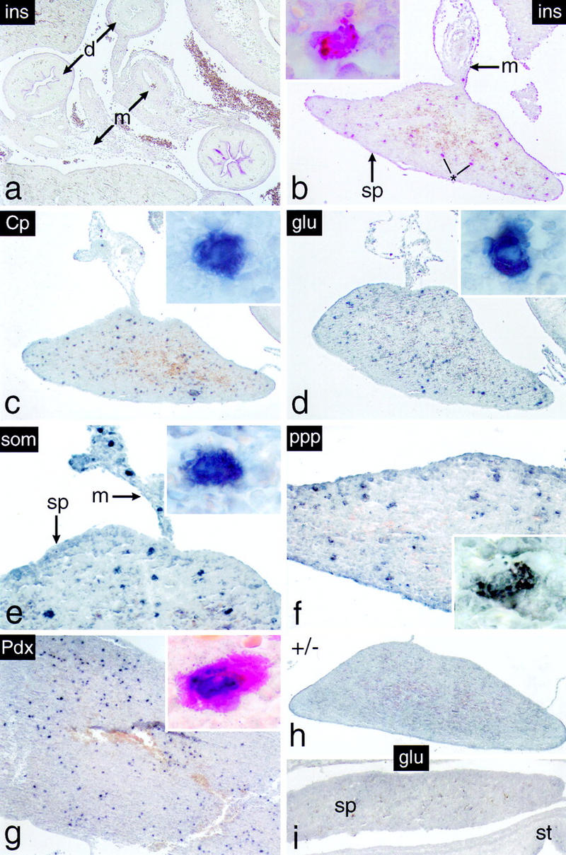
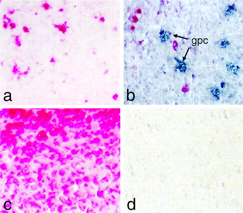
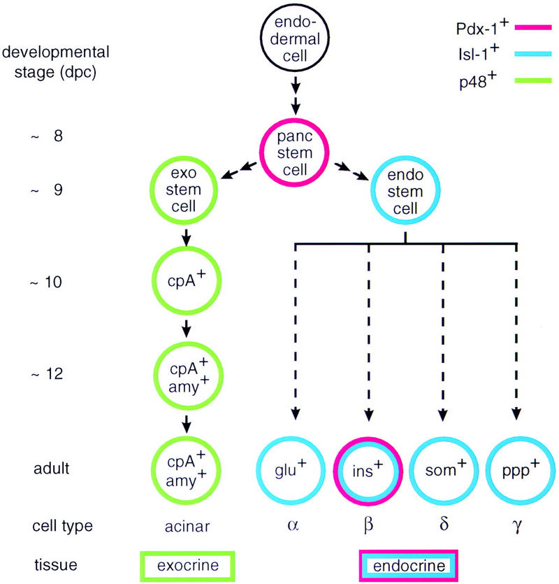
We have generated a mouse bearing a null allele of the gene encoding basic helix-loop-helix (bHLH) protein p48, the cell-specific DNA-binding subunit of hetero-oligomeric transcription factor PTF1 that directs the expression of genes in the exocrine pancreas. The null mutation, which establishes a lethal condition shortly after birth, leads to a complete absence of exocrine pancreatic tissue and its specific products, indicating that p48 is required for differentiation and/or proliferation of the exocrine cell lineage. p48 is so far the only developmental regulator known to be required exclusively for committing cells to an exocrine fate. The hormone secreting cells of all four endocrine lineages are present in the mesentery that normally harbors the pancreatic organ until day 16 of gestation. Toward the end of embryonic life, cells expressing endocrine functions are no longer detected at their original location but are now found to colonize the spleen, where they persist in a functional state until postnatal death of the organism occurs. These findings suggest that the presence of the exocrine pancreas is required for the correct spatial assembly of the endocrine pancreas and that, in its absence, endocrine cells are directed by default to the spleen, a site that, in some reptiles, harbors part of this particular cellular compartment.


Figures

Figure 1
Figure 1
Targeted inactivation of the p48 gene by homologous recombination and genotype analysis of heterozygote interbreeding. (A) Schematic maps of the targeting vector (a), the wild-type p48 locus (b) and the mutated allele (c). Exons are shown as hatched boxes and intronic or gene flanking sequences as heavy lines. Bacterial vector sequences (v) are designated by thin lines. The direction of transcription is indicated by bent arrows. The herpesvirus tk gene and the neo gene used for negative and positive selection, respectively, are in the opposite transcriptional orientation to the p48 gene. The neo cassette was inserted into exon 1 (E1) of the p48 gene upstream of the bHLH sequence. Cleavage sites for restriction endonucleases SacI (S) and NotI (N) were those used for construction and linearization of the targeting vector, respectively. Cleavage at EcoRI sites (R) was used to distinguish between wild-type and mutant alleles. The diagnostic EcoRI fragments are highlighted by double-headed arrows, and their size is shown. The origin of radiolabeled DNA probes used for filter hybridizations in B is indicated below the map of b. The position and polarity of oligonucleotide primers used for PCR amplification are represented by the small arrows below the map of c. (B) Southern analysis of EcoRI-digested genomic DNA of newborn littermates from a +/− interbreeding. DNA probes I and II both detect a 10.5-kb restriction fragment of the wild-type allele. In addition, probe I detects a 4-kb fragment, and probe II a 7.5-kb fragment, of the mutant allele. Subsequent genotype analysis was carried out by PCR on tail DNA with the gene-specific primers shown in A (data not shown).
Figure 2
Figure 2
The p48-deficient mouse lacks recognizable pancreatic structures. The +/− and −/− newborn mice shown in a are from the same litter of a +/− interbreeding. Both animals were alive when their pictures were taken (1 hr after birth). Note the slender body shape and the smooth texture and bluish color of the skin in the null mutant. The dissected intestinal tracts of these animals are compared in b. Only the relevant region of the intestine that harbors the pancreatic gland in wild-type mice is shown. In c, the abdominal region was examined in whole-mount sagittal sections of 18-dpc +/− and −/− embryos after staining with Giemsa. d, duodenum; m, mesentery; p, pancreas; sp, spleen; st, stomach. Abbreviations used are the same for Figs. 4–7. Photographs of +/− and −/− genotypes in a, b, and c are shown at the same relative magnification.
Figure 3
Figure 3
The p48-null mutant is deficient for exocrine pancreatic functions. (a) Western blot analysis for the immunodetection of pancreatic α-amylase in extracts containing total protein derived from dissected gastrointestinal tracts and the adhering spleens of newborn +/+, +/−, or −/− littermates. The prominent band at 56 kD in +/+ and +/− samples is α-amylase. (b) Northern blot analysis for the detection of exocrine and endocrine mRNAs. Total RNA of newborn (nb) and 16-dpc embryos was isolated from samples containing the tissues described in a. The relative accumulation of pancreatic mRNAs at steady state was estimated by comparison to the signal produced by the mRNA for intestinal fatty acid binding protein (Fab). (c) RT–PCR for the detection of mRNAs diagnostic for the exocrine and the four different endocrine cell lineages in two newborn +/− and −/− littermates each. Ethidium bromide-stained PCR products are shown in reverse contrast. Their size was estimated by comparison to a commercial 100-bp DNA ladder. The numbers on the right of the gel pictures show the actual size in base pairs as deduced from the DNA sequence. The specificity of each amplification reaction was monitored by including a no cDNA control (Co). Weak bands for carboxypeptidase A and elastase 1 were often seen in amplification reactions of −/− samples. Their presence suggests that cells expressing low levels of transcripts for these exocrine markers persist in the gastrointesinal tract even though we have never detected their protein products by immunohistochemistry.
Figure 4
Figure 4
The p48-null mutant lacks cells expressing exocrine functions. Sagittal tissue sections of whole-mount 12-dpc (a,b), 16-dpc (c,d) and 18-dpc (e–h) +/− or −/− embryos were analyzed for the presence of cells producing carboxypeptidase A (cpA; blue color) or α-amylase (amy; red or brown color). Arrows in b and d designate cell masses that stain positive for endocrine markers in butterfly sections shown in Fig. 6, b and e, f, and h), respectively. Cell nuclei in e and f were counterstained with hematoxylin. The insets of a, c and e emphasize the cytoplasmic location of the markers. All panels are shown at the same relative magnification. Tissue in this and all subsequent figures is oriented such that caudal is toward the top, distal toward the bottom, ventral toward the left and dorsal toward the right.
Figure 5
Figure 5
The endocrine pancreas of newborn mice bearing one inactive p48 allele shows a normal structural and functional organization. Tissue sections of dissected intestinal tracts derived from 18-dpc +/+ and +/− mice were analyzed for the expression of markers diagnostic for the endocrine (Li) compartment of the pancreas, such as insulin (ins; red color), glucagon (glu; dark blue color), C peptide (Cp; dark blue color), somatostatin (som; dark blue color), and pancreatic polypeptide (ppp; dark blue color). The pancreas in h was assayed for the simultaneous presence of insulin (red color) and Pdx-1 (dark blue color) in the β cell cytoplasm and nucleus (arrows), respectively. The pancreas of a was counterstained with hematoxylin. The inset of c highlights the cytoplasmic localization of the immunostain. Panels a–g are shown at the same relative magnification.
Figure 6
Figure 6
Cells expressing endocrine functions persist in the p48-null mutant embryo. The endocrine markers indicated (see Fig. 5 for abbreviations) were localized in whole-mount sections from 12-dpc a–d and 16-dpc e–h +/− and −/− embryos by immunostaining. Note that endocrine cells of −/− mice are closely associated with intramesenterial ductlike structures at both developmental stages. Arrows in d designate cellular aggregates that stain positive for glucagon in the adjacent serial section of b. Panels for a given developmental stage are shown at the same final magnification. (Insets) Detail at a higher magnification.
Figure 7
Figure 7
Cells expressing endocrine functions appear in the spleen of p48-deficient mice. Immunohistochemistry was carried out on dissected intestinal tracts of 18-dpc −/− embryos (b–f) or from spleen of newborn −/− mice (g). (a) The abdominal region of an 18-dpc −/− embryo showing, in this particular case, that insulin-producing cells are absent from the intestinal mesentery. Individual cells expressing the endocrine markers indicated were detected exclusively in spleens of −/− mice from 18 dpc onward. They were not observed in spleens of +/− mice (h) or 16-dpc −/− embryos (i) as exemplified by the absence of glucagon staining. (Insets) Single intrasplenic cells positive for the respective endocrine markers at a higher magnification. (Inset in g) Cell simultaneously expressing insulin in its cytoplasm (red color) and Pdx-1 in the nucleus (dark blue stain). Note that individual immunopositive cells are also present in the mesentery adjacent to the spleen (e.g., in e).
Figure 8
Figure 8
Intrasplenic cells expressing glucagon are not apoptotic. (a) Apoptotic nuclei in the spleen (red color) are visualized by chromogenic detection of anti-DIG antibody without treatment with DNase I. (b) Same as a but using, in addition, an antibody for the simultaneous detection of glucagon producing cells (gpc; blue color). (c) Same as a but including a DNase I digestion step prior to the incorporation of labeled nucleotide as a positive control. (d) Same as a but omitting terminal transferase as a negative control.
Figure 9
Figure 9
The origin of the exocrine and the endocrine cell lineages in the mammalian pancreas. The model for the exocrine cell lineage is based on results described in this paper but also incorporates data from other laboratories (Gittes and Rutter 1992; Guz 1995). The origin of the various endocrine cell lineages is shown for completeness and is based on data from the literature (e.g., see Larsson 1996). Arrows in the diagram are not meant to imply a precursor–product relationship between the different cells of a given lineage but rather suggest a sequential order of events deduced from the localization of cell-type-specific markers in the developing pancreas of wild-type and mutant mice. The early target genes of bHLH protein p48 and homeodomain proteins Pdx-1 or Isl-1 are not known.

Similar articles

Cited by

References

    1. Ahlgren U, Jonsson J, Edlund H. The morphogenesis of the pancreatic mesenchyme is uncoupled from that of the pancreatic epithelium in IPF1/PDX1-deficient mice. Development. 1996;122:1409–1416. - PubMed
    1. Ahlgren U, Pfaff SL, Jessell TM, Edlund T, Edlund H. Independent requirement for ISL1 in formation of pancreatic mesenchyme and islet cells. Nature. 1997;385:257–260. - PubMed
    1. Apelqvist A, Ahlgren U, Edlund H. Sonic hedgehog directs specialised mesoderm differentiation in the intestine and pancreas. Curr Biol. 1997;7:801–804. - PubMed
    1. Birkedahl-Hansen H. Proteolytic remodeling of extracellular matrix. Curr Opin Cell Biol. 1995;7:728–735. - PubMed
    1. Bradley A. Production and analysis of chimaeric mice. In: Robertson EJ, editor. Teratocarcinomas and embryonic stem cells: A practical approach. Oxford, UK: IRL Press; 1987. pp. 113–151.

Publication types

Substances