. 1998 Dec 1;12(23):3764-75.
doi: 10.1101/gad.12.23.3764.

BMP4 is essential for lens induction in the mouse embryo

Affiliations

BMP4 is essential for lens induction in the mouse embryo

Y Furuta et al. Genes Dev. .

Abstract

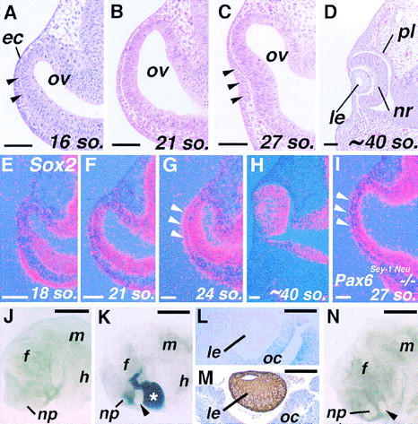
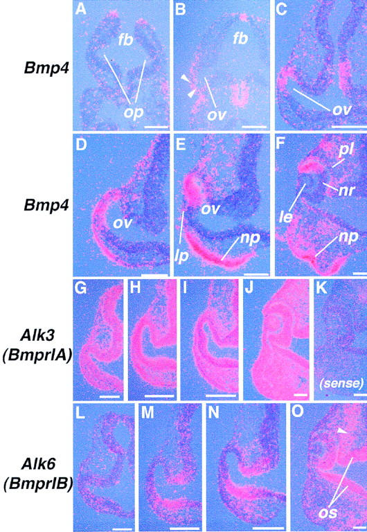
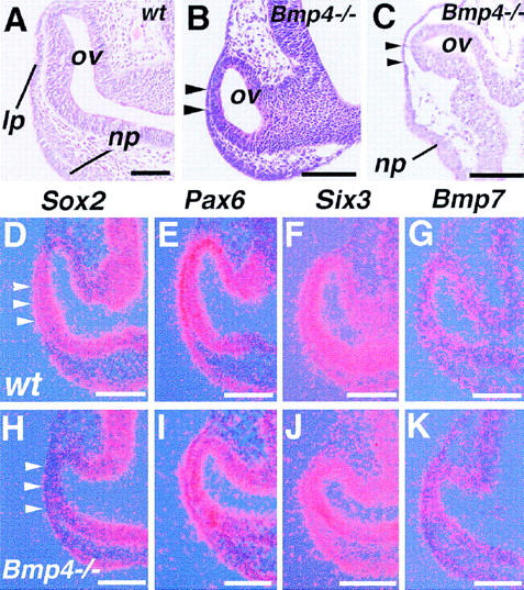
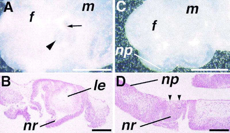
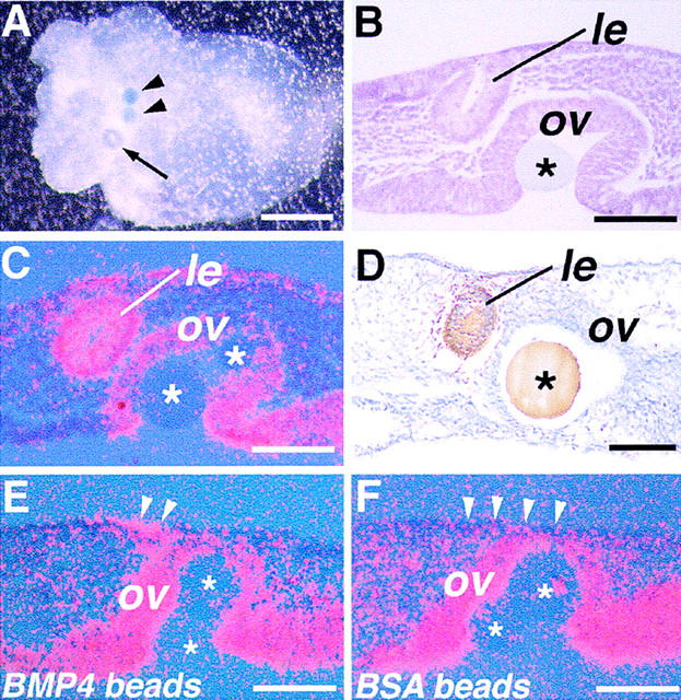
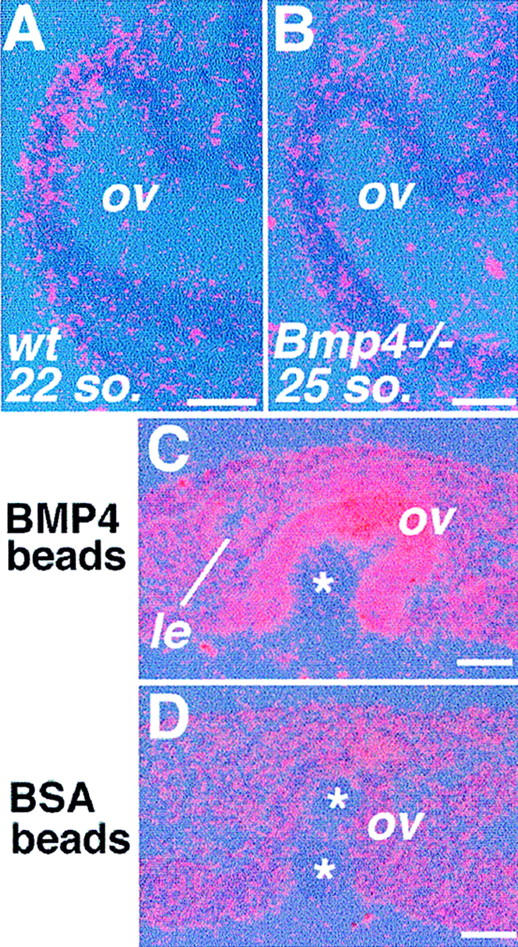
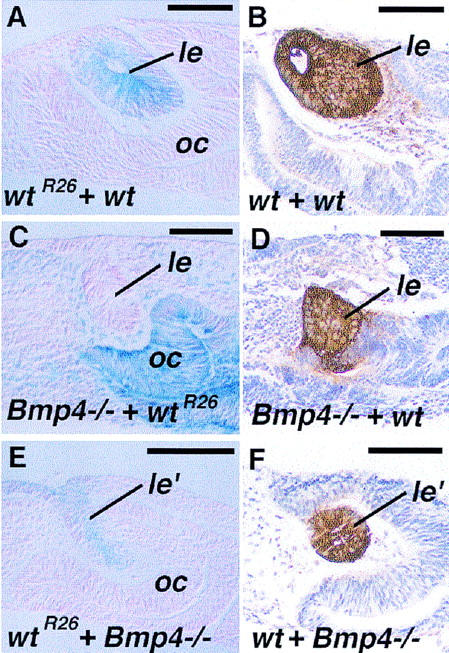
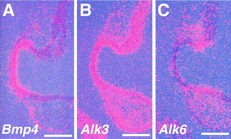
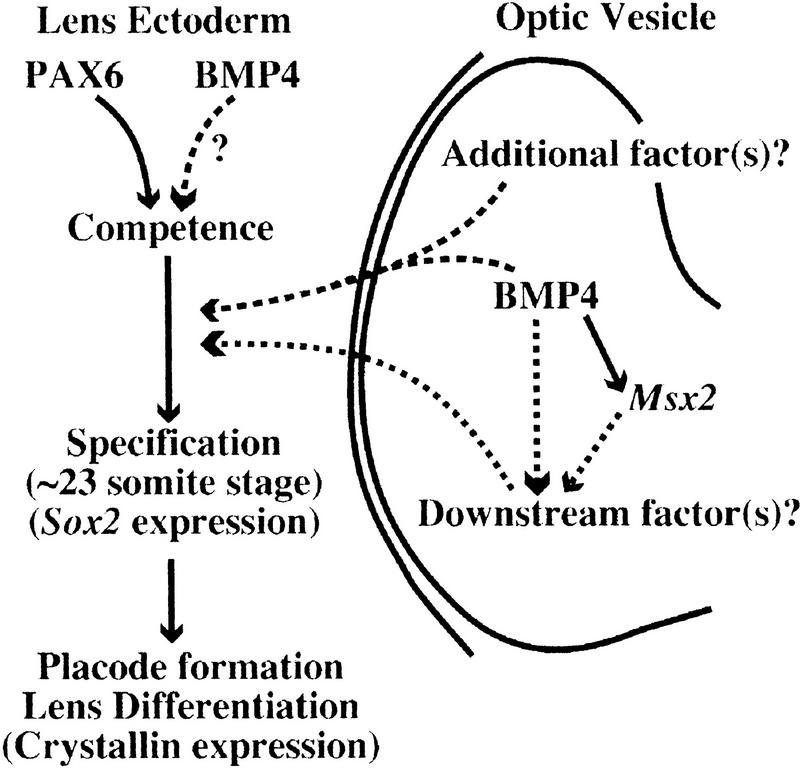
Vertebrate lens development is a classical model system for studying embryonic tissue interactions. Little is known, however, about the molecules mediating such inductive events. Here, we show that Bmp4, which is expressed strongly in the optic vesicle and weakly in the surrounding mesenchyme and surface ectoderm, has crucial roles during lens induction. In Bmp4(tm1) homozygous null mutant embryos, lens induction is absent, but the process can be rescued by exogenous BMP4 protein applied into the optic vesicle in explant cultures. This is associated with rescue of ectodermal expression of Sox2, an early lens placode marker. Substituting the optic vesicle in explant cultures with BMP4-carrying beads, however, does not lead to lens induction, indicating that other factors produced by the optic vesicle are involved. BMP4 appears to regulate expression of a putative downstream gene, Msx2, in the optic vesicle. No change in Pax6 expression is seen in Bmp4(tm1) mutant eyes, and Bmp4 expression appears unaffected in the eyes of homozygous Pax6(Sey-1Neu), suggesting that PAX6 and BMP4 function independently. Based on these results we propose that BMP4 is required for the optic vesicle to manifest its lens-inducing activity, by regulating downstream genes and/or serving as one component of multiple inductive signals.


Figures

Figure 1
Figure 1
Lens formation in the mouse embryo in vivo and in vitro. (A–D) Frontal sections of the eye primordia from the 16 (16 so.; 8.75–9.0 dpc) to ∼40-somite stage (10.5 dpc). Arrowheads in (A) indicate head mesenchyme cells that still exist between the ectoderm (ec) and optic vesicle (ov) at this stage, and in C, formation of the lens placode. (E–H) Expression of Sox2 in the developing eye detected by in situ hybridization. Red dots represent hybridization signals, and the blue background illuminates the tissues. Sox2 is expressed widely in the central nervous system, but its expression in the eye increases only from around 9.0 dpc (E,F). (G) In the presumptive lens ectoderm, significant upregulation of Sox2 occurs soon after contact between the ectoderm and optic vesicle (arrowheads), and its expression persists within the lens at later stages (H). (I) In homozygous Pax6Sey-1Neu mutant embryos no upregulation of Sox2 expression is observed at stages of lens placode formation. (J–N) Explant cultures of eye primordia of 8.75–9.0 dpc mouse embryos. (J) An explant from an 18-somite-stage embryo cultured for 4 days with the optic vesicle removed. No lens formation is observed. (np) Nasal placode; (f) forebrain; (m) midbrain; and (h) hindbrain. (K) Lens formation (arrowhead) in an explant taken from an embryo at the same stage as the one shown in J with the optic vesicle replaced with one from a ROSA26 embryo (asterisk). This allows easy distinction between host- and graft-derived tissues by β-Gal staining (blue). (L) The rescued lens (le) in such recombinants is derived from host ectoderm (no β-Gal staining), and β-Gal staining is present in the grafted optic cup (oc). (M) αA-crystallin expression in the lens formed in a recombinant detected by immunohistochemistry (brown). (N) In some explants from 21- to 23-somite-stage embryos with the optic vesile removed, lens formation is observed (arrowhead). In this particular sample, the optic vesicle was replaced with beads (blue dots) to inhibit possible reassociation between the ectoderm and remaining neuroectoderm. The beads have been dislocated by the growth of the lens. (nr) Neuroretinal layer of the optic cup; (pl) pigment layer of the optic cup. Bar, 50 μm (A–I); 500 μm (J,K,N); 100 μm (L,M).
Figure 2
Figure 2
Expression of Bmp4 and BMP type-I receptor genes during early eye development. (A–F) In situ hybridization using an antisense riboprobe for Bmp4 on transverse sections of 10- (A) and 14- (B) somite-stage embryos, and on frontal sections of 18- (C), 22- (D), 27- (E), and ∼40-somite-stage (10.5 dpc) (F) embryos. Arrowheads in B indicate early Bmp4 expression both in the ectoderm and distal optic vesicle. (G–O) In situ hybridization for BMP type-I receptor genes on frontal sections of embryos at the 18- (G,L), 22- (H, M), 26- (I,N), and ∼40-somite stages (10.5 dpc) (J,O). Arrowhead in O shows expression of Alk6 (BmprIB) in the neural crest mesenchyme. Hybridization with a sense control probe for Alk3 gives signals only at background levels in a section of a 10.5 dpc embryo (K). (lp) Lens placode; (op) optic pit; (os) optic stalk. Bar, 50 μm.
Figure 3
Figure 3
Eye phenotypes of late surviving Bmp4tm1 homozygous mutant embryos. (A) Morphology of the normal eye of a 27-somite-stage embryo (9.75 dpc). (ov) Optic vesicle; (lp) lens placode; and (np) nasal placode. (B) Histological section of a mutant embryo with 25 somites at 10.0 dpc. No sign of lens placode formation is seen at the site of contact between the ectoderm and the optic vesicle (arrowheads). (C) Another mutant embryo at 10.5 dpc showing no lens placode formation (arrowheads). Note that the nasal placode is formed. (D–K) Gene expression in the eyes of 22- to 24-somite-stage wild-type (wt) (D–G), and 9.5–10.0 dpc homozygous Bmp4tm1 mutant embryos (Bmp4−/−) which formed 22–25 somites (H–K). Note that, although upregulated in the wild-type presumptive lens ectoderm by the 24-somite stage (D, arrowheads), Sox2 fails to be induced in the mutant ectoderm and the optic vesicle (H). (E–G,I–K) Expression of other putative regulatory genes, including Pax6 (E,I), Six3 (F,J), and Bmp7 (G, K) is not apparently changed. Each panel is representative of results from at least three wild-type or five mutant embryos. Bar, 100 μm (A–C); 50 μm (D–K).
Figure 4
Figure 4
Morphology of eye explants in vitro. (A) A wild-type eye primordium taken at the 20-somite stage and cultured for 4 days. Not only the lens (arrowhead) but also surrounding retina and pigmented epithelium (arrow) are easily recognized under the dissection microscope. (B) Histological section of the eye explant shown in A. A well-differentiated lens (le) is seen, surrounded by the neuroretina (nr) of the optic cup. (C) An eye primordium from a 9.5-dpc mutant embryo cultured for 5 days. No lens formation is recognized macroscopically. (D) Histological section reveals that the lens is absent in the mutant eye primordium at the site of contact between the ectoderm and prospective neuroretina (arrowheads), whereas the nasal placode (np) is developed in the adjacent ectoderm. Bar, 500 μm (A,C); 100 μm (B,D).
Figure 5
Figure 5
Rescue of lens formation in explant cultures of Bmp4tm1 mutant eyes. (A) A mutant eye primordium cultured with BMP4-carrying beads (arrowheads) in the optic vesicle after 4 days of culture. A single vesicular structure is found macroscopically (arrow). (B) Histological section of the explant shown in A. The vesicular structure is a lens (le) derived from the ectoderm. (C) Pax6 is expressed in the formed lens vesicle. (D) αA-crystallin expression within the vesicle (arrowheads) detected by immunohistochemistry, confirming differentiation of lens cells. (E,F) Expression of Sox2 in mutant eye explants cultured for 1 day. (E) Sox2 is induced in both the surface ectoderm (arrowheads) and the optic vesicle of explants cultured with BMP4-carrying beads. (F) In contrast, although activated in the optic vesicle, expression of Sox2 is not detected in the ectoderm of the lens region (arrowheads) in the explants cultured with control BSA beads. Asterisks indicate the beads transplanted into the optic vesicle (ov). Bar, 250 μm (A); 50 μm (B–F).
Figure 6
Figure 6
Expression of Msx2 in the eye. (A) Msx2 is weakly expressed by the 22-somite stage in normal embryos. (B) In the eye of advanced mutants, Msx2 expression is not detected even at the 25-somite stage or later (n = 9). (C) Application of BMP4 in the mutant optic vesicle induces Msx2, and its expression is maintained after 4 days of culture. (D) In mutant explants with control beads, Msx2 expression is never detected throughout the culture period. Asterisks indicate implanted beads. (le) Rescued lens. Bar, 50 μm (A,B); 25 μm (C,D).
Figure 7
Figure 7
Tissue recombination explants between wild-type (wt) and Bmp4tm1 mutant (Bmp4−/−) eye tissues after 4-day culture. Wild-type tissues from the ROSA26 strain of mouse (wtR26) are revealed by blue β-Gal staining in (A,C,E). (A) Recombinant between wild-type ectoderm (wtR26) and optic rudiment (optic vesicle and surrounding mesenchyme). Formation of a relatively normal sized lens (le) is observed (blue staining), associated with formation of the optic cup (oc). (B) αA-crystallin expression in a wild-type–wild-type recombinant (brown). (C) Lens formation is also found in the mutant ectoderm when recombined with a wild-type optic rudiment. (D) Expression of αA-crystallin in such Bmp4−/−–wild-type recombinants. (E) Formation of the lentoid (le′) in a recombinant between a wild-type ectoderm and a Bmp4−/− optic rudiment. Such lentoids are often irregular in shape, but are surrounded by the retinal neuroectoderm, which forms an optic cup (oc). (F) Such lentoids are positive for αA-crystallin. Bar, 50 μm.
Figure 8
Figure 8
Expression of genes for BMP4 and its receptors in Pax6Sey-1Neu homozygous mutant eyes. Expression of Bmp4 (A), Alk3 (B), and Alk6 (C) in 21-somite-stage mutant embryos is essentially localized to the appropriate areas compared with normal embryos (see Fig. 2D,H,M). Bar, 50 μm.
Figure 9
Figure 9
A model for the possible roles of BMP4 during determination of the lens ectoderm. BMP4 may induce the optic vesicle factor(s) (downstream factors) that serve(s) as the signal(s) for lens induction. Transcription factors, such as MSX2, encoded by putative BMP4 downstream genes, may regulate expression of such optic vesicle factor(s). Alternatively, or in addition, BMP4 itself may function as a part of the inductive signal in synergy with other secreted factors (additional factors). PAX6 function in the ectoderm is essential for establishment of the competence to respond to the optic vesicle signal, and BMP4 may also be required independently from Pax6 for this process.

Similar articles

Cited by

References

    1. Chanut F, Heberlein U. Role of decapentaplegic in initiation and progression of the morphogenetic furrow in the developing Drosophila retina. Development. 1997;124:559–567. - PubMed
    1. de Iongh R, Lovicu FJ, Hanneken A, McAvoy JW. Spatio-temporal distribution of acidic and basic FGF indicates a role for FGF in rat lens morphogenesis. Dev Dyn. 1993;198:190–202. - PubMed
    1. Dewulf N, Verschueren K, Lonnoy O, Moren A, Grimsby S, Vande Spiegle K, Miyazono K, Huylebroeck D, Ten Dijke, P P. Distinct spatial and temporal expression patterns of two type I receptors for bone morphogenetic proteins during mouse embryogenesis. Endocrinology. 1995;136:2652–2663. - PubMed
    1. Dominguez M, Hafen E. Hedgehog directly controls initiation and propagation of retinal differentiation in the Drosophila eye. Genes & Dev. 1997;11:3254–3264. - PMC - PubMed
    1. Dudley AT, Lyons KM, Robertson EJ. A requirement for bone morphogenetic protein-7 during development of the mammalian kidney and eye. Genes & Dev. 1995;9:2795–2807. - PubMed

Publication types

MeSH terms