. 1998 Dec;180(24):6450-8.
doi: 10.1128/JB.180.24.6450-6458.1998.

Campylobacter fetus surface layer proteins are transported by a type I secretion system

Affiliations

Campylobacter fetus surface layer proteins are transported by a type I secretion system

S A Thompson et al. J Bacteriol. 1998 Dec.

Abstract

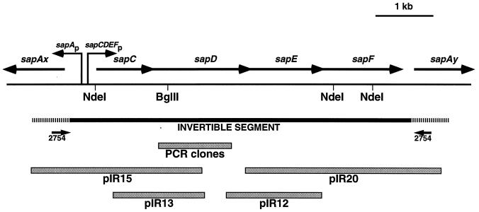
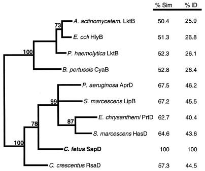
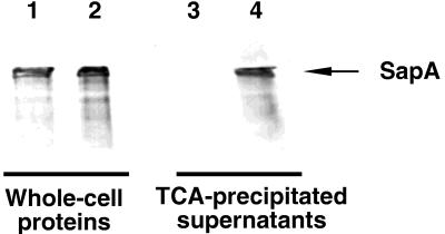
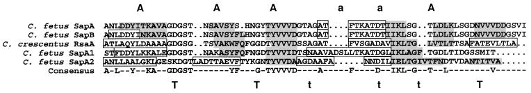
The virulence of Campylobacter fetus, a bacterial pathogen of ungulates and humans, is mediated in part by the presence of a paracrystalline surface layer (S-layer) that confers serum resistance. The subunits of the S-layer are S-layer proteins (SLPs) that are secreted in the absence of an N-terminal signal sequence and attach to either type A or B C. fetus lipopolysaccharide in a serospecific manner. Antigenic variation of multiple SLPs (encoded by sapA homologs) of type A strain 23D occurs by inversion of a promoter-containing DNA element flanked by two sapA homologs. Cloning and sequencing of the entire 6.2-kb invertible region from C. fetus 23D revealed a probable 5.6-kb operon of four overlapping genes (sapCDEF, with sizes of 1,035, 1,752, 1,284, and 1,302 bp, respectively) transcribed in the opposite direction from sapA. The four genes also were present in the invertible region of type B strain 84-107 and were virtually identical to their counterparts in the type A strain. Although SapC had no database homologies, SapD, SapE, and SapF had predicted amino acid homologies with type I protein secretion systems (typified by Escherichia coli HlyBD/TolC or Erwinia chrysanthemi PrtDEF) that utilize C-terminal secretion signals to mediate the secretion of hemolysins, leukotoxins, or proteases from other bacterial species. Analysis of the C termini of four C. fetus SLPs revealed conserved structures that are potential secretion signals. A C. fetus sapD mutant neither produced nor secreted SLPs. E. coli expressing C. fetus sapA and sapCDEF secreted SapA, indicating that the sapCDEF genes are sufficient for SLP secretion. C. fetus SLPs therefore are transported to the cell surface by a type I secretion system.


Figures

FIG. 1
FIG. 1
Schematic representation of the sapA invertible region showing the sapCDEF genes, the locations of the divergent sapA promoter and the putative sapCDEF promoter(s), and the clones (pIR15, pIR13, pIR12, and pIR20) from which the invertible region sequence was determined. The hatched areas represent the ca. 600-bp conserved regions at the 5′ ends of sapA homologs flanking the invertible region (here designated sapAx and sapAy), at which recombination occurs as the basis of sapA homolog rearrangements during SLP antigenic variation (22, 24).
FIG. 2
FIG. 2
Phylogram, generated by parsimony analysis, demonstrating the relatedness of the cytoplasmic membrane ABC proteins of type I secretion systems. Percentages of amino acid similarity (% Sim) and identity (% ID) with the C. fetus protein are shown at the right. The bold numbers adjacent to the phylogram branches indicate the percentages of 1,000 bootstrap replicates supporting the clustering of those branches. Branches without bootstrap values were clustered in less than 70% of the bootstrap replicates.
FIG. 3
FIG. 3
Phenotypes of C. fetus wild-type (23D), spontaneous S mutant (23B), and sapD mutant (97-205) cells. (A) Serum susceptibilities of C. fetus strains. Serial dilutions of 103 to 105 CFU/ml were suspended in either 40% NHS or HINHS (56°C, 30 min) for 60 min. The amount of killing relative to that of the time zero inoculum was determined and is presented as mean log10 killing ± the standard error of the mean. (B) Western blot of C. fetus strains. Cell surface water extracts and whole-cell lysates were probed with polyclonal serum against C. fetus SLPs. Lanes: 1, strain 23D (S+); 2, strain 23B (S); 3, strain 97-205 (sapD mutant). The positions of molecular mass markers are shown at the left.
FIG. 4
FIG. 4
Primer extension analysis of sapA-specific mRNA in wild-type and sapD mutant C. fetus strains. The levels of sapA transcription were determined by reverse transcription of standardized 40-μg RNA samples by using primer 2754 (59). The sequencing ladder was generated by using primer 2754 and plasmid pIR15, which contains the sapA promoter and 5′ region (Fig. 1). Lanes: 1, strain 23D (wild type); 2, strain 97-205 (sapD mutant). The +1 site for strain 23D is identical to that determined previously (59).
FIG. 5
FIG. 5
Secretion of SapA from E. coli expressing C. fetus sapCDEFA, detected by immunoblotting with antiserum to C. fetus SLPs. One microgram of whole-cell proteins (lanes 1 and 2) or the amount of TCA-precipitated protein present in 250 μl of culture supernatant (lanes 3 and 4) was analyzed. Lanes: 1 and 3, C600(pSPORT1)(pBGYC1); 2 and 4, C600(pIR100)(pBGYC1).
FIG. 6
FIG. 6
Conserved primary and secondary structures of the C termini of four C. fetus SLPs (SapA, SapA1, SapA2, and SapB) and RsaA, the SLP of C. crescentus, predicted by GCG PILEUP and by the algorithm of Garnier et al. (32). The consensus line indicates amino acid residues that were conserved in at least three of the five SLPs. Secondary-structure features shown are regions of predicted α-helix (boxed), β-sheet (light shading), random coil (no shading), and strong or weak amphipathicity (A or a, respectively) and strong or weak turn-forming residues (T or t, respectively).

Similar articles

Cited by

References

    1. Ausubel F M, Brent R, Kingston R E, Moore D D, Seidman J G, Smith J A, Struhl K, editors. Current protocols in molecular biology. Vol. 1. New York, N.Y: John Wiley & Sons, Inc.; 1997.
    1. Awram P, Smit J. The Caulobacter crescentus paracrystalline S-layer protein is secreted by an ABC transporter (type I) secretion apparatus. J Bacteriol. 1998;180:3062–3069. - PMC - PubMed
    1. Bachman B J. Derivations and genotypes of some mutant derivatives of Escherichia coli K-12. In: Neidhardt F C, Ingraham J L, Low K B, Magasanik B, Schaechter M, Umbarger H E, editors. Escherichia coli and Salmonella typhimurium: cellular and molecular biology. Vol. 2. Washington, D.C: American Society for Microbiology; 1987. pp. 1190–1219.
    1. Binet R, Wandersman C. Cloning of the Serratia marcescens hasF gene encoding the Has ABC exporter outer membrane component: a TolC analogue. Mol Microbiol. 1997;22:265–273. - PubMed
    1. Binet R, Wandersman C. Protein secretion by hybrid bacterial ABC-transporters: specific functions of the membrane ATPase and the membrane fusion protein. EMBO J. 1995;14:2298–2306. - PMC - PubMed

Publication types

Associated data

LinkOut - more resources