. 1998 Dec;180(24):6571-80.
doi: 10.1128/JB.180.24.6571-6580.1998.

Cytochrome bd biosynthesis in Bacillus subtilis: characterization of the cydABCD operon

Affiliations

Cytochrome bd biosynthesis in Bacillus subtilis: characterization of the cydABCD operon

L Winstedt et al. J Bacteriol. 1998 Dec.

Abstract

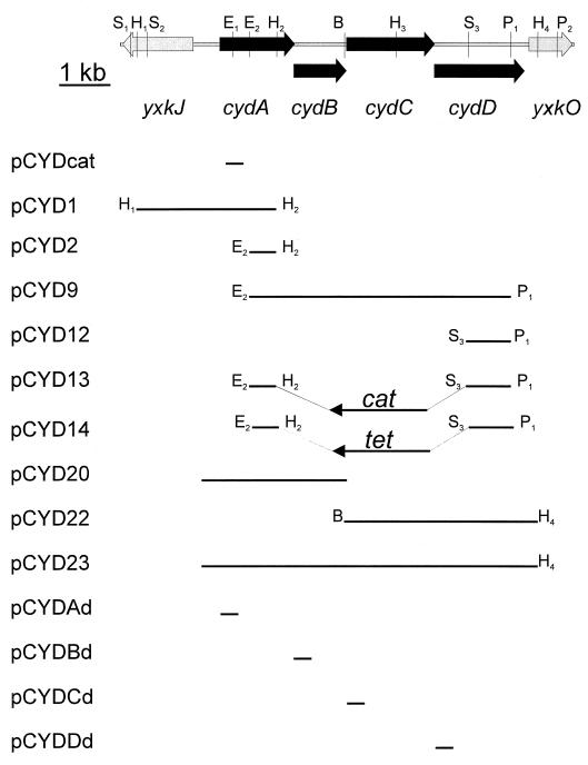
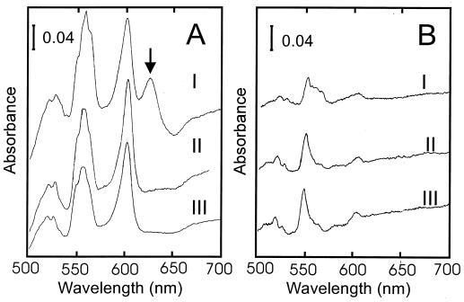
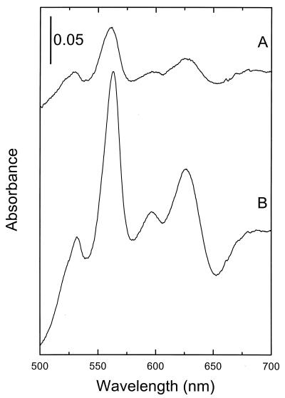
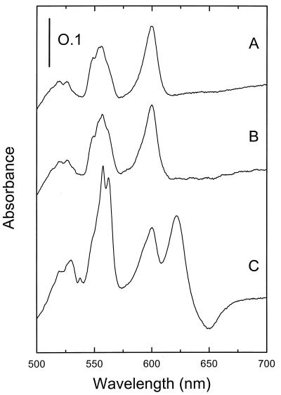
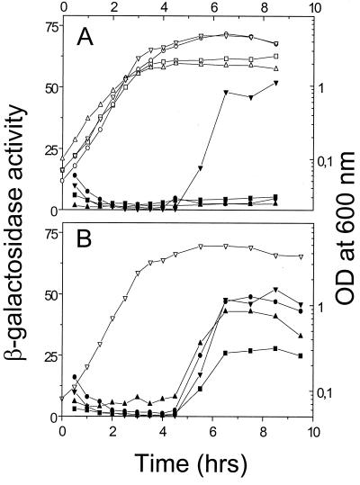
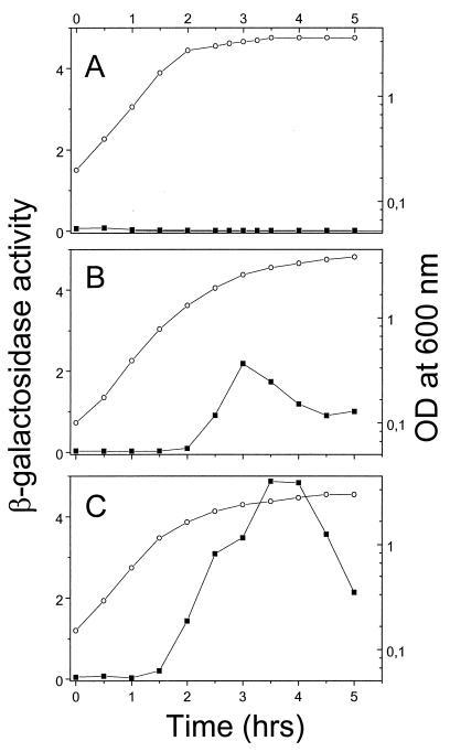
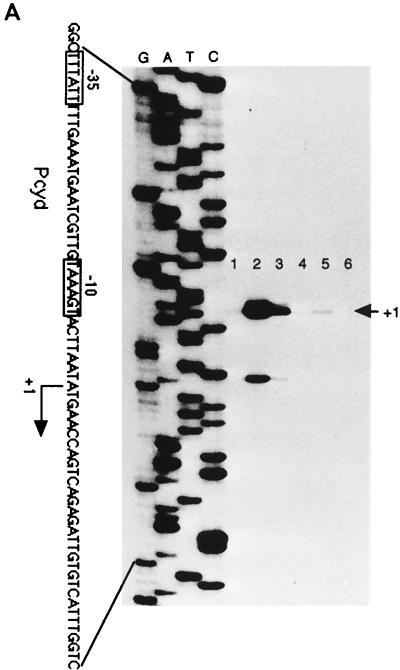
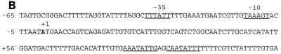
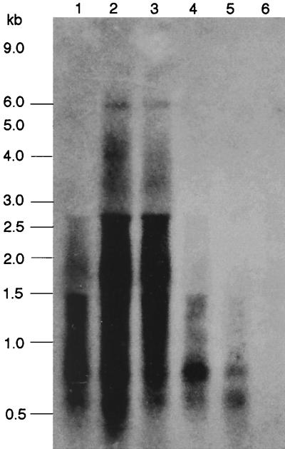
Under aerobic conditions Bacillus subtilis utilizes a branched electron transport chain comprising various cytochromes and terminal oxidases. At present there is evidence for three types of terminal oxidases in B. subtilis: a caa3-, an aa3-, and a bd-type oxidase. We report here the cloning of the structural genes (cydA and cydB) encoding the cytochrome bd complex. Downstream of the structural genes, cydC and cydD are located. These genes encode proteins showing similarity to bacterial ATP-binding cassette (ABC)-type transporters. Analysis of isolated cell membranes showed that inactivation of cydA or deletion of cydABCD resulted in the loss of spectral features associated with cytochrome bd. Gene disruption experiments and complementation analysis showed that the cydC and cydD gene products are required for the expression of a functional cytochrome bd complex. Disruption of the cyd genes had no apparent effect on the growth of cells in broth or defined media. The expression of the cydABCD operon was investigated by Northern blot analysis and by transcriptional and translational cyd-lacZ fusions. Northern blot analysis confirmed that cydABCD is transcribed as a polycistronic message. The operon was found to be expressed maximally under conditions of low oxygen tension.


Figures

FIG. 1
FIG. 1
Restriction map of the B. subtilis cyd region and plasmids carrying different parts of this region. The sequence of this region was determined previously (54). The cyd genes are oriented in the same direction as replication of the B. subtilis chromosome. The insert carried by each plasmid is shown as a solid line. Restriction sites used for subcloning fragments of the cyd region are abbreviated as follows: B, BspEI; E, EcoRI; H, HindIII; P, PstI; S, SphI. Plasmids pCYD1 and pCYD2 are derivatives of pBluescript II KS(−) (Stratagene, Inc.); plasmid pCYDcat is a derivative of pT7Blue(R) (Novagen) that contains the chloramphenicol acetyltransferase gene of pHV32 (35) on a 2,000-bp HindIII-SalI fragment; plasmid pCYD9 is a derivative of pUC18 (53); plasmids pCYD12 and pCYD13 are derivatives of pHV32; pCYD20, pCYD22, and pCYD23 are derivatives of the E. coli/B. subtilis shuttle vector pHP13 (19); pCYD14 is a derivative of pCYD13 in which the cat gene (HindIII-BamHI) has been replaced with the tet gene of plasmid pDG1515 (carried on a 2,142-bp HindIII-BamHI fragment) (18); pCydAd, pCydBd, pCydCd, and pCydDd are derivatives of pMutin2 (lacZ lacI amp ery) (48).
FIG. 2
FIG. 2
Low-temperature (77 K) light absorption difference spectra of B. subtilis membranes. I, wild-type strain 168A; II, cydD mutant LUW9; III, cydA mutant LUW3. Membranes (10 mg of protein ml−1) were reduced in the sample cuvette with sodium dithionite (A) or sodium ascorbate (B) and oxidized in the reference cuvette with potassium ferricyanide. The arrow indicates the 622-nm maximum of reduced cytochrome d of the cytochrome bd complex. The absorbance scales are indicated by the bars.
FIG. 3
FIG. 3
Light absorption difference spectra of B. subtilis membranes recorded at room temperature. Membrane suspensions (4 mg of protein ml−1) were reduced in the sample cuvette with sodium dithionite and oxidized in the reference cuvette with potassium ferricyanide. (A) LUH17/pHP13; (B) LUH17/pCYD20. Strain LUH17 has cytochromes aa3 and caa3 deleted. The vertical bar indicates the absorption scale.
FIG. 4
FIG. 4
Difference spectra of B. subtilis LUW20 membranes isolated from cells harboring plasmid pHP13 (vector alone) (A), pCYD20 (cydAB) (B), or pCYD23 (cydABCD) (C). Strain LUW20 has cydABCD deleted. The spectra were recorded at 77 K on membrane suspensions containing 10 mg of protein ml−1. Membranes were reduced in the sample cuvette with sodium dithionite and oxidized in the reference cuvette with potassium ferricyanide. The vertical bar indicates the absorption scale.
FIG. 5
FIG. 5
β-Galactosidase expression from cyd-lacZ fusions as a function of growth. (A) Activities for the cydA-lacZ fusion are shown for cells grown in DSM (solid squares), NSMP (solid triangles), DSM supplemented with glucose (solid circles), and NSMPG (solid inverted triangles). The optical densities for cells grown in each medium are shown by the corresponding open symbol. (B) β-Galactosidase expression from transcriptional fusions to lacZ integrated by single-crossover recombination at the cydA (solid inverted triangles), cydB (solid circles), cydC (solid triangles), or cydD (solid squares) locus. β-Galactosidase activity is expressed as nanomoles of 2-nitrophenyl-β-d-galactopyranoside hydrolyzed per minute and milligram of protein. The growth (optical densities) of the cydA-lacZ fusion strain is shown (open inverted triangles). The four strains were grown in NSMPG and showed similar growth properties. Data from a single experiment are presented. Each experiment was repeated at least twice, with similar results.
FIG. 6
FIG. 6
Effect of aeration on cydA-lacZ expression. B. subtilis LUW48 was grown in NSMPG in a bioreactor. (A) High aeration, maintained during growth by manually increasing the stirring speed from 250 to 800 rpm. (B) Medium aeration, maintained by constant stirring at 250 rpm. (C) Low aeration, maintained by constant stirring at 250 rpm. The vessel was sparged with sterile air at a flow rate of 1 (A and B) or 0.5 (C) volume of air per volume of liquid per minute. β-Galactosidase activity (solid squares) is expressed as nanomoles of 4-methylumbelliferyl-β-d-galactoside hydrolyzed per minute per OD600 unit. Optical densities are shown as open circles.
FIG. 7
FIG. 7
Primer extension mapping of the cydA promoter. (A) Identification of the transcriptional initiation site of the cydA promoter (Pcyd). A known nucleotide sequence ladder was used to estimate the size of the extended product, which is indicated by an arrow. The end-labeled primer was annealed to total RNA isolated from B. subtilis 1A1 cultured in NSMPG (lanes 1 to 3) or in NSMP (lanes 4 to 6). Samples were taken in the exponential-growth phase (lanes 1 and 4), at the time of transition from the exponential-growth to the stationary phase (lanes 2 and 5), and in the stationary phase (lanes 3 and 6). Boxes, −10 and −35 promoter regions of cydA. This promoter is likely to be recognized by RNA polymerase containing the vegetative sigma factor, ςA. (B) Nucleotide sequence of the yxkJ-cydA intergenic region. The apparent transcription start site is indicated as +1. The −10 and −35 promoter regions of the cydA gene are indicated. Also shown is a putative operator sequence, the 16-bp palindrome starting at +80 (underlined). The coordinates are given with respect to the cydA transcription start point. A likely initiation codon (ATG) of cydA is at position +194.
FIG. 7
FIG. 7
Primer extension mapping of the cydA promoter. (A) Identification of the transcriptional initiation site of the cydA promoter (Pcyd). A known nucleotide sequence ladder was used to estimate the size of the extended product, which is indicated by an arrow. The end-labeled primer was annealed to total RNA isolated from B. subtilis 1A1 cultured in NSMPG (lanes 1 to 3) or in NSMP (lanes 4 to 6). Samples were taken in the exponential-growth phase (lanes 1 and 4), at the time of transition from the exponential-growth to the stationary phase (lanes 2 and 5), and in the stationary phase (lanes 3 and 6). Boxes, −10 and −35 promoter regions of cydA. This promoter is likely to be recognized by RNA polymerase containing the vegetative sigma factor, ςA. (B) Nucleotide sequence of the yxkJ-cydA intergenic region. The apparent transcription start site is indicated as +1. The −10 and −35 promoter regions of the cydA gene are indicated. Also shown is a putative operator sequence, the 16-bp palindrome starting at +80 (underlined). The coordinates are given with respect to the cydA transcription start point. A likely initiation codon (ATG) of cydA is at position +194.
FIG. 8
FIG. 8
Northern blot analysis of B. subtilis 1A1 total RNA using a cydA probe labeled with 32P. RNA was isolated from cells grown in NSMPG (lanes 1 to 3) or in NSMP (lanes 4 to 6). Samples were taken in the exponential-growth phase (lanes 1 and 4), at the time of transition from the exponential-growth to the stationary phase (lanes 2 and 5), and in the stationary phase (lanes 3 and 6). The positions of RNA size standards are indicated on the left. In addition to cyd-specific transcripts, the probes hybridized nonspecifically to 16S (1,553 bases) and 23S (2,928 bases) rRNA.

Similar articles

Cited by

References

    1. Atkinson M R, Wray L V, Jr, Fisher S H. Regulation of histidine and proline degradation enzymes by amino acid availability in Bacillus subtilis. J Bacteriol. 1990;172:4758–4765. - PMC - PubMed
    1. Cole S T, Brosch R, Parhill J, Garnier T, Churcher C, Harris D, Gordon S V, Eiglmeier K, Gas G, Barry III C E, Tekaia F, Badcock K, Basham D, Brown D, Chillingwort T, Conner R, Davies R, Devlin K, Feltwell T, Gentles S, Hamlin N, Holroyd S, Hornsby T, Jagels K, Krogh A, McLean J, Moule S, Murphy L, Oliver K, Osborne J, Quail M A, Rajandream M-A, Rogers J, Rutter S, Seeger K, Skelton J, Squares R, Squares S, Sulston J E, Taylor K, Whitehead S, Barrell B G. Deciphering the biology of Mycobacterium tuberculosis from the complete genome sequence. Nature. 1998;393:537–544. - PubMed
    1. Cook G M, Membrillo-Hernandez J, Poole R K. Transcriptional regulation of the cydDC operon, encoding a heterodimeric ABC transporter required for assembly of cytochromes c and bd in Escherichia coli K-12: regulation by oxygen and alternative electron acceptors. J Bacteriol. 1997;179:6525–6530. - PMC - PubMed
    1. Cunningham L, Pitt M, Williams H D. The cioAB genes from Pseudomonas aeruginosa code for a novel cyanide-insensitive terminal oxidase related to the cytochrome bd quinol oxidases. Mol Microbiol. 1997;24:579–591. - PubMed
    1. Dahl M K, Meinhof C-G. A series of integrative plasmids for Bacillus subtilis containing unique cloning sites in all three open reading frames for translational lacZ fusions. Gene. 1994;145:151–152. - PubMed

Publication types

LinkOut - more resources