. 1998 Dec;180(24):6625-34.
doi: 10.1128/JB.180.24.6625-6634.1998.

Topological analysis of the aerobic membrane-bound formate dehydrogenase of Escherichia coli

Affiliations

Topological analysis of the aerobic membrane-bound formate dehydrogenase of Escherichia coli

S Benoit et al. J Bacteriol. 1998 Dec.

Abstract

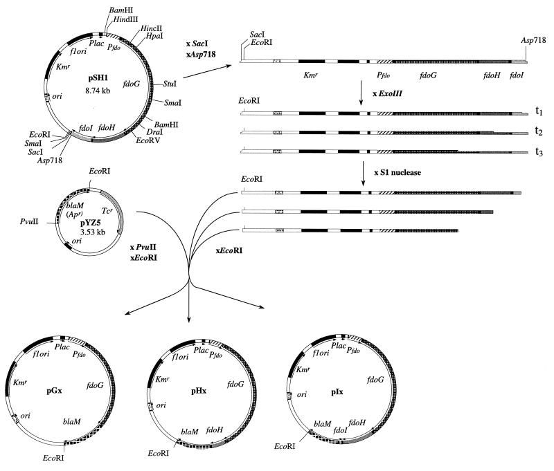
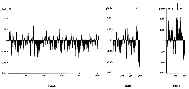
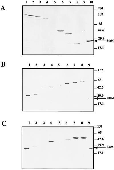
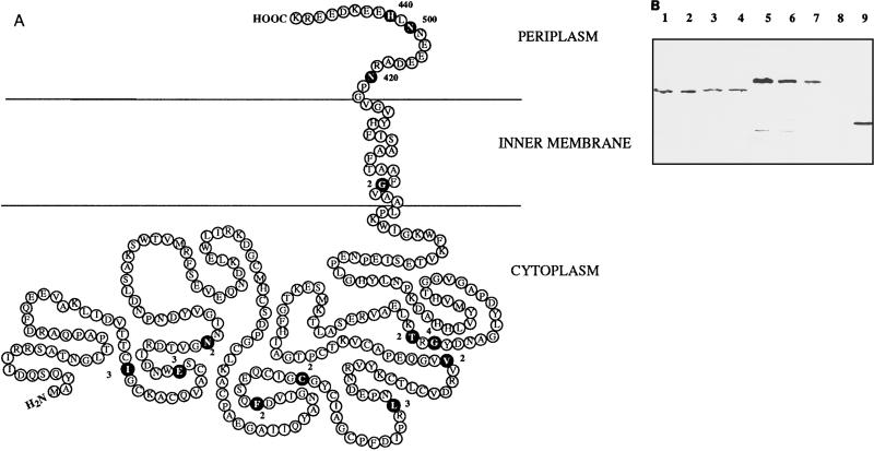
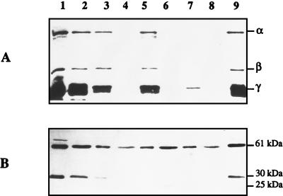
Besides formate dehydrogenase N (FDH-N), which is involved in the major anaerobic respiratory pathway in the presence of nitrate, Escherichia coli synthesizes a second isoenzyme, called FDH-O, whose physiological role is to ensure rapid adaptation during a shift from aerobiosis to anaerobiosis. FDH-O is a membrane-bound enzyme complex composed of three subunits, alpha (FdoG), beta (FdoH), and gamma (FdoI), which exhibit high sequence similarity to the equivalent polypeptides of FDH-N. The topology of these three subunits has been studied by using blaM (beta-lactamase) gene fusions. A collection of 47 different randomly generated Fdo-BlaM fusions, 4 site-specific fusions, and 3 sandwich fusions were isolated along the entire sequence of the three subunits. In contrast to previously reported predictions from sequence analysis, our data suggested that the alphabeta catalytic dimer is located in the cytoplasm, with a C-terminal anchor for beta protruding into the periplasm. As expected, the gamma subunit, which specifies cytochrome b, was shown to cross the cytoplasmic membrane four times, with the N and C termini exposed to the cytoplasm. Protease digestion studies of the 35S-labelled FDH-O heterotrimer in spheroplasts add further support to this model. Consistently, prior studies regarding the bioenergetic function of formate dehydrogenase provided evidence for a mechanism in which formate is oxidized in the cytoplasm.


Figures

FIG. 1
FIG. 1
Map of plasmid pSH1 and construction of random fdo′-blaM fusions. Plasmid pSH1 was made by ligation of the 5-kb HindIII-SacI fragment from plasmid pHA3 (1) into the corresponding sites in vector pYZ4 (49). The cloned fragment carries the entire fdo locus with its own promoter. Random β-lactamase fusions were subsequently constructed by digesting from the 3′ end of the fdoI gene with exonuclease III and ligating the blunt-ended DNA with the truncated blaM gene.
FIG. 2
FIG. 2
Hydropathy analysis of the FdoG, FdoH, and FdoI subunits. The hydropathy plot was derived by using the algorithm of Kyte and Doolittle (23) with a window size of 11 residues. Arrows indicate putative transmembrane segments.
FIG. 3
FIG. 3
Western blots of FdoG-BlaM, FdoH-BlaM, and FdoI-BlaM fusion proteins. Equal amounts of cell extracts were electrophoresed on SDS–12.5% polyacrylamide gels, and β-lactamase fusion proteins were visualized by immunoblotting with polyclonal antibodies against BlaM. (A) Cell extracts of strain NM522 containing fdoG′-blaM fusion plasmids. Amino acids at which fusions occur: lane 1, Asp967; lane 2, Phe876; lane 3, Arg776; lane 4, Asn698; lane 5, Arg423; lane 6, Val155; lane 7, Val86; lane 8, Arg36; lane 9, none (parental NM522). Lane 10, TEM β-lactamase from pBR322 (29 kDa). (B) Cell extracts of NM522 containing fdoH′-blaM fusion plasmids. Lane 1, Ile40; lane 2, Asn61; lane 3, Cys136; lane 4, Val168; lane 5, Thr205; lane 6, Gly264; lane 7, Asn282; lane 8, Asn290; lane 9, TEM β-lactamase from pBR322. (C) Cell extracts of NM522 containing fdoI′-blaM fusion plasmids. Lane 1, Lys2; lane 2, Phe23; lane 3, Gly34; lane 4, Gly105; lane 5, Val123; lane 6, Leu150; lane 7, Ala191; lane 8, Tyr199; lane 9, TEM β-lactamase from pBR322.
FIG. 4
FIG. 4
Model for the topological organization of FdoH and protease susceptibility of FdoH-BlaM hybrid proteins in spheroplasts. (A) Model based on the properties of the FdoH–β-lactamase fusions and the hydropathy plot. Fusions are indicated by black circles, and the adjacent numbers correspond to MICs in micrograms of ampicillin per milliliter. (B) Visualization by Western blotting of the sensitivity of two FdoH-BlaM fusion proteins to proteinase K digestion in spheroplasts. Lanes 1 to 4, strain NM522 carrying the plasmid-encoded Thr205FdoH-BlaM gene fusion; lanes 5 to 8, strain NM522 carrying the plasmid-encoded Asn290FdoH-BlaM gene fusion; lane 9, strain NM522(pBR322) expressing wild-type TEM β-lactamase. Proteinase K was added at a final concentration of 0 (lanes 1 and 5), 200 (lanes 2 and 6), 500 (lanes 3 and 7), or 1,000 (lanes 4 and 8) μg/ml.
FIG. 5
FIG. 5
Model for the topological organization of FdoI, based on the properties of β-lactamase fusions, the hydropathy plot, and the positive-inside rule (44). β-Lactamase fusions are indicated by black circles, and the adjacent numbers correspond to MICs in micrograms of ampicillin per milliliter. Conserved histidine residues that are proposed to act as heme iron ligands are indicated by shaded circles (8). The positions of positively and negatively charged residues are shown.
FIG. 6
FIG. 6
Protease susceptibility of the 35S-labelled fdo gene products. (A) Proteins were labelled with l-[35S]methionine-cysteine in K38 harboring pGP1-2 after induction at 42°C and addition of rifampin by the method of Tabor and Richardson (41). Cells were converted to spheroplasts and incubated with or without proteinase K or trypsin. Lane 1, untreated cell extracts; lane 2, untreated spheroplasts; lane 3, spheroplasts with 2.5 mg of trypsin/ml; lane 4, trypsin-solubilized fraction; lane 5, spheroplasts with 300 μg of proteinase K/ml; lane 6, proteinase K-solubilized fraction; lane 7, lysed spheroplasts with 300 μg of proteinase K/ml; lane 8, proteinase K-solubilized fraction (lysed spheroplasts); lane 9, lysed spheroplasts. (B) The same samples were subjected to Western blotting using anti-HYD2 antibodies. The three subunits, α, β, and γ, of FDH-O, as well as the large subunit (61 kDa), the small subunit (30 kDa), and the trypsin-released small-subunit derivative (25 kDa) of HYD2 are indicated on the right.

References

    1. Abaibou H, Pommier J, Benoit S, Giordano G, Mandrand-Berthelot M A. Expression and characterization of the Escherichia coli fdo locus and a possible physiological role for aerobic formate dehydrogenase. J Bacteriol. 1995;177:7141–7149. - PMC - PubMed
    1. Augier V B, Guigliarelli M, Asso P, Bertrand C, Frixon G, Giordano G, Chippaux M, Blasco F. Site-directed mutagenesis of conserved cysteine residues within the β subunit of Escherichia coli nitrate reductase. Physiological, biochemical, and EPR characterization of the mutated enzymes. Biochemistry. 1993;32:2013–2023. - PubMed
    1. Ballantine S P, Boxer D H. Isolation and characterisation of a soluble active fragment of hydrogenase isoenzyme 2 from the membranes of anaerobically grown Escherichia coli. Eur J Biochem. 1986;156:277–284. - PubMed
    1. Bartolomé B, Jubete Y, Martinez E, de la Cruz F. Construction and properties of a family of pACYC184-derived cloning vectors compatible with pBR322 and its derivatives. Gene. 1991;102:75–78. - PubMed
    1. Begg Y A, Whyte J N, Haddock B A. The identification of mutants of Escherichia coli deficient in formate dehydrogenase and nitrate reductase activities using dye indicators plates. FEMS Microbiol Lett. 1977;2:47–50.

Publication types

MeSH terms

LinkOut - more resources