. 1998 Dec;180(24):6655-60.
doi: 10.1128/JB.180.24.6655-6660.1998.

Functional similarities between the Listeria monocytogenes virulence regulator PrfA and cyclic AMP receptor protein: the PrfA* (Gly145Ser) mutation increases binding affinity for target DNA

Affiliations

Functional similarities between the Listeria monocytogenes virulence regulator PrfA and cyclic AMP receptor protein: the PrfA* (Gly145Ser) mutation increases binding affinity for target DNA

Y Vega et al. J Bacteriol. 1998 Dec.

Abstract

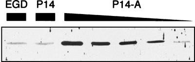
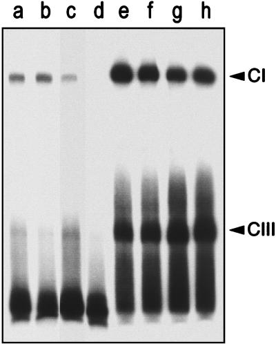
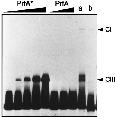
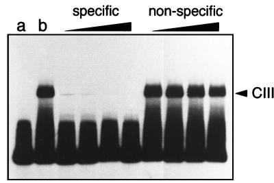
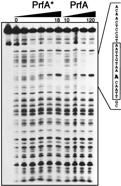
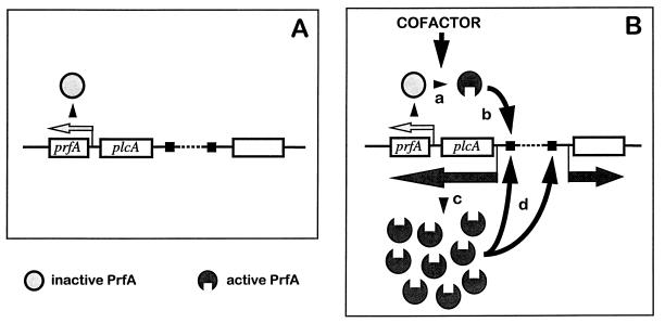
Most Listeria monocytogenes virulence genes are positively regulated by the PrfA protein, a transcription factor sharing sequence similarities with cyclic AMP (cAMP) receptor protein (CRP). Its coding gene, prfA, is regulated by PrfA itself via an autoregulatory loop mediated by the upstream PrfA-dependent plcA promoter. We have recently characterized prfA* mutants from L. monocytogenes which, as a result of a single amino acid substitution in PrfA, Gly145Ser, constitutively overexpress prfA and the genes of the PrfA virulence regulon. Here, we show that about 10 times more PrfA protein is produced in a prfA* strain than in the wild type. Thus, the phenotype of prfA* mutants is presumably due to the synthesis of a PrfA protein with higher promoter-activating activity (PrfA*), which keeps its intracellular levels constantly elevated by positive feedback. We investigated the interaction of PrfA and PrfA* (Gly145Ser) with target DNA. Gel retardation assays performed with a DNA fragment carrying the PrfA binding site of the plcA promoter demonstrated that the PrfA* mutant form is much more efficient than wild-type PrfA at forming specific DNA-protein complexes. In footprinting experiments, the two purified PrfA forms interacted with the same nucleotides at the target site, although the minimum amount required for protection was 6 to 7 times lower with PrfA*. These results show that the primary functional consequence of the Gly145Ser mutation is an increase in the affinity of PrfA for its target sequence. Interestingly, similar mutations at the equivalent position in CRP result in a transcriptionally active, CRP* mutant form which binds with high affinity to target DNA in the absence of the activating cofactor, cAMP. Our observations suggest that the structural similarities between PrfA and CRP are also functionally relevant and support a model in which the PrfA protein, like CRP, shifts from transcriptionally inactive to active conformations by interaction with a cofactor.


Figures

FIG. 1
FIG. 1
Determination of PrfA protein in L. monocytogenes P14 (wild type), P14-A (prfA* [Gly145Ser] mutant from P14), and EGD (control wild-type strain). Total cell extracts from these strains were subjected to SDS-PAGE in a 12% acrylamide gel (protein amounts loaded: P14 and EGD, 30 μg; P14-A [from left to right], 30, 15, 10, 5, and 2.5 μg) and analyzed by Western immunoblotting with an anti-PrfA hyperimmune serum. The PrfA protein is detected as a 27-kDa band. Note that equivalent amounts of PrfA protein are present in 30 μg of the P14 and EGD extracts and 2.5 μg of the P14-A extract.
FIG. 2
FIG. 2
Electrophoretic mobility shift assays with a 136-bp DNA fragment containing the PrfA-box of the plcA-hly promoter region and L. monocytogenes cell extracts. Lanes: a and b, P14 (30 and 60 μg, respectively); c, EGD (30 μg); d, ΔprfA mutant from EGD (30 μg); e to h, P14-A (30, 15, 10, and 5 μg, respectively). Lanes b and h contain equal amounts of PrfA protein (Fig. 1). CI and CIII, respectively, low- and high-mobility specific PrfA-DNA complexes. (See text and references and for details.)
FIG. 3
FIG. 3
Binding of the purified PrfA proteins to the plcA-hly promoter fragment. Various amounts of PrfA preparation were used (from left to right: PrfA* [Gly145Ser], 0.5, 1.5, 3, 6, and 18 ng; PrfA, 120, 240, and 480 ng). Lanes a and b: low-mobility CI-specific protein-DNA complex formation by purified PrfA* and PrfA proteins (50 ng each), respectively, in the presence of a PrfA-free L. monocytogenes cell extract (30 μg) from EGD ΔprfA. CIII, high-mobility specific PrfA-DNA complexes. (See text and references and for details.)
FIG. 4
FIG. 4
Specificity of the interaction of PrfA* (8 ng) with target DNA resulting in CIII complex formation. Competition assays with (from left to right) 50-, 100-, 200-, and 400-fold molar excess of specific (unlabeled 136-bp plcA-hly promoter fragment) and nonspecific DNA (from herring sperm). Lanes: a, control with the labeled 136-bp plcA-hly promoter fragment alone; b, control with the labeled probe plus 8 ng of purified PrfA*.
FIG. 5
FIG. 5
DNA footprinting experiments with various amounts of the purified PrfA proteins (PrfA*, 0, 0.5, 1.5, 3, 6, and 18 ng; PrfA, 10, 20, 40, and 120 ng) and the plcA-hly promoter fragment. The protected sequences, identical for the two PrfA proteins, are indicated on the right. The palindromic PrfA binding site is boxed, and numbers indicate the nucleotide position with respect to the transcription start site of the hly mRNA. The hypersensitive nucleotide (A) at position −40, close to the center of the palindrome, is in boldface. To the left is shown the uncleaved probe.
FIG. 6
FIG. 6
Model for PrfA-mediated gene regulation (29). Central to this model is the assumption that PrfA has two functional conformations, inactive and active, and shifts from one to the other on interaction with a hypothetical activating cofactor, the intracellular concentrations of which depend on the environmental conditions. A key element is also that prfA can be expressed in two different ways: (i) constitutively and at low levels, from monocistronic transcripts driven by promoters in the plcA-prfA intergenic region (represented by a small light-gray arrow above prfA on panels A and B); and (ii) dependent on PrfA, from bicistronic transcripts originating from the plcA promoter (dark gray arrow below plcA and prfA on panel B), thereby creating an autoregulatory circuit. The regulation mechanism would be as follows. Under normal conditions, there is no cofactor, and, thus, the PrfA protein is synthesized at low, basal levels from the monocistronic transcripts (A). However, if L. monocytogenes senses a suitable combination of activating environmental signals (a temperature of 37°C and a particular composition of the extracellular medium), the intracellular concentration of the hypothetical cofactor increases (B). This cofactor interacts with the inactive PrfA protein synthesized from monocistronic transcripts (a), causing a conformational change that results in a significant increase in the binding affinity of PrfA for its target DNA (b) (PrfA sites are indicated by black squares). The transcriptionally active PrfA causes the synthesis of more PrfA (in active conformation) by positive feedback (c), which boosts the transcription of all the PrfA-dependent genes (d) (dark gray arrows; the empty rectangle represents any PrfA-dependent gene). The PrfA regulon remains switched on as long as there are sufficiently high levels of the cofactor in the bacterial cytoplasm, but the system is rapidly switched off if the activating environmental signals cease and the concentration of the cofactor drops. A second level of regulation is provided by the differential response of the PrfA-dependent promoters according to the structure of the PrfA target site, which affects the binding affinity of PrfA. (See references , , , , and for details about this cis-acting control mechanism.) Evidence for a negative autoregulation mechanism involving a putative PrfA-binding site in the plcA-prfA intergenic region has been also presented (11, 12), which would add complexity to the transcriptional control mediated by PrfA. The proposed regulatory model is highly versatile and makes possible an immediate, fine-tuned adaptive response to rapidly changing environmental conditions, such as those encountered by the soil bacterium L. monocytogenes during its transition from free to parasitic life and within the various compartments and tissues of the infected host.

References

    1. Baichoo N, Heyduk T. Mapping conformational changes in a protein: application of a protein footprinting technique to cAMP-induced conformational changes in cAMP receptor protein. Biochemistry. 1997;36:10830–10836. - PubMed
    1. Blazy B, Ullmann A. Properties of cyclic AMP-independent catabolite gene activator proteins of Escherichia coli. J Biol Chem. 1986;261:11645–11649. - PubMed
    1. Böckmann R, Dickneite C, Middendorf B, Goebel W, Sokolovic Z. Specific binding of the Listeria monocytogenes transcriptional regulator PrfA to target sequences requires additional factor(s) and is influenced by iron. Mol Microbiol. 1996;22:643–653. - PubMed
    1. Bohne J, Kestler H, Uebele C, Sokolovic Z, Goebel W. Differential regulation of the virulence genes of Listeria monocytogenes by the transcriptional activator PrfA. Mol Microbiol. 1996;20:1189–1198. - PubMed
    1. Bohne J, Sokolovic Z, Goebel W. Transcriptional regulation of prfA and PrfA-regulated virulence genes in Listeria monocytogenes. Mol Microbiol. 1994;11:1141–1150. - PubMed

Publication types

MeSH terms