. 1998 Dec;180(24):6704-12.
doi: 10.1128/JB.180.24.6704-6712.1998.

New small, acid-soluble proteins unique to spores of Bacillus subtilis: identification of the coding genes and regulation and function of two of these genes

Affiliations

New small, acid-soluble proteins unique to spores of Bacillus subtilis: identification of the coding genes and regulation and function of two of these genes

I Bagyan et al. J Bacteriol. 1998 Dec.

Abstract

Eleven small, acid-soluble proteins (SASP) which are present in spores but not in growing cells of Bacillus subtilis were identified by sequence analysis of proteins separated by acrylamide gel electrophoresis of acid extracts from spores which lack the three major SASP (alpha, beta, and gamma). Six of these proteins are encoded by open reading frames identified previously or by analysis of the complete sequence of the B. subtilis genome, including two minor alpha/beta-type SASP (SspC and SspD) and a putative spore coat protein (CotK). Five proteins are encoded by short open reading frames that were not identified as coding regions in the analysis of the complete B. subtilis genomic sequence. Studies of the regulation of two of the latter genes, termed sspG and sspJ, showed that both are expressed only in sporulation. The sspG gene is transcribed in the mother cell compartment by RNA polymerase with the mother cell-specific sigma factor for RNA polymerase, sigmaK, and is cotranscribed with a downstream gene, yurS; sspG transcription also requires the DNA binding protein GerE. In contrast, sspJ is transcribed in the forespore compartment by RNA polymerase with the forespore-specific sigmaG and appears to give a monocistronic transcript. A mutation eliminating SspG had no effect on sporulation or spore properties, while loss of SspJ caused a slight decrease in the rate of spore outgrowth in an otherwise wild-type background.


Figures
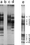
FIG. 1
FIG. 1
Low-pH PAGE analysis of minor SASP from B. subtilis. Acetic acid and HCl extracts from dormant spores and growing cells of strain PS482 were prepared and redissolved as described in Materials and Methods, aliquots were run on PAGE at low pH, either the gel was stained with Coomassie blue (lanes a to d) or proteins were transferred to polyvinylidene difluoride paper, and the paper was stained with Coomassie blue (lane e). The samples (and amounts) of redissolved extract run in the various lanes were as follows: a and e, acetic acid extract of spores (20 μl); b, acetic acid extract of growing cells (20 μl); c, HCl extract of spores (20 μl); and d, HCl extract of growing cells (5 μl). The numbers adjacent to lane e denote protein bands present in spores but not in growing cells and are the numbers of the bands analyzed in Table 2.
FIG. 2
FIG. 2
Nucleotide sequence of the sspG-yurS (A) and sspJ (B) regions. (A) The sequence shown includes nucleotides 3352955 to 3353300 in the B. subtilis genome. The sspG start and stop codons are in bold and underlined. The start codon for yurR is underlined, and that for yurS is in bold. Sequences corresponding to −10 and −35 promoter elements and the sspG and yurS ribosome binding sites (RBS) as well as those corresponding to the start of transcription of sspG-yurS (+1) are underlined. The consensus −35 and −10 sequences for ςK-dependent promoters (10, 48) are shown in bold below these elements in the sspG sequence; the abbreviation used in the −35 consensus sequence is H for A or C. The boxed residues denote putative GerE binding sites (32); one is from positions 3353061 to 3353050 on the strand transcribed to give sspG, and the other is from positions 3353013 to 3353024 on the nontranscribed strand. (B) The sequence shown includes nucleotides 3420842 to 3420491 in the B. subtilis genome (note that the direction of transcription of sspJ is counterclockwise). The sspJ start and stop codons are in bold and underlined; the start codon for yvsG is underlined. Sequences corresponding to −10 and −35 promoter elements and the sspJ ribosome binding site (RBS) as well as those corresponding to the start of transcription (+1) are underlined. The consensus −10 and −35 sequences for ςG-dependent promoters (10) are shown in bold below these elements in the sspJ sequence. The designations in the consensus sequence are H for A or C, R for A or G, and X for A or T. The two underlined G residues upstream of the −10 consensus sequence show the position of the two G residues in promoters recognized primarily by ςF (44). The apposed arrows denote the putative transcriptional terminator of sspJ.
FIG. 3
FIG. 3
Expression of the translational sspG-lacZ fusion in various spo mutants. Strains with a PY79 background (A) and a CU267 background (B) were sporulated by the resuspension method, and β-galactosidase was assayed as described in Materials and Methods. Time 0 is when sporulation was initiated. The symbols used for the various strains are as follows. (A) •, IB470 (spo+); ○, IB469 (spoIVCB), (B) •, IB492 (spo+); ■, IB494 (gerE36); ▵, IB498 (this strain does not contain an sspG-lacZ fusion but rather has a cotA-lacZ fusion in a spo+ background).
FIG. 4
FIG. 4
Expression of the translational sspJ-lacZ fusion in various spo mutants. Strains with a PY79 background were sporulated by the resuspension method, and β-galactosidase was assayed as described in Materials and Methods. Time 0 is when sporulation was initiated. The symbols used for the various strains are as follows. (A) •, IB475 (spo+); ▵, IB474 (spoIVCB), (B) □, IB471 (spoIIAC); ■, IB472 (spoIIGB); ○, IB473 (spoIIIG).
FIG. 5
FIG. 5
Induction of expression in vegetative growing cells of sspG-lacZ in cells expressing ςK (A) or sspJ-lacZ in cells expressing ςF (B) or ςG (C). Cells of strains IB502 (sspG-lacZ PspacK) (A), IB480 (sspJ-lacZ PspacF) (B), or IB481 (sspJ-lacZ PspacG) (C) were grown at 37°C in 2× YT medium. An OD600 of 0.25 (time 0 in the figure), the cultures were divided in half, one-half was made 2 mM in IPTG, incubation was continued, and samples were taken from both cultures for assay of β-galactosidase. ■, without IPTG; □, with IPTG. Note the different scales in panels B and C.
FIG. 6
FIG. 6
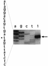
Primer extension analysis of the start site for transcription of sspG and yurS. RNA from cells of strain IB464 was isolated 7.5 h into sporulation, and the primer extension product was obtained and analyzed as described in Materials and Methods. The primer used is yurS-140, which anneals only to yurS. Lanes a, g, c, and t are DNA sequencing reactions with the same primer and plasmid pIB517; lane 1 is a primer extension reaction with sporulating-cell RNA. The primer extension product is marked with an arrow, and the transcription start site on the sspG upstream sequence to the left of the figure is marked with a square. Note that the sequence shown is the complement of the mRNA sequence.
FIG. 7
FIG. 7
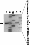
Primer extension analysis of the sspJ transcription start site. RNA from cells of strain IB465 was isolated 5 h into sporulation, and primer extension products were obtained and analyzed as described in Materials and Methods. The primer used is prot3-50, which anneals only to sspJ mRNA. Lanes a, g, c, and t are DNA sequencing reactions with the same primer and plasmid pIB460; lane 1 is the primer extension reaction with sporulating-cell RNA. The primer extension product is marked with an arrow, and the transcription start site on the sspJ upstream sequence to the right of the figure is marked with a square. Note that the sequence shown is the complement of the mRNA sequence.

Similar articles

Cited by

References

    1. Anagnostopoulos C, Spizizen J. Requirements for transformation in Bacillus subtilis. J Bacteriol. 1961;81:741–748. - PMC - PubMed
    1. Bagyan I, Casillas-Martinez L, Setlow P. The katX gene, which codes for the catalase in spores of Bacillus subtilis, is a forespore-specific gene controlled by ςF, and KatX is essential for hydrogen peroxide resistance of the germinating spore. J Bacteriol. 1998;180:2057–2062. - PMC - PubMed
    1. Bagyan I, Noback M, Bron S, Paidhungat M, Setlow P. Characterization of yhcN, a new forespore-specific gene of Bacillus subtilis. Gene. 1998;212:179–188. - PubMed
    1. Cabrera-Hernandez, A., J.-L. Sanchez-Salas, M. Paidhungat, and P. Setlow. 1998. Unpublished results. - PubMed
    1. Carillo-Martinez Y, Setlow P. Properties of Bacillus subtilis small, acid-soluble spore proteins with changes in the sequence recognized by their specific protease. J Bacteriol. 1994;176:5357–5363. - PMC - PubMed

Publication types

LinkOut - more resources